MeSH: pectus excavatum; Brugada phenocopie
Introduction
Brugada phenocopies (BrP) are new clinical entities characterized by an ECG pattern that is identical to type 1 or type 2 Brugada pattern, despite the absence of the true congenital Brugada syndrome (BrS).1 BrP are caused by various factors such as mechanical mediastinal compression, myocardial ischemia, pulmonary embolism, pericardial diseases and metabolic conditions.2 However, only few cases have been reported of patients with pectus excavatum and BrP. They have an ECG showing right bundle branch block, but also mild ST-segment elevation in the right precordial leads, mimicking the ECG patterns of type 2 Brugada syndrome.3
Case
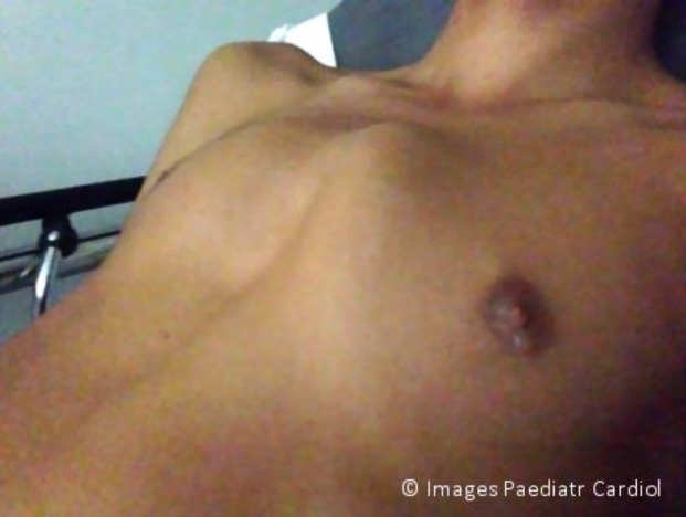
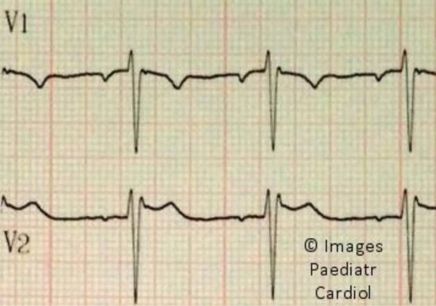
A 13-year old adolescent with pectus excavatum of the thorax (Figure 1) was admitted to the emergency department with an electrocardiogram (ECG) on admission showing a Brugada type 2 pattern (Figure 2). The patient had no personal or familial history of Brugada syndrome, sudden cardiac death, cardiac arrest, or non-vagal syncope. Moreover, he was known to paediatricians for a history of tachycardia and had several ECGs before his adolescence, without ST-segment modification.
Figure 1.
Pectus excavatum
Figure 2.
Brugada type 2 ECG pattern
Discussion
This case illustrates with typical images a rare association of symptoms. We speculate that the Brugada-type ECG observed in our patient was probably caused by long-term mechanical injury to the right ventricular free wall, as a result of chronic compression by the pectus excavatum (as it appeared only after his adolescence).4 Our patient was discharged from the emergency department without any evidence of true Brugada syndrome and a normal 24h Holter ECG recording. No additional investigation was done.
References
- 1. Baranchuk A, Nguyen T, Ryu MH, Femenía F, Zareba W, Wilde AA, et al. Brugada phenocopy: new terminology and proposed classification. Ann Noninvasive Electrocardiol. 2012;17:299-314. [DOI] [PMC free article] [PubMed] [Google Scholar]
- 2. Anselm DD, Baranchuk A. Brugada Phenocopy: redefinition and updated classification. Am J Cardiol. 2013;111:453-456. [DOI] [PubMed] [Google Scholar]
- 3. Kataoka H. Electrocardiographic patterns of the Brugada syndrome in 2 young patients with pectus excavatum. J Electrocardiol. 2002;35:169-171. [DOI] [PubMed] [Google Scholar]
- 4. Cartoski MJ, Nuss D, Goretsky MJ, Proud VK, Croitoru DP, Gustin T, et al. Classification of the dysmorphology of pectus excavatum. J Pediatr Surg. 2006;9:1573-1581. [DOI] [PubMed] [Google Scholar]


