Abstract
Fluorodeoxyglucose (FDG) positron emission tomography-computed tomography (PET-CT) scan is fast becoming a very useful tool in diagnosing and staging of several malignancies that affect the human body. We report three cases of ocular choroidal malignant melanoma, wherein FDG PET-CT scan did not show as good uptake as seen in other cancers.
Keywords: Choroidal melanoma, fluorodeoxyglucose, positron emission tomography-computed tomography
Introduction
Uveal malignant melanoma is the most common primary intraocular malignant tumor in Caucasians though it is relatively rare in Asian Indian eyes.[1] It can affect any part of the uveal tract, but posterior choroidal melanoma are most common (86.3%), followed by iris and ciliary body melanomas.[2] In the last few years, fluorodeoxyglucose (FDG) positron emission tomography- computed tomography (PET-CT) scan has emerged as a new imaging modality for the detection and staging of cancer affecting any organ of the body.[3] It has been also tested in choroidal melanoma with equivocal results. We report three cases of choroidal malignant melanoma in Asian Indian eyes, where FDG PET-CT scan was not very reliable in giving a clear diagnosis.
Case Reports
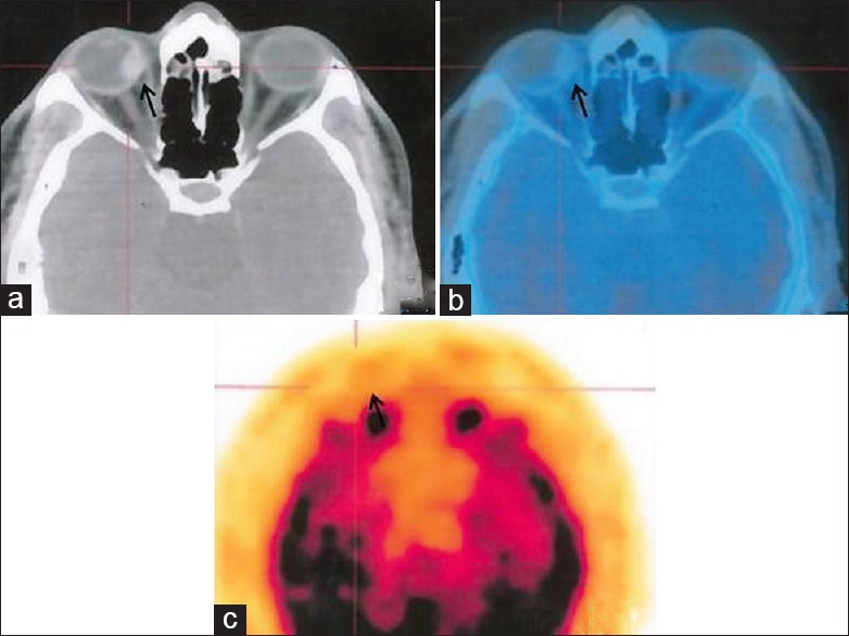
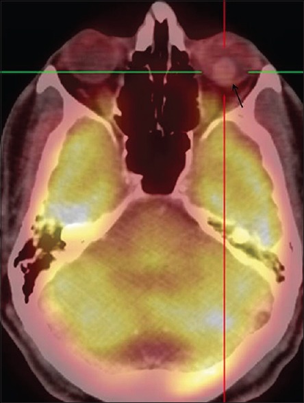
Three eyes of three patients, who presented with a pigmented intraocular mass lesion, were included in the study. All patients were males with a mean age of 54 years. Clinically, all eyes had a macula sparing choroidal mass lesion with a mean largest basal diameter of 14.1 mm and mean apical diameter of 9.4 mm. Complete ocular examination was done including best-corrected visual acuity assessment, slit lamp examination, indirect ophthalmoscopy and B-scan ultrasonography before doing the FDG PET-CT scan. The baseline characteristics of the cases are given in Table 1. All the lesions were classified according to Collaborative Ocular Melanoma Study group classification.[4] Whole body PET scan for all three cases showed a low to moderately activity with minimal to moderate FDG uptake for the intraocular tumors [Figures 1 and 2]. The mean standardized uptake value (SUV) was 2.3 (range 1.3–3.4). None had any systemic metastasis.
Table 1.
Baseline characteristics of the cases with PET scan values

Figure 1.

(a) Transaxial computed tomography scan image of case 3 showing the intraocular mass in right eye (black arrow). (b) Fused transaxial image and (c) positron emission tomography transaxial images showing poor fluorodeoxyglucose uptake with maximum standardized uptake value 1.3
Figure 2.

Fused positron emission tomography-computed tomography image of case 1 showing moderate fluorodeoxyglucose uptake in the intraocular mass in left eye (black arrow) with maximum standardized uptake value 3.4
Treatment given was enucleation for case 1 while other two cases underwent ophthalmic brachytherapy using I-125 seeds with good regression. Histopathological examination of the enucleated eye showed mixed cell type of choroidal melanoma. The mean followup was 14 months (range 12–18) and none developed any systemic metastasis.
Discussion
Like many intraocular tumors, diagnosis of choroidal melanoma can be made clinically with an accuracy of 99.5%[4] and seldom biopsy is required for confirmation. FDG PET-CT scan is fast becoming the standard of care for diagnosis and staging any form of malignancy.[3] In choroidal melanomas, though it has shown to be quite accurate in picking distant metastasis,[5] it is not very accurate in diagnosing the primary intraocular mass especially the small and medium sized ones.[6] In our series one tumor was medium sized while the other two were large tumors and none showed good FDG uptake. In another study the authors concluded that FDG PET-CT scan is a good tool in diagnosing nodular melanomas but not the diffuse infiltrating types.[7] In our series, all three were nodular, and all showed poor activity on PET-CT scan. Some authors[8] have kept SUV ≥ 4 as a risk factor for metastasis for choroidal melanomas while others have kept the cut off as 2.5.[9] However none is universally accepted. The mean SUV for our study was 2.3 and none developed any systemic metastasis till the mean final follow-up of 14 months. Loss of chromosome 3 in the tumor has been associated with high risk of metastasis and positive FDG uptake.[10] Lack of chromosome 3 analysis and small sample size are the drawbacks of our study. In summary, although FDG PET-CT is fast becoming the gold standard for diagnosis and staging any form of malignancy, caution is advised in interpreting the results of a low uptake of FDG in setting of clinically suspected intraocular choroidal melanomas, especially in Asian Indian eyes.
Footnotes
Source of Support: Nil
Conflict of Interest: None declared.
References
- 1.Shah PK, Selvaraj U, Narendran V, Guhan P, Saxena SK, Dash A. Indigenous (125) I brachytherapy source for the management of intraocular melanomas in India. Cancer Biother Radiopharm. 2013;28:21–8. doi: 10.1089/cbr.2011.1123. [DOI] [PubMed] [Google Scholar]
- 2.Jovanovic P, Mihajlovic M, Djordjevic-Jocic J, Vlajkovic S, Cekic S, Stefanovic V. Ocular melanoma: An overview of the current status. Int J Clin Exp Pathol. 2013;6:1230–44. [PMC free article] [PubMed] [Google Scholar]
- 3.Gallamini A, Zwarthoed C, Borra A. Positron Emission Tomography (PET) in oncology. Cancers (Basel) 2014;6:1821–89. doi: 10.3390/cancers6041821. [DOI] [PMC free article] [PubMed] [Google Scholar]
- 4.Accuracy of diagnosis of choroidal melanomas in the Collaborative Ocular Melanoma Study. COMS report no 1. Arch Ophthalmol. 1990;108:1268–73. doi: 10.1001/archopht.1990.01070110084030. [DOI] [PubMed] [Google Scholar]
- 5.Finger PT, Kurli M, Reddy S, Tena LB, Pavlick AC. Whole body PET/CT for initial staging of choroidal melanoma. Br J Ophthalmol. 2005;89:1270–4. doi: 10.1136/bjo.2005.069823. [DOI] [PMC free article] [PubMed] [Google Scholar]
- 6.Reddy S, Kurli M, Tena LB, Finger PT. PET/CT imaging: Detection of choroidal melanoma. Br J Ophthalmol. 2005;89:1265–9. doi: 10.1136/bjo.2005.066399. [DOI] [PMC free article] [PubMed] [Google Scholar]
- 7.Matsuo T, Ogino Y, Ichimura K, Tanaka T, Kaji M. Clinicopathological correlation for the role of fluorodeoxyglucose positron emission tomography computed tomography in detection of choroidal malignant melanoma. Int J Clin Oncol. 2014;19:230–9. doi: 10.1007/s10147-013-0538-5. [DOI] [PubMed] [Google Scholar]
- 8.Finger PT, Chin K. Iacob CE 18-Fluorine-labelled 2-deoxy-2-fluoro-D-glucose positron emission tomography/computed tomography standardised uptake values: A non-invasive biomarker for the risk of metastasis from choroidal melanoma. Br J Ophthalmol. 2006;90:1263–6. doi: 10.1136/bjo.2006.097949. [DOI] [PMC free article] [PubMed] [Google Scholar]
- 9.Lee CS, Cho A, Lee KS, Lee SC. Association of high metabolic activity measured by positron emission tomography imaging with poor prognosis of choroidal melanoma. Br J Ophthalmol. 2011;95:1588–91. doi: 10.1136/bjo.2010.198085. [DOI] [PubMed] [Google Scholar]
- 10.McCannel TA, Reddy S, Burgess BL, Auerbach M. Association of positive dual-modality positron emission tomography/computed tomography imaging of primary choroidal melanoma with chromosome 3 loss and tumor size. Retina. 2010;30:146–51. doi: 10.1097/IAE.0b013e3181b32f36. [DOI] [PubMed] [Google Scholar]


