Abstract
Major vascular invasion is one of the worst prognostic factors of hepatocellular carcinoma (HCC). Fludeoxyglucose F 18 (18 F-FDG) positron emission tomography/computed tomography (PET/CT) method is succesfully being used in HCC patients for the detection of particularly long-distance metastasis. Major vascular invasion is shown by radiological methods [particularly dynamic CT and/or magnetic resonance imaging (MRI)]. A male patient aged 60 years was diagnosed with HCC, according to biopsy after the detection of a mass in the liver. His medical examinations that were performed for the evaluation in terms of liver transplantation were dynamic CT and dynamic MRI; invasion in the intrahepatic branches of the portal vein and in main portal vein was also detected. PET/CT was performed to investigate the distant metastases. Moreover, diffuse 18 F-FDG uptake in the intrahepatic branches of the portal vein and in the main portal vein was observed.
Keywords: Diffuse fludeoxyglucose F 18 (18 F-FDG) uptake, hepatocellular carcinoma (HCC), intrahepatic branches of the portal vein
Introduction
Hepatocellular carcinoma (HCC) ranks sixth in the incidences of cancer worldwide. Of all cancers, HCC is the third in cancer mortality globally.[1]
Ultrasonography (USG) is the suggested sacanning modality for periodic surveillance of HCC. Dynamic and multiphase contrast-enhanced computed tomography (CT) and magnetic resonance imaging (MRI) are the standard diagnostic methods for HCC. Fludeoxyglucose F 18 (18F-FDG) positron emission tomography/computed tomography (PET/CT) method is succesfully being used in HCC patients for detecting particularly long-distance metastasis.[2] In the literature, we could not find any study that utilized 18F-FDG PET/CT for detecting particularly intrahepatic vascular invasion. This paper is the first case presentation that shows increased diffuse 18F-FDG uptake in the right portal vein and its intrahepatic branches.
Case Report
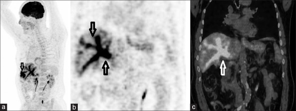
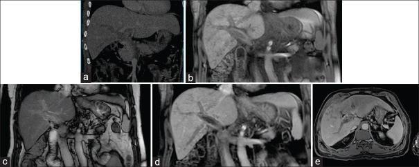
A male patient age of 60, which is diagnosed as having hepatocellular carcinoma according to biopsy after detected a mass in the liver. His medical examinations which was performed for evaluation in terms of liver transplantation are. PET/CT was performed to investigate the distant metastases. And in intrahepatic branches of the portal vein and in main portal vein diffuse 18F-FDG uptake was observed [Figure 1]. 18F-FDG PET/CT was performed 1 hour after intravenous injection of 370 MBq 18F-FDG following 8-hour fasting with a blood glucose of 101 mg/dL. Histopathological examination of the biopsy from removed lesion confirmed diagnosis of hepatocellular carcinoma. Vascular invasion were visualized with dynamic CT and dynamic MR [Figure 2].
Figure 1.
18F-FDG PET [maximum intensity projection (MIP)] images showed focal increased uptake in the lesion (SUVmax: 29) located in the right liver lobe as continuation of this lesion (a) Coronal PET images of distal branches of the right portal vein, the main trunk of the right portal vein, the main trunk of the left portal vein, and the main portal vein showing diffuse 18F-FDG (SUVmax: 18.9) uptake (b and c)
Figure 2.
An enhanced coronal CT image (hepatic venous phase) (a) coronal unenhanced T1 and T2 weighted image (b and c) Coronal enhanced T1 weighted image (equilubrium phase) (d) and axial enhanced T1 weighted images (equilubrium phase) reveal heterogeneously enhanced lesion fulfilling the portal vein and increasing in caliber of portal veins (e)
Discussion
The general approach for the diagnosis of HCC includes the use of contrast USG, high-resolution contrast CT, and MRI. This method allows better detection of HCC.[3] 18F-FDG PET can be benefical in the detection of extrahepatic metastases that are not seen in CT or MRI. However, it has a low sensitivity for small and/or well-differentiated HCCs located within the liver, secondary to the high background liver uptake of FDG.[4] PET images were not observed in 30-50% of the HCC lesions.[5]
One of the most significant criteria in terms of negative prognosis of HCC is major vascular invasion. Invasion into the hepatic vein or portal vein is a risk factor for systemic metastasis and it reduces the survival rate of patients.[6] In HCC, while microvascular invasion can be solely detected by histological methods, macrovascular invasion can be detected by gross medical inspection, CT, or MRI.[7,8]
However, in recent papers, it has been stated that PET can be used to predict microvascular invasions in patients with HCC and with 18F-FDG uptake.[9] It should be taken into consideration that the 18F-FDG uptake should be kept in mind that it might belong in the vascular structure.
Footnotes
Source of Support: There has been no support, either financial or of any other kind.
Conflict of Interest: The authors have no conflict of interest to declare.
References
- 1.Ferlay J, Shin HR, Bray F, Forman D, Mathers C, Parkin DM. Estimates of worldwide burden of cancer in 2008: GLOBOCAN 2008. Int J Cancer. 2010;127:2893–917. doi: 10.1002/ijc.25516. [DOI] [PubMed] [Google Scholar]
- 2.Kawaoka T, Aikata H, Takaki S, Uka K, Azakami T, Saneto H, et al. FDG positron emission tomography/computed tomography for the detection of extrahepatic metastases from hepatocellular carcinoma. Hepatol Res. 2009;39:134–42. doi: 10.1111/j.1872-034X.2008.00416.x. [DOI] [PubMed] [Google Scholar]
- 3.Rode A, Bancel B, Douek P, Chevallier M, Vilgrain V, Picaud G, et al. Small nodule detection in cirrhotic livers: Evaluation with US, spiral CT, and MRI and correlation with pathologic examination of explanted liver. J Comput Assist Tomogr. 2001;25:327–36. doi: 10.1097/00004728-200105000-00001. [DOI] [PubMed] [Google Scholar]
- 4.Ho CL, Chen S, Yeung DW, Cheng TK. Dual-tracer PET/CT imaging in evaluation of metastatic hepatocellular carcinoma. J Nucl Med. 2007;48:902–9. doi: 10.2967/jnumed.106.036673. [DOI] [PubMed] [Google Scholar]
- 5.Sørensen M, Frisch K, Bender D, Keiding S. The potential use of 2-[18 F] fluoro-2-deoxy-D-galactosis as a PET/CT tracer for detection of hepatocellular carcinoma. Eur J Nucl Med Mol Imaging. 2011;38:1723–31. doi: 10.1007/s00259-011-1831-z. [DOI] [PMC free article] [PubMed] [Google Scholar]
- 6.Llovet JM, Brú C, Bruix J. Prognosis of hepatocellular carcinoma: The BCLC staging classification. Semin Liver Dis. 1999;19:329–38. doi: 10.1055/s-2007-1007122. [DOI] [PubMed] [Google Scholar]
- 7.Sumie S, Kuromatsu R, Okuda K, Ando E, Takata A, Fukushima N, et al. Microvascular invasion in patients with hepatocellular carcinoma and its predictable clinicopathological factors. Ann Surg Oncol. 2008;15:1375–82. doi: 10.1245/s10434-008-9846-9. [DOI] [PubMed] [Google Scholar]
- 8.Chu KK, Chan SC, Fan ST, Chok KS, Cheung TT, Sharr WW, et al. Radiological prognosticators of hepatocellular carcinoma treated by hepatectomy. Hepatobiliary Pancreat Dis Int. 2012;11:612–7. doi: 10.1016/s1499-3872(12)60232-x. [DOI] [PubMed] [Google Scholar]
- 9.Cheung TT, Chan SC, Ho CL, Chok KS, Chan AC, Sharr WW, et al. Can positron emission tomography with the dual tracers [11C] acetate and [18F] fluorodeoxyglucose predict microvascular ınvasion in hepatocellular carcinoma? Liver Transpl. 2011;17:1218–25. doi: 10.1002/lt.22362. [DOI] [PubMed] [Google Scholar]


