Fig 1.
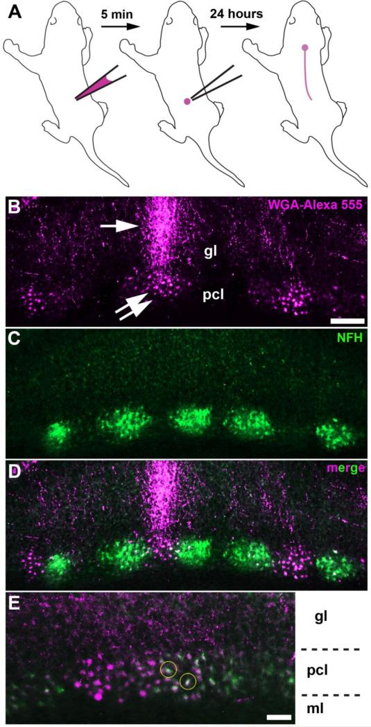
a Schematic illustrating the injection of WGA-Alexa 555 into the spinal cord of a pup. b WGA-Alexa 555 tracing of spinocerebellar mossy fibers (arrow) and transynaptic tracing of neuronal somata (double arrow) in the anterior cerebellum. c, d NFH staining revealed a pattern of zones that was complementary to a subset of traced Purkinje cells. e, Some of the WGA-Alexa 555 traced neuronal somata were co-labeled with NFH, a marker for Purkinje cells (yellow circles). The layers of the cerebellar cortex are indicated by ml (molecular layer), pcl (Purkinje cell layer), and gl (granular layer). The scale bar = 100 μm in B (applies to C, D) and 50 μm in E.

