Neural Regeneration Research logoLink to Neural Regeneration Research
. 2015 Dec;10(12):1953–1975. doi: 10.4103/1673-5374.172314

Repair, protection and regeneration of spinal cord injury

PMCID: PMC4730820  PMID: 26889184
Neural Regen Res. 2015 Dec;10(12):1953–1975.

Reading guide


1954 Nanodrug-coated three-dimensional microelectronic stent used in axon regeneration after spinal cord injury

1955 Macrophage polarization and spinal cord injury repair

1957 Inhibiting the RhoA signaling pathway promotes regeneration of axons and myelin sheath

1958 Microsurgical lysis of spinal cord nerve roots for the treatment of spinal cord injury

1959 Local manipulation of the growth cone cytoskeleton: new strategies to promote axon regeneration

1961 Interaction of miR-21 and TGF-β/SMADs signaling pathway affects the formation of fibrotic scars after spinal cord injury

1962 Elucidating the molecular mechanisms of CSPG-mediated axon growth inhibition through phosphoproteomics analysis

1963 The repair of spinal cord injury with multifunctional, three-dimensional, electroactive scaffolds

1964 A strategy for treating spinal cord injury: targeting the variation of immune cells in the local microenvironment

1965 Epidemiological data is critical for decreasing the incidence of spinal cord injury

1966 The importance of olfactory ensheathing cells for spinal cord injury

1967 Optogenetics: a new method to repair urination dysfunction after spinal cord injury?

1968 Effects of zinc on the recovery of neurological function after spinal cord injury

1969 Durotomy and dural grafting to treat lower cervical spine injuries with extensive spinal cord edema

1971 Reconstruction of artificial micturition reflex arc for neurogenic bladder after spinal nerve injury

1972 Old drugs, new tricks: the strategy for new drug development in spinal cord injury

First authors and corresponding authors:

Xiao-jian Cao* Department of Spine Surgery, First Affiliated Hospital of Nanjing Medical University

Shi-qing Feng* Department of Orthopedics, Tianjin Medical University General Hospital

Chang-feng Fu* Department of Spine Surgery, First Hospital of Jilin University

Kai Gao# Department of Orthopedics, First Affiliated Hospital of Liaoning Medical University

Jia-song Guo* Department of Histology and Embryology, Southern Medical University; Key Laboratory of Tissue Construction and Detection of Guangdong Province; Institute of Bone Biology, Academy of Orthopedics of Guangdong Province

Xiao-dong Guo* Department of Orthopedics, Union Hospital, Tongji Medical College, Huazhong University of Science and Technology

Xi-jing He* Department of Spine Surgery, The Second Hospital of Xi’an Jiaotong University

Zi-wei Huang# Department of Orthopedics, the First Affiliated Hospital, Orthopedic Institute, Soochow University

Zhong-hai Li#, * Department of Orthopedics, the First Affiliated Hospital of the PLA General Hospital, Orthopedics Institute of PLA

Ling Liu# Institute of Neuroscience, the Fourth Military Medical University of Chinese PLA

Rong-han Liu# Department of Spine Surgery, Jinan Central Hospital Affiliated to Shandong University

He-Zuo Lü#, * Anhui Key Laboratory of Tissue Transplantation, Bengbu Medical College, Bengbu, Anhui Province, China

Xi-fan Mei* Department of Orthopedics, First Affiliated Hospital of Liaoning Medical University

Bin Ning* Department of Spine Surgery, Jinan Central Hospital Affiliated to Shandong University

Guang-zhi Ning# Department of Orthopedics, Tianjin Medical University General Hospital

Chang-hui Qian# Department of Histology and Embryology, Southern Medical University

Jie Qin# Department of Spine Surgery, The Second Hospital of Xi’an Jiaotong University

Yan-zhen Qu# Department of Orthopedics, Union Hospital, Tongji Medical College, Huazhong University of Science and Technology

Saijilafu* Department of Orthopedics, the First Affiliated Hospital, Orthopedic Institute, Soochow University

Bo Shi# Department of Spine Surgery, First Hospital of Jilin University

Tao Sui# Department of Spine Surgery, First Affiliated Hospital, Nanjing Medical University

Tian-sheng Sun* Institute of Trauma Orthopedic Surgery of Chinese PLA, General Hospital of Beijing Military Region

Jian Wang* Institute of Neuroscience, the Fourth Military Medical University

Jin-kun Wen# Department of Histology and Embryology, Southern Medical University

Jian Xiao* Molecular Pharmacology Research Center, School of Pharmacy, Wenzhou Medical University

Bin Xu* Department of Orthopedics, Jinling Hospital, Nanjing University (Nanjing General Hospital of Nanjing Military Command)

Hai-dong Xu# Department of Orthopedics, Jinling Hospital, Nanjing University (Nanjing General Hospital of Nanjing Military Command)

Pan-pan Yu#, * Guangdong-Hongkong-Macau Institute of CNS Regeneration (GHMICR), Jinan University

Zhi-cheng Zhang# Institute of Trauma Orthopedic Surgery of Chinese PLA, General Hospital of Beijing Military Region

Yong Zhou# Department of Orthopedics, Tianjin Medical University General Hospital

Yu-long Zhou# Department of Orthopedics, the Second Affiliated Hospital, Wenzhou Medical University; Molecular Pharmacology Research Center, School of Pharmacy, Wenzhou Medical University

Note: Arranged vertically in alphabetical order by their last name; #First author; *Corresponding author.

Neural Regen Res. 2015 Dec;10(12):1953–1975.

Nanodrug-coated three-dimensional microelectronic stent used in axon regeneration after spinal cord injury

Hai-dong Xu 1, Bin Xu 1,*

Nerve repair after spinal cord injury (SCI) requires orderly growth, migration, and transmission of neuronal signals in the regenerated nerve fibers at the injury site. Neuronal apoptosis occurs at the site of injury, so a regional “barrier” forms, and the nerve fibers cannot be reconstructed in an orderly manner. The biggest obstacle in the repair of SCI is the formation of a glial scar, and the subsequent disorderly axon regeneration. Here, we report our recent construction of a tissue-engineered, nano-coated, three-dimensional repair scaffold, designed to improve the nerve fiber stretching of axons during nerve regeneration.

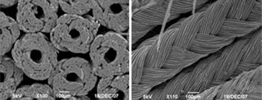
The stent material was polyglycolide-lactide (9:1 polyglycolide:polylactide ratio), prepared at the Laboratory of Textile Materials, Donghua University, Shanghai, China. The polyglycolide-lactide was melted at 250°C, drawn using a Textile weaving machine, and woven into a microtubule (100 µm outer diameter) using a knitting machine. The microtubule was coated with 3.5% chitosan. Fifty microtubules were then woven into a stent (3 mm outer diameter), which was coated again with chitosan and sterilized using ethylene oxide. Multiple sets of stents were woven, Drug coated, vacuum packed, and stored at −4°C until use. The three-dimensional structure of the stent was observed using a scanning electron microscope (Figure 1) to ensure the diameter of the tube met the criteria.

Figure 1.

Figure 1

Scanning electron micrograph of repair materials used in three-dimensional stents.

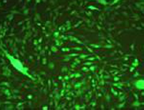
The stents were then subjected to tissue engineering and bioelectric function processing. Tissue engineering was carried out using Schwann cells infected with a lentivirus carrying neurotrophin-3 (Figure 2), which were implanted into prepared three-dimensional stents. Neurotrophic factors promote neuronal survival and growth in mammals with central nervous system damage. Then the brain-derived neurotrophic factor-transfected fibroblasts were transplanted into the injury site, which contributed to nerve regeneration in the rubrospinal tract. Furthermore, neurotrophin-3 and glial cell line-derived neurotrophic factor might promote axon growth in models of dorsal root ganglion injury, and neurotrophin-3 has a strong and specific effect on axon growth in sensory neurons.

Figure 2.

Figure 2

Schwann cells transfected with a lentivirus carrying the neurotrophin-3 gene (immunofluorescence staining).

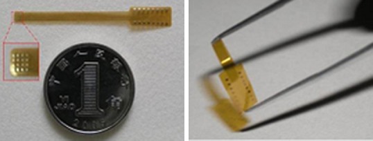
Microelectronic and bioelectric functional processing was carried out at the Shanghai Institute of Microsystem and Information Technology, Chinese Academy of Sciences, Shanghai, China. We prepared a flexible 4 × 4 microelectrode array, which provided a good local bioelectric effect, and used microelectronic processing to make a sieve-like bioelectrode chip (Figure 3) with three parts: a polyimide-substrate electrode; a current pulse generator comprising a power supply, pulse signal generator and output power amplifier; and a wireless magnetic induction system to supply energy. The system emitted continual pulses of current.

Figure 3.

Figure 3

Prepared microelectrode chip.

The chip comprised a biological scaffold, microelectrode, and Schwann cells transfected with a neurotrophin-3-carrying lentivirus.

We constructed rat models of isometric sciatic nerve defects, into which we implanted the three-dimensional tissue-engineered stents. Using light and electron microscopy, we studied the morphological characteristics in the junction between the nerve stump and new material (Figure 4), neuronal survival at the proximal and distal ends of the defect, and survival of the motor end plate of the distal skeletal muscle. We also calculated the numbers and proportion of myelinated and unmyelinated nerve fibers. Electrophysiology was used to quantify behavioral recovery in the hindlimb.

Figure 4.

Figure 4

Morphological characteristics in the junction between the sciatic nerve stump and new material.

Images from light microscope (A1, A2) and electron microscope (B1, B2).

Our previous studies and others’ indicate that the most important obstacle to the repair and reconstruction of a nerve after injury is the glial scar. Glial scars are principally composed of reactive astrocytes, microglia, macrophages, and extracellular matrix. The extracellular matrix mainly comprises chondroitin sulfate proteoglycans and myelin proteins. However, despite successful construction of a tissue-engineered stent and simulation of a microenvironment of bioelectric stimulation using microelectrodes, functional recovery was not satisfactory. Furthermore, the electromyogram indicated that the extent and range of nerve fiber regeneration did not meet the expected satisfactory results of nerve repair.

Our group's research focus now is the prevention of glial scar formation by blocking extracellular or intracellular inhibitory signaling pathways. The current research focuses on three directions targeting different exogenous inhibitory signals. I: Chondroitin sulfate proteoglycans, which elevate neuron-specific protein expression after SCI and promote axon regeneration. Chondroitin sulfate and its enzymatic hydrolysates are thought to play important roles in nerve regeneration and repair. II: Extracellular matrix and myelin protein. Blocking the neuronal receptor Nogo-66 improves nerve repair, and Nogo-66 receptor knockout animals show enhanced synaptic plasticity and good functional recovery after injury. III: Astrocytes and extrinsic signaling pathways. For example, when astrocytes are transfected with herpes simplex virus, selective elimination of the virus using ganciclovir and inhibiting the growth of astrocytes can increase the speed of axon growth. Knockout of the STAT gene, which mediates the glial reaction, causes an inflammatory reaction and expands the range of injury; therefore, astrocytes can be considered to improve repair during the acute stage of injury. However, once the glial scar is formed, axon regeneration is blocked. Control of the timing of astrocyte activity might improve nerve regeneration and repair. Endogenous regeneration can be promoted by neurotrophic factors and signaling pathways. Our ongoing investigations include the role of endogenous signaling pathways, such as cAMP, Rho/ROCK, PTEN/mTOR and MAPK, in nerve regeneration.

In summary, we report the use of a three-dimensional stent and microelectrode chip to simulate electrical stimulation and neurotrophic factors. The regenerative capacity of the central nervous system following SCI is limited, so it is necessary to combine the use of a number of methods to improve nerve regeneration and repair. Calcium channels have a notably inhibitory effect on matrix protein synthesis and secretion in the scar. The calcium antagonist verapamil was added to nanospheres and used to coat three-dimensional stents. In future, we will investigate the effectiveness of verapamil in nerve repair. Applying calcium antagonists in drug-coated stents is one of simple and feasible solutions of the above methods. Pictures have shown that animal experiments have good repair.

Neural Regen Res. 2015 Dec;10(12):1953–1975.

Macrophage polarization and spinal cord injury repair

Ling Liu 1, Kun Zhang 1, Jian Wang 1,*

In addition to the poor regenerative ability of neurons, the local microenvironment is not conducive to the regeneration of injured nerves following spinal cord injury (SCI). Neuroinflammation is the most important pathological process affecting the local microenvironment, and infiltrating peripheral macrophages and spinal microglia (macrophage/microglia) are the major effector cells (Burda et al., 2014; Gensel et al., 2015). The polarization state of these cells determines their fate as either neuroprotective or neurotoxic, as well as their structural reconstruction and functional recovery after SCI (Murray et al., 2014; Gensel et al., 2015; Hu et al., 2015). Current clinical studies have shown very promising results that may provide a new strategy and method for the treatment of SCI (Shechter et al., 2013). In this short review, we just give our opinions about the polarization of macrophages in spinal cord injury repairing.

Macrophages are widely distributed in the tissue and circulation, and they are the resident macrophage of the nerve tissue. Microglia account for 5–20% of cells in the central nervous system (CNS) of rodents (Casano et al., 2015). These cells share the same origin as macrophages; they are derived from erythro-myeloid progenitors from the embryonic yolk sac between embryonic day 9.0 and 9.5. Microglia expresses numerous macrophage-associated molecular markers, such as cluster of differentiation (CD) molecule 11b, CD14, and F4/80. Therefore, in the CNS, microglia are generally considered to be the resident macrophage where they can maintain CNS homeostasis and are thus regarded as the most important innate immune cell (Casano et al., 2015; Hu et al., 2015).

SCI initiates a chronic inflammation that involves the activation of spinal microglia and the infiltration of neutrophils, monocytes/macrophages, and lymphocytes into the CNS. Following SCI, activated microglia and infiltrating macrophages cannot be distinguished based on their morphology or marked antigen molecules, and thus form a specific macrophage/microglial population in the CNS (Gensel et al., 2015; Casano et al., 2015). Under physiological conditions in the CNS, microglia exhibit a resting state and maintain homeostasis. Under these conditions, the microglial surface expresses low levels of CD45, CD11b+, F4/80+, and major histocompatibility complex (MHC) class II. However, after SCI, the blood vessels and blood-brain barrier are damaged and cells undergo necrosis. Macrophages/microglia are activated by pattern-recognition receptors, such as Toll-like receptors, and these receptors play a role in inflammatory signaling following SCI (Gensel et al., 2015; Hu et al., 2015). Resting microglia can be distinguished from activated macrophages/microglia by their low expression of CD45; however, fully activated cells exhibit a high expression of CD45 (CD45high). Furthermore, these cells have processes that are retracted and thus, their morphology resembles that of amebae. Overall, macrophages/microglia express the following surface markers after SCI: CD45high, CD11b+, F4/80+ and MHC-IIhigh (Gensel et al., 2015; Hu et al., 2015).

Macrophages can exhibit different functional phenotypes according to the cytokines in their microenvironment (Novak et al., 2013; Gensel et al., 2015; Hu et al., 2015). They can polarize into two kinds of subpopulations: classically activated macrophages (M1) and alternatively activated macrophages (M2). Classical activation is the activation of macrophages by gamma interferon and lipopolysaccharide. M1 macrophages produce high levels of oxidative metabolites, such as nitric oxide and superoxide, and generate proinflammatory mediators. The most typical feature of M1 macrophages is their ability to secrete high levels of interleukin (IL) 12 and the production of low levels of IL-10. M1 can also secrete IL-1β, IL-15, IL-18, tumor necrosis factor (TNF)-α, and the chemotactic factors CCL15 and CCL20. Moreover, M1 cells highly express MHC-II and the costimulatory molecules CD80 and CD86. Overall, M1 cells are important for antigen presentation. M1 acts as a defense in the body by eliminating invasive pathogenic microorganisms and tumor cells, but it also causes injury to normal cells and tissue. Alternative activation is the activation of macrophages by IL-4 and IL-13. In contrast to M1, the most typical feature of M2 macrophages is their secretion of high levels of both IL-10 and tumor growth factor (TGF)-β and the production of low levels of IL-12. In addition, further research has shown that M2 cells can further differentiate into subtypes—M2a, M2b, or M2c. Under the stimulation of IL-4, IL-13, IL-1, or Toll-like receptor, macrophages can polarize into M2a or M2b and exert immunomodulatory effects. In the presence of IL-10, macrophages can differentiate into M2c and affect immunosuppression and tissue remodeling (Murray et al., 2014; Gensel et al., 2015).

SCI causes injury to the blood-brain barrier and the infiltration of microglia, neutrophils, monocytes/macrophages, natural killer cells, and lymphocytes. Neutrophil infiltration is the earliest event, occurring 3–24 hours at the injury site following SCI. Monocytes/macrophages infiltrate the injury site 2–3 days following injury, peaking at 1–2 weeks. In rats, guinea pigs, and cats, activated macrophages/microglia are visible at the injury site within 6 months after SCI. The number of infiltrating neutrophils is less than that of monocytes, macrophages/microglia. However, in mice and rats, infiltrating neutrophils can still be observed at the injury site 6 weeks and 6 months after SCI, respectively. Taken together, inflammatory signaling occurs as a result of SCI, with macrophages/microglia participating in this response (Shechter et al., 2013; Gensel et al., 2015; Hu et al., 2015).

Macrophages are distributed throughout the body, and their activation affects the immune response, tissue homeostasis, disease occurrence, and the regulation of inflammation (Novak et al., 2013). During the different stages of wound healing, the polarized states of macrophages can promote the removal of necrotic tissue and initiate cell proliferation, angiogenesis, collagen deposition, and extracellular matrix remodeling (Novak et al., 2013). However, inappropriate polarization of macrophages negatively impacts the healing process. The healing process from tissue injury consists of three stages: (1) inflammation: phagocytes remove necrotic tissue; (2) cell proliferation: angiogenesis, revascularization, and extracellular matrix deposition; (3) remodeling: wound contraction, decrease in inflammation, tissue replacement, and restoration of tissue homeostasis (Novak et al., 2013; Shechter et al., 2013). Different polarized macrophages play an important role during this process. M1 and M2a macrophages are present during the inflammation stage. M1 macrophages secrete the proinflammatory mediators IL-1β, IL-12, TNF-α, and IL-6. M2a macrophages highly express arginase-1 (Arg1) and Ym1. During the proliferation stage, macrophages occupy the M2b state and secrete more IL-10. At the remodeling stage, M2c macrophages are present and highly express TGF-β and CD206, while expressing low levels of Arg1. A previous study has shown that M2 macrophages only appear at the injury site during the early stage of SCI and then disappear. However, M1 macrophages are present throughout all stages of SCI. An abnormal transformation of macrophages from M1 to M2 can lead to a chronic inflammatory state and thereby affect functional recovery. Furthermore, poor macrophage polarization can lead to the production of a large number of reactive oxygen and nitrogen species. Increased production of free radicals affects polyunsaturated fatty acids and generates oxidized phosphatidylcholine, 4-hydroxynonenal, and acrolein. These molecules also interfere with cell membrane permeability, cell metabolism, and the ion transport system, while severely affecting cell survival and axonal regeneration (Novak et al., 2013).

A reduced transformation of macrophages from M1 to M2 after SCI causes a chronic inflammatory response by M1 macrophages. Macrophage heterogeneity is evidenced by their classification as either M1 or M2. Furthermore, the M2 subtypes show different effects. Therefore, future studies should investigate the expression and roles of the various isoforms during SCI (Novak et al., 2013; Shechter et al., 2013). Many transcription factors, such as nuclear factor kappa B, signal transducer and activator of transcription (STAT)-1, and interferon regulatory factor (IRF)-5, contribute to macrophage polarization towards M1. However, STAT6, IRF4, and peroxisome proliferator-activated receptors contribute to polarization towards M2; although the regulatory effects of these transcription factors on the M2 subtypes remain unclear (Murrary et al., 2014; Gensel et al., 2015). The way in which the expression and activity of these transcription factors are effectively regulated may therefore play a role in deciding on the transformation of macrophages (Shechter et al., 2013; Hu et al., 2015). Many different types of cells can be seen at the injury site, including intrinsic mature neural cells (for neurons, oligodendrocytes, astrocytes, NG2-positive oligodendrocyte precursor cells, neural stem/progenitor cells, and ependymal cells), endogenous non-neuronal cells (microglia, perivascular fibroblasts, pericytes, endothelial cells, and progenitor cells), leukocytes (monocytes/macrophages, neutrophils, basophils, natural killer cells, and T and B lymphocytes), and bone marrow-derived cells (platelets, fibroblasts, bone marrow mesenchymal stem cells). Macrophages/microglia and astrocytes play key roles in regulating cell number and the degree of inflammation. Therefore, the interaction between macrophages and microglia, and these cells with astrocytes, can determine if the environment is suitable for axonal regeneration, re-myelination, and functional recovery after SCI, in addition to determining the direction of polarization and the degree of astrocytic activation (Shechter et al., 2013; Burda et al., 2014; Gensel et al., 2015).

Neural Regen Res. 2015 Dec;10(12):1953–1975.

Inhibiting the RhoA signaling pathway promotes regeneration of axons and myelin sheath

Chang-hui Qian 1,#, Jin-kun Wen 1,#, Jia-song Guo 1,2,3,*

The successful treatment of spinal cord injury (SCI) depends on the regeneration of axons and myelin sheath, and the restoration of neural pathways at the injury site. One way to achieve these is by improving the microenvironment at the injury site, using a number of approaches such as hormone therapy, nutrient supply, elimination of inhibitory factors, the use of biomaterials or cell transplants, or peripheral nerve grafting.

Results from our laboratory, and from numerous other previous studies, have shown that such therapeutic strategies promote nerve regeneration and functional recovery to varying degrees, but that none lead to satisfactory functional recovery of the spinal cord. Therefore, we are now approaching SCI treatment from another angle, directing our attention to the regulation of the intrinsic regenerative capacity of neurons and myelin-forming cells. Here, we focus on the inhibitory RhoA signaling pathway as a novel pharmacological target in the treatment of SCI.

RhoA, and other proteins in the Rho family, control the structural changes of the cytoskeleton, influencing cell morphology, polarity, cell adhesion, metastasis, signal transduction, and apoptosis. Since 1985, 21 Rho family members have been identified. RhoA, Rac1 and Cdc42 are the most widely studied, and their functions are the most well defined. Whereas Rac1 and Cdc42 contribute to nerve regeneration, RhoA is one of the most important factors that prevent it, and our previous studies confirmed that mechanical trauma increased RhoA expression in neurons and gliocytes in the spinal cord (Kang et al., 2013).

Following SCI, in addition to axon guidance signaling molecules (semaphorins and ephexin), a large number of factors that inhibit nerve regeneration accumulate at the injury site, such as neurite outgrowth inhibitor (Nogo), myelin-associated glycoprotein, oligodendrocyte myelin glycoprotein, and chondroitin sulfate proteoglycans. The inhibitory factors are considered to be the most important contributors to the failure of regeneration after SCI. A number of studies have investigated their elimination using antibodies or enzymes, with limited results. Because there are so many inhibitory factors, focusing on just one or a few is not enough to solve the fundamental problem. Interestingly, we found that these inhibitory factors exerted their effects by activating RhoA. RhoA can bind to GDP or GTP. When binding to GDP, RhoA is not active; however, the inhibitory factors can convert GDP into GTP by a series of reactions, activating the bound RhoA. Thus, RhoA serves as a molecular switch when inhibitory factors block SCI repair. Its activation induces a downstream signaling cascade involving Rho-associated protein kinase and Diaphanous (Dia), phosphorylation of myosin II, remodeling of terminal actin, and collapse of growth cones, causing axons to fail to regenerate. Furthermore, activation of the RhoA signaling pathway can cause neuronal apoptosis in the spinal cord, which permanently stops axon regeneration. In addition to its action on neurons and axons, RhoA also affects oligodendrocytes and inhibits remyelination(Fujita and Yamashita., 2014).

Our team has used a self-assembling peptide nanofiber scaffold to fill and repair the injured cavity in a model of complete spinal cord transection. We then added a specific siRNA or RhoA inhibitor according to the sustained-release capacity of the scaffold. Our results showed that suppressing the RhoA signaling pathway markedly promoted nerve regeneration and functional recovery in the injured spinal cord (Jiang et al., 2011; Zhang et al., 2012). Scholars have proposed that RhoA inhibition reduces oligodendrocyte death following SCI, enhances the maturation and differentiation of oligodendrocyte precursor (NG2) cells, and promotes neurite extension and myelination (Xing et al., 2011; Mar et al., 2015).

In summary, promoting the regeneration of axons and myelin sheath by inhibiting the RhoA signaling pathway is a promising new avenue in the treatment of SCI.

However, there is a long way to go before this treatment plan can be applied in the clinic. We suggest the following research strategies: (1) Identifying the mechanism underlying the action of RhoA in secondary injury and regeneration after SCI; in particular, its effects on neuron and oligodendrocyte survival, degeneration, apoptosis, necrosis, and regeneration; as well as on axon extension and remyelination. (2) Screening new compounds for a safe and specific pharmacological inhibitor of the RhoA signaling pathway to decrease RhoA protein expression. Although this can also be achieved by gene knockout, RNA interference, and epigenetic inheritance, pharmacological inhibitors are the most promising from the perspective of safety, convenience, and cost. However, to date, no inhibitor has been identified that acts specifically on the RhoA signaling pathway. (3) Exploring prospective methods to local deliver drugs into the lesion site of SCI. Conventionally, drugs used for CNS deseases are systemically administered, which are associated with lots of problems such as the need for higher doses, at a higher cost, and often with serious side effects. The RhoA signaling pathway plays an important role in various cells in vivo. Systemically administration of RhoA inhibitors must raise up unexpected outcomes in non-target tissues, therefore, local RhoA inhibitors use is preferred for treating SCI. Due to SCI often leads to tissue loss and cavitation at the injury site, biomaterials are always utilized for repairing local tissue which is critical important for supporting and bridging the axonal regeneration and cell migration. So, we think designing the tissue-engineered materials to carry sustained-release inhibitors or drugs have good prospects for local administration.

This study was funded by Chinese National Ministry of Science and Technology 973 Project, No. 2014CB542202; the National National Natural Science Foundation of China, No. 81371354, 81571182; the Natural Science Foundation of Guangdong Province of China, No. S2013010014697; the Science and Technology Foundation of Guangdong Province of China, No. 2015A020212024.

Neural Regen Res. 2015 Dec;10(12):1953–1975.

Microsurgical lysis of spinal cord nerve roots for the treatment of spinal cord injury

Zhong-hai Li 1,*

Until now, there has been no convincing method to restore the function of the completely injured spinal cord. Experiments and clinical studies have confirmed that some or most of the functions lost as a result of incomplete nerve root injury can be restored by microsurgical lysis (Prinjha et al., 2000; Guerra et al., 2007). Anatomically, most spinal nerve roots emanating from the spinal cord distribute obliquely downward. The original plane of the spinal cord is higher than that of the nerve roots; 1–2 segments of the lower cervical spine and 2–4 segments of the upper lumbar spine. External force-induced spinal fractures often crush and stretch nerve roots 1–3 segments above the fracture plane. Due to extrusion swelling, the injured nerve roots are often completely dysfunctional. Some or most functions can be restored with the disappearance of acute trauma. Unfortunately, at 1–3 months after injury, especially in patients with a retracted spinal cord stump, scars surrounding the spinal cord stump adhere to nerve roots causing disorders in nerve root conduction, blocking functional recovery of nerve roots. However, microsurgical lysis used to save nerve root function can improve the quality of life of young patients with complete spinal cord injury (SCI) in the lower neck and lumbar spine (Zhang et al., 2000).

In this study, 27 patients with SCI underwent microsurgical lysis from July 2011 to September 2014. Two cases (7.4%) were lost to follow-up. Finally, 20 males and 5 females were included, with an average age of 38.2 ± 9.9 years. The causes of injury included traffic accident, falling injury, bruising, and stab injury. All patients received primary decompression and internal fixation in different hospitals, but none were subjected to probing of the spinal cord. Injury plane included C5–7 in 9 cases and T12–L2 in 16 cases. Grading according to the American Spinal Injury Association classification revealed grade A in 16 cases, grade B in 6 cases, grade C in 1 case, and grade D in 2 cases. Neuropathic pain classification revealed segmental pain in 16 cases and diffuse pain in 9 cases. The nature of pain included mechanical in 12 cases, burning in 6 cases, and complex in 7 cases. Pain frequency was intermittent in 7 cases and persistent in 18 cases. Time from injury to microsurgical lysis ranged from 10 months to 3 years (average of 18 months). In surgery, a median incision was made on the back (or original incision). The vertebral plate was completely exposed, and laminectomy for decompression was conducted until the dural sac was seen at the proximal and distal ends. Next, we carefully explored the epidural scar under a microscope, resected scars to expose the dura, cut open the dura, probed pia mater and dorsal root entry zone, isolated cord-like scars on the surface of the spinal cord, loosened adhesions of dentate ligaments and fibrous scaring between the anterior and posterior branches of nerve roots, relieved scar oppression, loosened adhered fibrous scaring, and finally, repaired dura mater. The incision was closed layer by layer. All patients were followed up for 1–4 years (average of 2.4 years). Sensory recovery was detected in 1–3 nerve root segments in 14 cases (56%). Recovery of motor function was observed in 1–1.5 nerve root segments in 9 cases (36%). Muscle strength innervated by the nerve roots of the above segments was restored from grade 0 preoperatively to grade II in 1 case, from grade I preoperatively to grade III in 6 cases, and from grade II preoperatively to grade IV in 2 cases. Neuropathic pain was relieved completely in 10 cases (40%), mostly in 9 cases (36%), and was not resolved in 6 cases (24%). After microsurgical lysis, significant differences in the degree of pain reduction were found between segmental pain and diffuse pain (P < 0.05), between mechanical pain and burning and complex pain (P < 0.05). No significant difference in the degree of pain reduction was found between persistent and intermittent pain.

The pia mater is attached to the surface of the spinal cord and is soft and blood vessel-rich. It penetrates deeply into the anterior median fissure and is close to nerve roots. It is connected to the dura mater, together with nerve roots, through the subarachnoid space. Two rows of triangular ligaments on both sides of the spinal cord, the denticulate ligaments, form a double-deck sheet-like extension from the foramen magnum to the first lumbar vertebra, to the conus medullaris. A total of 19–21 tooth-like projections, present on the outer margin, extend outwardly from the pia mater. The tips of denticulate ligaments push arachnoid to the outside. Denticulate ligaments attach to the inner wall of the dura mater between the upper and lower spinal nerve roots to fix the spinal cord in place. The diaphragm is partially connected to the dura mater via the posterior median sulcus. Thus, the pia mater, arachnoid, two adjacent nerve roots, denticulate ligaments between nerve roots, and the dura mater constitute a relatively independent unit. During SCI, hemorrhage appears in the dural sac, and material may accumulate and form fibrous scars that adhere to the spinal cord. In the current study, different degrees of cord-like scars formed at injury sites in 15 patients, and caused compression with subsequent adhesion, accompanied by deformation of the spinal cord caused by poor traction of dentate ligaments. Simultaneously, anterior and posterior radicular arteries and nerve roots compensated for loss of spinal cord blood supply due to anterior and posterior median artery injury. When anterior and posterior median arteries and radicular arteries are injured or compressed, a disorder of blood circulation often occurs, and SCI is worsened as a result. Scar tissue formed as a result of injury is often thin and occupies a small area, but it can have a large impact on the recovery of neurological function. However, it cannot be revealed by CT, or sagittal or coronal MRI, and thus it is difficult to provide radiological imaging of the injury (Delamarter et al., 1995; Zhang et al., 2003).

After excluding bone compression and instability factors during early SCI and after the microcirculation disturbance and edema stage, neurological function begins to return. However, fibrous scars gradually form and peak at 3 months. If scar tissue impinges upon the spinal cord, recovery may terminate, or even reverse (Zhang et al., 1993; Delamarter et al., 1995). Subsequently, 3 months later, scar tissue begins to soften and is partly absorbed. This results in a secondary peak of recovery. If recovery of neurological function is completely terminated, scar tissue attached to the spinal cord cannot be removed by the body, and intradural lysis may be considered as a treatment option. We believe that the main reason for lack of effective improvement in SCI patients is the presence of obstruction to the spinal cord in the dural sac in patients without apparent bony oppression, spinal stenosis, or spinal instability, and where recovery of neurological function is terminated. Although scar tissue is often thin and small and cannot be revealed by MRI or CT, scar tissue is usually close to the spinal cord where epidural fat or cerebrospinal fluid is absent, making contact more direct and more dangerous for the patient. During surgery, transverse or oblique girdle scars form in the dural sac and contact the spinal cord. Spinal cord pulsation can be seen at the proximal end of contact, but disappears at the distal end. After removal of girdle scars, pulsation at the distal end occurs. Taken together, microsurgical lysis in the dura mater is an effective method in such cases.

Statistically, 10–30% of patients with SCI and paraplegia have permanent disability and post-traumatic chronic neuropathic pain. The pain can appear suddenly, paroxysmally worsen, and occur persistently (Spaiζ et al., 2002; Milan et al., 2007). Pain can be located at the site of SCI, be caused by nerve root injury, and can also appear below the plane of injury. Following SCI, neuropathic pain often occurs chronically, and has a poor response to treatment. Moreover, an overall assessment for expected results and side effects of treatment before use and during follow-up is needed. Maynard et al considered that neuropathic pain in injured nerve root segments was induced by syringomyelia secondary to SCI in nerve roots (Maynard et al., 1997). Pain below the plane is often induced by spinal cord trauma or disease. Based on this, Haisma and Finnerup considered that obstruction of nerve roots could worsen the degree of pain (Haisma et al., 2007; Finnerup et al., 2012).

In this study, microsurgical lysis was performed according to the degree of scaring and adhesion seen under the microscope, until the remaining spinal cord could move below the arachnoid or blood supply was fully restored to the spinal cord. The range of microsurgical lysis should be from cephalic to caudal sides and probe the normal spinal cord. Duraplasty should be conducted during dura mater suture to avoid secondary damage to the spinal cord. Follow-up results verified that microsurgical lysis as a treatment for adhesions to the spinal cord was effective in improving neuropathic pain in patients with SCI. Furthermore, postoperative immediate symptoms were improved, and no significant differences could be identified during follow-up.

Neural Regen Res. 2015 Dec;10(12):1953–1975.

Local manipulation of the growth cone cytoskeleton: new strategies to promote axon regeneration

Zi-wei Huang 1, Feng-quan Zhou 2, Saijilafu 1,*

Thousands of spinal cord injuries occur worldwide as a result of traumatic or non-traumatic injuries. Spinal cord injury usually results in axonal damage, leading to permanent functional deficits and paralysis. Hence, axonal regeneration is the most important step for reconnecting neurons with their original targets, and subsequent effective functional recovery after spinal cord injuries. However, it is well known that injured neurons in the mammalian central nervous system (CNS) cannot regenerate injured axons that transverse lesion sites. Two important reasons for the failure of axonal regeneration in the mammalian adult CNS are: (1) muted response of CNS neurons per se to axonal injury, and (2) an inhibitory environment containing multiple axon growth inhibitors. During the past decades, extensive efforts have been made towards understanding extrinsic axon-growth inhibitory factors in the mammalian CNS (Filbin, 2003; Silver and Miller, 2004; Yiu and He, 2006; Giger et al., 2010). There are two major classes of inhibitory proteins in the injured mammalian CNS environment: myelin-associated inhibitors (including Nogo, myelin-associated glycoprotein (MAG), and oligodendrocyte myelin glycoprotein (OMgp)) and reactive glia-associated chondroitin sulfate proteoglycans (CSPGs). Myelin-associated inhibitors and their downstream signaling molecules have been broadly studied. Several receptors have been identified that mediate the inhibitory effects of myelin-associated inhibitors, including Nogo receptors, TROY, and Lingo-1. Although numerous attempts have been made to promote CNS axonal regeneration by antagonizing these inhibitory molecules and their receptors, genetic studies to date have shown that axons still regenerate poorly in the spinal cord of mice lacking Nogo, MAG, and OMgp, or their receptors (Zheng et al., 2005; Steward et al., 2008; Lee et al., 2010). Altogether, these studies indicate that removing one or a few inhibitory molecules may not be an effective way to promote axonal regeneration due to the presence of other inhibitory molecules and potentially unidentified inhibitors.

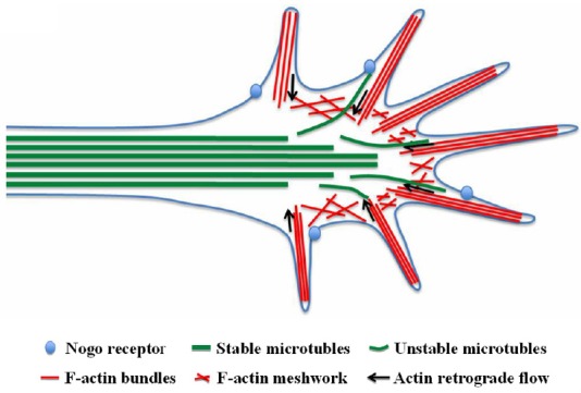
Growth cones are highly motile structures found at the tips of active growing axons during development and regeneration. The highly dynamic nature of growth cones allows them to respond to various environmental signals, including both axon growth promoting cues, which drive axon extension, and inhibitory signals that impede axon growth. Nonetheless, much less attention has been paid to promoting axonal regeneration after spinal cord injury via local regulation of the axonal cytoskeleton at the growth cone. Indeed, no study to date has provided concrete evidence that direct manipulation of the growth cone cytoskeleton promotes mammalian CNS axon regeneration in vivo. The growth cone cytoskeleton is composed of microtubules and actin filaments, which not only provide the anatomical structure of the growth cone, but are also involved in driving axon extension (Figure 1). It is widely accepted that axon growth is achieved through microtubule assembly and subsequent protrusion towards the growth cone. Therefore, in addition to the axon growth machinery, the cytoskeletal structure of growth cones are also common targets of most, if not all, axon growth inhibitors. For instance, myelin-based inhibitory molecules bind to the Nogo-66 receptor and activate the Rho-Rho kinase (ROCK) signaling pathway, which in turn induces growth cone collapse through disruption of the actin-myosin system (Sivasankaran et al., 2004). Additionally, an earlier study demonstrated that disorganized microtubules in the growth cone underlie the formation of retraction bulbs and failure of axonal regeneration after CNS injury (Ertürk et al., 2007). Importantly, stabilization of microtubules with taxol is sufficient to prevent retraction bulb formation in vivo and promote axon regeneration over inhibitory molecules such as myelin in vitro (Ertürk et al., 2007). Thus, local manipulation of the growth cone cytoskeleton may enable neurons to overcome multiple axon growth inhibitors and promote axonal regeneration. Previous studies have shown that microtubule protrusion towards the growth cone leading edge is impeded by actin retrograde flow, which is powered by myosin II (Hur et al., 2011b). As a result, actin retrograde flow driven by myosin II may be a major negative factor of microtubule protrusion in the neuronal growth cone, and consequently, axonal growth. Blocking myosin II activity may promote microtubule extension and subsequent axon regeneration. Indeed, in one of our published studies (Hur et al., 2011b), we found that regenerating sensory neurons (which show very strong intrinsic axonal growth) maintained much shorter axon growth when cultured on CSPG-coated substrate. However, axon growth was strikingly rescued back to control levels when myosin II activity was blocked by blebbistatin, a specific inhibitor of myosin II ATPase activity, not only with CSPG but also myelin-based inhibitors. To confirm this pharmacological data, we also used siRNAs against myosin IIA/B. Consistently, downregulation of endogenous myosin IIA/B in adult sensory neurons enhanced axonal regeneration on CSPG to the same extent as blebbistatin. Although several CSPG receptors have recently been identified, we still know very little about the downstream mechanism by which CSPG inhibits axonal regeneration. A previous study found that CSPG activates Rho GTPase, and inhibition of ROCK is able to partially rescue axon growth inhibited by CSPGs (Sivasankaran et al., 2004). Furthermore, many studies have implicated the myosin light chain kinase, which activates myosin II activity downstream of ROCK. We therefore determined whether ROCK inhibition, using its inhibitor Y27632, is able to promote axon growth using CSPGs. We found that Y27632 had little effect compared with blebbistatin, indicating that myosin II inhibition promotes axonal growth on CSPGs via a distinct molecular mechanism. We also found that inhibiting myosin II activity with blebbistatin markedly promotes microtubule protrusion into the growth cone periphery (Hur et al., 2011b). Thus, we believe that myosin II inhibition enhances axonal regeneration over CNS inhibitory molecules via promoting microtubule protrusion in the growth cone. Further studies with detailed high-resolution live cell imaging are needed to test this hypothesis. More importantly, further animal studies will be necessary to determine if inhibition of endogenous myosin IIA/B activity can promote axonal regeneration and functional recovery after spinal cord injury in vivo.

Figure 1.

Figure 1

Schematic representation of the growth cone cytoskeleton.

In addition to myosin II, there are many other proteins that strictly regulate growth cone microtubules, such as microtubule-based motor proteins, dyneins, and kinesins. Regulation of these microtubule-based motor molecule activities might also promote axonal regeneration after spinal cord injury. The microtubule-associated deacetylase, HDAC6, is reported to promote axon regeneration on inhibitory molecules such as CSPGs (Rivieccio et al., 2009). Furthermore, many of these microtubule-regulating proteins are affected by small molecules. Recently, cytoskeletal proteins became the most successful targets of cancer chemotherapy. Our previous study found that glycogen synthase kinase 3 regulates axon regeneration via microtubules plus the end-binding protein, CLASP (a cytoplasmic linker protein) (Hur et al., 2011a). Thus, local regulation of the growth cone cytoskeletal machinery may provide a novel and effective approach to promote axonal regeneration after spinal cord injury. Because cytoskeletal reorganization in the growth cone is the conversion point of both axon promoting and axon inhibiting signals, we believe that proper regulation of the growth cone cytoskeleton will not only ensure fast and efficient axon assembly but also allow growing axons to overcome multiple inhibitory signals in both the CNS and peripheral nervous system. It should be noted that disruption of the growth cone cytoskeleton might render regenerating axons insensitive to environmental axon guidance cues, which are important for regenerating axons to accurately reach the correct targets and ensure successful functional recovery. Precise temporal control of pharmacological treatments may provide a solution for such problems.

Successful axonal regeneration is achieved via elevated intrinsic axon growth ability in the soma and efficient cytoskeleton assembly at the nerve growth cone. Reduced intrinsic axon growth ability or muted response of CNS neurons to axonal injury is another major reason for mature CNS axon regeneration failure. During the past 10 years, tremendous progress has been made towards determining the molecular mechanisms underlying neuronal intrinsic axon growth ability, leading to the identification of a growing list of novel molecules and pathways that intrinsically regulate neuronal axon growth ability, such as Pten, KLF4, and SOCS3. Manipulation of these genes, either alone or in combination, in adult CNS neurons has produced by far the strongest effects on CNS axon regeneration (Park et al., 2008; Moore et al., 2009; Smith et al., 2009). However, regenerating axons induced by these approaches are still strongly inhibited by environmental CNS inhibitors. For instance, only a small percentage of regenerating corticospinal tract axons induced by Pten knockout can pass the injury site (Liu et al., 2010). In addition, many regenerating optic nerve axons induced by manipulation of multiple intrinsic factors are turning back at the optic chiasm, likely due to the presence of inhibitory molecules. Therefore, we believe that a combinatory approach addressing both up-regulation of neuronal intrinsic axon growth ability and manipulation of the local growth cone cytoskeleton is the best approach for long distance CNS axon regeneration.

This work was supported by a grant from the Priority Academic Program Development of Jiangsu Higher Education Institutions, Jiangsu Provincial Special Program of Medical Science of China, No. BL2012004; the Research Grants from Soochow University to Saijilafu.

Neural Regen Res. 2015 Dec;10(12):1953–1975.

Interaction of miR-21 and TGF-β/SMADs signaling pathway affects the formation of fibrotic scars after spinal cord injury

Rong-han Liu 1, Bin Ning 1,*

Scarring after spinal cord injury (SCI) consists of glial scar formation followed by fibrotic scar formation. A glial scar is formed by reactive astrocytes during the acute phase of SCI. Reactive astrocytes are formed by astrocyte activation, proliferation and hypertrophy; they highly express glial fibrillary acidic protein, can fill the injured region, limit inflammatory diffusion, secrete and release factors promoting nerve regeneration, and improve the microenvironment surrounding the injury site (Sabelstrom et al., 2013). Consequently, fibroblasts from the dura or blood vessels invade the damaged area and form a fibrotic scar (Hellal et al., 2011). Fibrotic scarring is involved in biological inhibition and acts as a mechanical barrier in axon regeneration and functional recovery. Fibrotic scars can secrete various axon growth inhibitory molecules including NG2 proteoglycan, tenascin C, Semaphorin 3A and EphB2, forming a biological barrier that hinders the recovery of neurological function. The extracellular matrix proliferates and secretes factors that include type IV collagen, fibronectin and laminin, and forms a mechanical barrier that hinders axon growth. Moreover, following SCI, glial limiting membrane is formed together with astrocytes which severely blocks axon regeneration and functional recovery. Glial scars produce physical barriers and impact axon regeneration, but the suppression of fibrotic scar formation can obviously accelerate axon regeneration (Hellal et al., 2011; Sabelstrom et al., 2013).

Transforming growth factor β (TGF-β) is a main catalyst of resting fibroblast activation and its signaling pathway can activate fibroblasts allowing them to migrate, proliferate and secrete extracellular matrix in order to promote the process of fibrosis. In the TGF-β signaling pathway, SMADs protein, a direct substrate of TGF-β receptor (TGF-βR), participates in and controls intracellular signal transduction. SMADs protein can be divided into nine types. Of them, receptor modulating type SMAD2, SMAD3, co-regulatory type SMAD4 and inhibitory type SMAD7 are involved in TGF-β signal transduction. Currently, the mechanism of TGF-β/SMADs signaling pathway in the process of fibrosis following SCI has not been reported.

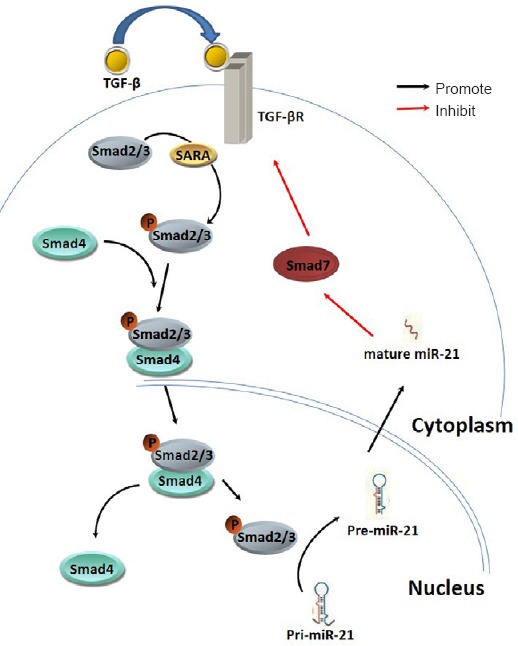
microRNA (miR-21) has been shown to be involved in the fibrosis of many tissues or organs (Thum et al., 2008; Liu et al., 2010). miR-21 and TGF-β/SMADs signaling pathways have a complex and diverse crosstalk which is known to greatly affect fibrosis and is probably an important factor in tissue fibrosis regulation. By the SMAD anchor for receptor activation (SARA) protein, intracellular SMAD2/3 protein binds to TGF-β complex, and then phosphorylates. The phosphorylated SMAD2/3 protein binds to SMAD4 protein to form a complex. The complex enters the nucleus, binds to p68, and forms a Drosha complex with DNA helicase. This Drosha complex promotes the conversion of pri-miR-21 to pre-miR-21, and accelerates miR-21 maturation. The mature miR-21 can reversely regulate the secretion of inhibitory protein SMADs (SMAD7). SMAD7 can act on TGF-βR, suppress TGF-β signaling pathway, and constitute a crosstalk between miR-21 and TGF-β/SMADs signaling pathways. Simultaneously, miR-21 can regulate signaling pathways by directly regulating the expression of TGF-β, TGF-βRII, TGF-βRI or SMAD2/3 (Butz et al., 2012). The interaction of information in the miR-21 and TGF-β/SMADs signaling pathway has a positive feedback effect, and presents a cascade effect (Figure 1). Our previous studies verified that miR-21 is highly expressed at the injury site and surrounding it, and its expression showed an increasing trend. Ultimately, investigating the mechanism of crosstalk in fibrotic scar formation following SCI can provide new insights into novel treatments.

Figure 1.

Figure 1

Interaction of TGF-β/Smad signaling and miR-21.

TGF: Transforming growth factor.

A previous study analyzed the inhibitory effects of astrocytes on neuronal growth, and confirmed that glial scars are formed by reactive astrocytes that have transformed from TGF-β1 (10 ng/mL, 5 days) stimulated astrocytes. After the model of glial scar was formed, cerebellar granule neurons were plated on the confluent monolayer of astrocytes and co-culture for 24 hours (Yu et al., 2012). As mentioned above, it is not glial scarring that plays a major role in physical barrier formation, but fibrotic scarring. Therefore, the first step to inhibit the activation, invasion, proliferation and secretion of fibroblasts is to restrict fibrotic scar formation. In a co-culture model of astrocytes with meningeal fibroblasts, fibroblasts showed a strong effect of secretion and proliferation, and surrounded astrocytes in clusters, similar to the fibrotic scar formation after SCI (Hellal et al., 2011). Accordingly, in this model, altering miR-21 expression by lentiviral transfection or siRNA interference may affect fibrotic scar formation.

It is possible that the crosstalk between miR-21 and TGF-β/SMADs signaling pathway may be a main regulatory network that regulates fibrosis after SCI, but it is not the only pathway. PI3K/AKT signaling pathway can be suppressed by PTEN protein, and PTEN protein is a direct target protein of miR-21. In fibrotic scars, through the PI3K/AKT signaling pathway, the extracellular matrix can be regulated by mTORC1 expression. ERK/MAPK signaling is also one of the key pathways that could be involved. Simultaneously, inhibitory protein Spry in the ERK/MAPK pathway can be directly regulated by miR-21 (Thum et al., 2008). PI3K/AKT and ERK/MAPK signaling pathways can regulate the secretion of matrix metalloproteinases, which can degrade extracellular matrix. The above two pathways can regulate miR-21 maturation via nuclear factor-κB signal.

In summary, investigating the important role and mechanism of miR-21 in the formation of fibrotic scars may lead to a therapeutic target for axon regeneration and functional recovery after SCI.

Neural Regen Res. 2015 Dec;10(12):1953–1975.

Elucidating the molecular mechanisms of CSPG-mediated axon growth inhibition through phosphoproteomics analysis

Pan-pan Yu 1,*

Chondroitin sulfate proteoglycans (CSPGs) are key components of the extracellular matrix in the central nervous system (CNS). High levels of CSPGs are expressed in the embryonic mammalian CNS, where they play important roles in axon guidance and pathfinding. In the adult CNS, CSPGs are found in the perineuronal net, a specialized structure that surrounds the cell bodies and proximal neurites of a specific subgroup of neurons, and is responsible for stabilizing existing synapses and restricting plasticity. After CNS injury in the adult, this pattern changes: CSPG levels are reduced in the perineuronal net but upregulated in the glial scar, where they act as a barrier to prevent axon extension and regrowth. Because the scar is largely responsible for the failure of axon regeneration after CNS injury, CSPGs are considered an attractive therapeutic target for CNS repair following injury.

CSPGs comprise a protein core and one or more chondroitin sulfate glycosaminoglycan side chains (CS-GAGs). The inhibitory action of CSPGs is largely due to the GAG chains. These are long unbranched polysaccharides composed of repeating disaccharide units of D-glucuronic acid (GlcA) and N-acetyl-D-galactosamine (GalNAc). CS-GAGs are covalently attached to a serine residue on the core protein via a tetrasaccharide linker. Axonal regeneration after injury can be improved by selective removal of these chains using chondroitinase. The inhibitory actions of CSPGs can be attributed to specific sulfation patterns in CS-GAG chains (Wang et al., 2008). Sulfation can occur at various positions including C2 of GlcA, C4 of GalNAc and/or C6 of GalNAc, and imparts a negative charge and specific binding properties to GAGs. Unlike nucleic acids and proteins, the synthesis of GAG side chains is not template driven, resulting in considerable heterogeneity in CSPGs: the number of GAG chains on each CSPG core protein and the length of each chain are variable, and each chain may be sulfated at discrete positions along the GAG backbone. This heterogeneity promotes interactions with many biologically important proteins such as growth factors, cytokines, cell adhesion molecules, and other extracellular matrix molecules. Most recently, several cell-surface proteins, including receptor protein tyrosine phosphatase σ (PTPσ), leukocyte common antigen-related phosphatase (LAR), Nogo receptor 1 (NgR1) and NgR3 have been identified as receptors for CSPGs (Sharma et al., 2012). Based on their binding properties, CSPGs are thought to influence axon growth either directly, via their receptors, or indirectly, by modulating the activity of other factors.

Several signaling pathways activated downstream of receptors have been implicated in the mediation of CSPG inhibition, including RhoA/Rho Kinase (ROCK), protein kinase C (PKC), Akt and glycogen synthase kinase 3β (GSK3β). Signals from these pathways converge to modify cytoskeletal dynamics underlying growth cone motility. We and others found an important role of non-muscle myosin II in controlling growth cone behavior in response to CSPGs (Hur et al., 2011b; Yu et al., 2012). However, the exact mechanisms by which CSPGs inhibit axon growth remain incompletely understood. Therefore, the main focus of our research is to identify candidate proteins and pathways affected by CSPGs, which will motivate potential strategies for CNS injury repair by overcoming CSPG inhibition. We have been using in vitro models of reactive astrogliosis as well as a CSPG boundary assay to mimic the inhibition from accumulated CSPGs in the glial scar formed after CNS injury. When an extending axon encounters the boundary of a CSPG-rich region (in our case a spot of immobilized CSPGs), the growth cone senses the repulsive signal from CSPGs and turns away. We found that growth cone turning requires myosin II activity, as blocking it using a selective inhibitor or siRNA promotes axonal crossing into the CSPG-rich field (Yu et al., 2012). However, how CSPGs regulate myosin II activity requires further investigation.

In general, intracellular signaling pathways are transduced and amplified via reversible protein phosphorylation of signal transducers at specific sites. To gain further insight into the molecular mechanisms underlying axon growth inhibition by CSPGs, we performed iTRAQ-based quantitative phosphoproteomics to profile global protein phosphorylation changes in primary cerebellar granule neurons after exposure to CSPGs. Using this in combination with strong cation exchange chromatography fractionation, immobilized metal affinity chromatography and LC-MS/MS, we quantified over 2000 phosphorylation sites, among which around 100 proteins showed significant changes in phosphorylation after CSPG exposure (Yu et al., 2013). Gene ontology revealed that the most overrepresented protein category was cytoskeleton binding proteins, involved in the regulation of cell morphology, assembly, and organization, and many of which have been implicated in regulating axon growth. The quick response of growth cone turning against CSPGs requires coordination of cytoskeleton dynamics likely driven by a locally triggered signaling cascade. Our screening result indicated that this rapid turning response might be achieved by regulating the phosphorylation of cytoskeletal proteins.

A number of RNA binding proteins, responsible for posttranscriptional modification, were also regulated by CSPGs, suggesting that CSPGs alter gene expression. Pathway analysis revealed a number of signaling pathways regulated by CSPGs, including those involved in synaptic vesicle trafficking, semaphorin-guided axon growth, and integrin, cadherin and epidermal growth factor receptor signaling (Yu et al., 2013). Given that CSPGs can bind to a variety of different proteins, it is not surprising that many intracellular signaling pathways are altered by CSPGs. Together, evidence from this large-scale phosphoproteomics study has greatly expanded our knowledge addressing CSPG signaling and opens up new areas for investigation.

The multiplicity of binding partners (receptors, growth factors, cytokines and extracellular matrix molecules) and signal transduction pathways activated by CSPGs suggests that the actions of CSPGs are context-dependent. Both the microenvironment and CSPG GAG chain composition differ between the developing CNS, the healthy adult CNS and the injured CNS. Future studies will need to address these issues directly in simplified model systems, where each parameter can be controlled independently. Only then will we really understand the functions of this fascinating group of molecules.

Neural Regen Res. 2015 Dec;10(12):1953–1975.

The repair of spinal cord injury with multifunctional, three-dimensional, electroactive scaffolds

Bo Shi 1, Chang-feng Fu 1,*

Current clinical therapies for spinal cord injury (SCI) include large doses of methylprednisolone, surgical, and symptomatic treatments followed by rehabilitation (Shi et al., 2014). However, relevant advances in biomaterials research are increasing. Many materials with unique properties have been developed and applied to experimental research. The biological materials that have been applied to SCI repair are usually combined with drugs, cells, or both. Thus, combinations of neurotrophic factors and biodegradable electroactive scaffolds are a novel strategy for SCI repair.

Since 2013, we have treated complete SCI using neurotrophin-3 (NT-3)-loaded polymeric scaffolds. After a long development process in animal experiments, the locomotor function of most experimental animals can now be improved. NT-3 induces a positive effect on the axon regeneration of nerve cells, particularly by promoting axonal germination in the location of corticospinal tract damage and sensory axonal regeneration (Tuszynski et al., 2003).

Electroactive tissue-engineered scaffolds are beneficial in the fields of cell axon regeneration and nerve conduction pathways. However, traditional conducting polymers have large molecular weights and cannot be engulfed by macrophages. Therefore, they remain in the body for a long time, potentially triggering an inflammatory response and must be removed during a second surgery. Multiple operations not only increase the patient pain, but also add to the operating costs. We believe the development of conductive polymers that are biodegradable is necessary for such tissue-engineered applications. We demonstrated the first use of a biodegradable electroactive material for the field of SCI repair. Oligoanilines, which are environmentally stable, biocompatible, and have good electroactivity and reversible redox properties, have been widely applied as tissue-engineered scaffolds. Oligoanilines coupled with polyethylene glycol hydrogels show improved biocompatibility and corrosion resistance and a three-dimensional (3D) porous structure. In addition, the properties of this type of composite can be controlled by varying the concentration of the hydrogel to maintain the gelling temperature at 10–30°C. In addition, external electrical stimulation can be used as an effective signal for controlling cell behavior and promoting tissue regeneration. In summary, we developed multifunctional scaffolds based on oligoanilines. The relevant research shows that such scaffolds can localize and deliver uniform electric stimulation that can be effectively controlled. Analysis of the ultraviolet spectra and cyclic voltammetry also supported the finding that the complex material has good electroactive properties. After subcutaneous injection, the electroactive 3D hydrogel was shaped in situ, and hematoxylin-eosin staining showed satisfactory biocompatibility of the scaffold in vivo. NT-3 was dispersed into this electroactive hydrogel to create a multifunctional 3D scaffold (Figure 1). Such scaffolds provide sufficient space for the regeneration and repair of nerve axons and release their carrier drugs slowly. These features help inhibit the formation of glial scars, prolong the NT-3 metabolic effect time, and improve the therapeutic outcome.

Figure 1.

Figure 1

Schematic showing the therapy for spinal cord injury with neurotrophin (NT)-3-loaded three-dimensional electroactive hydrogels.

We believe that 3D electroactive hydrogel scaffolds combined with drugs that can be uniformly dispersed may be useful for clinical applications. The drug release and biodegradation properties of the scaffolds were systematically characterized both in vitro and in vivo. To verify the effects of the scaffold on the recovery of axon regeneration and motor function, spinal cord T9–10 transection models were created in Sprague-Dawley rats after 3 days of adaptive feeding. The experimental animals were randomly assigned to the electroactive hydrogel group, blank hydrogel group, NT-3-loaded blank hydrogel group, or NT-3-loaded electroactive hydrogel group. The 3D hydrogel scaffolds were implanted into the injury site of the SCI model rats. At the same time, electrodes were embedded into the rostral and caudal skin of the rats, and low-intensity microcurrent stimulation was applied each day to the animals in all of the groups. An SCI injury score, the Basso, Beattie, and Bresnahan locomotor scale, was evaluated (0 is completely paralyzed, 21 is normal) weekly for 12 weeks to assess the recovery of motor function. The electrophysiology of the spinal cord was determined, and treatment effect was observed at the third week after surgery. Pathological and immunohistochemical analyses of the normal and repaired spinal cord tissues were also performed after treatment, and the thickness of the myelin sheaths was assessed using transmission electron microscopy. Thus, we observed the functional recovery of the SCI repair model using tissue pathology and immunohistochemistry to determine the therapeutic effect of the electroactive hydrogel that slowly releases nerve repair drugs. These studies may eventually lead to the replacement of the widely used simple spinal fixation combined with neurotrophic drugs and hormones for treating SCI. The multifunctional 3D scaffold used here provides an appropriate microenvironment for neuron repair and promotes axon regeneration, suggesting a great potential for clinical application.

Regeneration of damaged spinal cord remains one of the most difficult challenges in neuroscience. Based on the industrious works published over the past 20 years describing the fundamentals of the damage of SCI at the molecular level, the regeneration of the central nerve is no longer regarded as impossible (Mortazavi et al., 2015). Many studies have been published on the treatment of SCI with biomaterials (Haggerty et al., 2013). However, few data are available concerning the treatment of nerve injury using oligoanilines. Although the prospects for the study of the oligoanilines in the field of SCI repair remain unknown, several problems are worth researching further. First, we need to improve the biocompatibility of the electroactive material to minimize the host response. Second, we also need to understand the mechanism of SCI repair using the electroactive scaffold and determine the dose-effect relationship between the concentration of oligoanilines in the composite scaffold and nerve regeneration. Third, we need to determine the special properties of the material and influence of the material on the activities of an organism caused by external stimulation. Then we would be able to improve the scaffold based on those features. Experimental studies have suggested many different techniques for rescuing nerve tissue from SCI, reconnecting the spinal cord, and reactivating healthy circuitry after damage. Whether or not these methods are ready for clinical translation depends on the strength of the preclinical evidence, the mechanistic clarity of the treatment, the choice of suitable animal models, and the results from testing in large animals. Solving these problems will improve the clinical outcomes of this treatment in the future.

This work was financially supported by Graduate Innovation Fund of Jilin University in China, No. 450060523297; and Fund of China Scholarship Council, No. 201306175113.

Neural Regen Res. 2015 Dec;10(12):1953–1975.

A strategy for treating spinal cord injury: targeting the variation of immune cells in the local microenvironment

He-Zuo Lü 1,*

Conventional wisdom holds that the central nervous system (CNS) is an immune privileged site. CNS and the immune system are anatomically isolated, with antigens hidden from immune cells. CNS injury can damage the blood-brain barrier, causing CNS antigens to drain into adjacent lymph nodes, activating corresponding immune cells. These activated immune cells entering the CNS can produce a specific immune or autoimmune response, including monocyte-macrophage accumulation and lymphocyte infiltration. The autoimmune reaction after CNS injury can therefore aggravate secondary damage, and is not conducive to protecting the nerve. Recent research has revealed a number of different functional subsets in locally infiltrated immune cells after CNS injury. Some can induce neurological damage, while others may be neuroprotective. The outcome of CNS repair may be determined by the balance among these different cell subsets (Plemel et al., 2014). Over the last few decades, work in our laboratory has focused on investigating the variation in reactive immune cell subsets in the injured microenvironment, in a rat model of spinal cord injury (SCI), to identify “good” or “bad” immune cell subsets and provide a new strategy for immunotherapy of SCI.

Immune cells in the injured spinal cord microenvironment consist of activated microglia, infiltrated peripheral blood neutrophils, monocytes, macrophages, and lymphocytes. These cells can be functionally divided into different subsets, and damage the nerve either directly or indirectly (by producing inflammatory factors, cytotoxic substances and oxygen free radicals), or they can have neuroprotective effects (removing tissue fragments at the injury site, generating anti-inflammatory factors, and scavenging cytotoxic substances or oxygen free radicals). These cells regulate the local microenvironment, but their functional differentiation can also be affected by local microenvironment signals (Bowes and Yip, 2014).

Under normal circumstances, T lymphocytes can be divided into CD4+ and CD8+ cells. Among CD4+ T cells, helper T (Th) cells are strongly associated with SCI and repair. According to the different types of secreted cytokines, CD4+ T cells can be divided into Th1, Th2, Th9, Th17, Th22 and regulatory T cells (Treg). The roles of the various CD4+ T cell subsets in the pathogenesis of SCI still need further clarification. There is no doubt that by releasing cytokines, these cells regulate the immune response, and affect the repair of the injured spinal cord. CD8+ T cells in the pathological processes of SCI are generally considered to play a “cytotoxic” role, but removal of CD8+ T cells has also been shown to affect remyelination in the CNS. In fact, CD8+ T cells can be divided into cytotoxic T cells (Tc) and suppressor T cells (Ts). The former have a cytotoxic effect, while the latter play a role in immune regulation. Whether the two types of CD8+ T cells have subsets with different functions in the repair of CNS injury remains to be determined.

According to their different functions, macrophages can be divided into M1 and M2 subsets. M1 type macrophages are the classical activated macrophages, with phagocytic and bactericidal activity, releasing inflammatory mediators, presenting antigens, and starting the adaptive immune response. They are the body's major defense against foreign invasion. M2 type macrophages are alternatively activated, heterogeneous macrophages with anti-inflammatory effects. After CNS injury, local microglia and infiltrated monocytes and macrophages respond to different inflammatory signals in the local microenvironment, converting into M1 and M2 cells. A large number of M1 cells are distributed around the blood vessels of the CNS. M1 cells can secrete proinflammatory cytokines, further injuring the host cells. M2 cells modulate the immune inflammatory response, clear necrotic debris, and promote angiogenesis, tissue remodeling and repair. Theoretically, there is a relationship between the severity of secondary pathological SCI and the proportion of M1/M2. Conversely, SCI itself may also affect the proportion of M1/M2. Further investigation is required into the local variation of M1 and M2 cell subsets after SCI.

In our previous studies, we detected temporal and spatial expression patterns in Th1/Th2 and M1/M2 cells at the injury site after SCI, using immunohistochemical methods and flow cytometry. Our results indicated that without any intervention, a large number of Th1 and M1 cells (nerve damage) were detected at the injury site, but the proportion of Th1 and M2 cells (nerve protection) were low (Hu et al., 2012; Chen et al., 2015). These findings suggest that under natural conditions, the local immune microenvironment is not conducive to repair. Immunologic intervention, such as adaptive immunity of neuroprotective cells, can elevate the proportion of Th2 and M2 cells at the injury site, produce anti-inflammatory cytokines, improve the immune microenvironment and achieve functional repair (Hu et al., 2012; Ma et al., 2015).

On the basis of our previous studies and international developments, we believe that Th2 and M2 cells play an important protective role in the pathological process of SCI. Targeting Th2 and M2 cells may reduce local cellular and molecular factors that are not conducive to nerve repair. Therefore, a technique that can promote Th2 and M2 cell polarization can be used as a treatment strategy for immunotherapy of SCI. Adoptive reinfusion of cells cultured in vitro elevates the proportion of Th2 and M2 cells at the injury site. However, cell culture takes time, so the use of this method in a clinical setting may mean the neuroprotective time window is missed. Taking Th1/Th2 and M1/M2 as targets, the discovery of small molecule drugs for use in the early stages of injury is a new approach to exploring therapeutic strategies for SCI.

This study was supported by grants from the National Natural Science Foundation of China, No. 81071268, 81271363, 81571194.

Neural Regen Res. 2015 Dec;10(12):1953–1975.

Epidemiological data is critical for decreasing the incidence of spinal cord injury

Yong Zhou 1,#, Guang-zhi Ning 1,#, Shi-qing Feng 1,*

Because of variations in socioeconomic conditions in different countries, and in their populations’ living standards and habits, incidences of spinal cord injury (SCI) correspondingly appear to differ. Lee et al. (2014) reported that average global and regional incidences of traumatic SCI cases per million people in different regions were as follows: 23 worldwide, 40 in North America, 16 in Western Europe, 15 in Australia, 25 in central Asia, 21 in southern Asia, 19 in the Caribbean, 24 in Latin America, 29 in the central part of Sub-Saharan Africa, and 21 in the eastern part of Sub-Saharan Africa. Furlan et al. (2013) stated that the global incidence of traumatic SCI ranged between 8 and 246 cases per million people. Our epidemiologic study indicated that the incidence of traumatic SCI was 23.7 cases per million people in Tianjin, China (Ning et al., 2011).

The procedure we used to conduct our epidemiological study on traumatic SCI patients in Tianjin City in China is depicted as a flow diagram in Figure 1. In our study, we investigated the age of the patient at the onset of SCI, sex, occupation, cause of injury, combined injuries, length of hospitalization, injured segments, injury severity, complications during hospitalization, and therapeutic conditions. The research team designed an integrated questionnaire. Hospital medical records were reviewed in medical record statistics room in 15 tertiary hospitals in Tianjin, China. In accordance with the International Classification of Diseases, Version 10, SCI cases were identified and screened out using diagnostic code. The research group leader conducted the whole research according to the research plan. The detailed data needed were double entered by two researchers, respectively, and then cross-checked two times to assure data accuracy. We found that male patients were the main sufferers of traumatic SCI. This may be because more males are engaged in high-risk jobs in industries. However, the number of females suffering from injuries reveals a rising trend. With socioeconomic development and improved living standards, women have increasingly begun to work outside the home. Simultaneously, high-risk forms of entertainment and sport such as square dancing and climbing are on the rise. Moreover, the increased occurrence of injuries among women is probably because they are more prone to osteoporotic vertebral compression fractures. Proportions of farmers and workers are high in relation to occupational distribution. This may be because in recent years, a large number of farmers have moved to urban areas. These migrant farmers are primarily engaged in the construction industry. Most have low education levels and poor awareness regarding self-protection measures. Consequently, their risk of experiencing accidents is high. The primary causes of their injuries are traffic accidents and falling from low (< 1 m) as well as high (≥ 1 m) levels. These findings are consistent with results from a previous study (Thompson et al., 2015). More recently, Tianjin's highway transportation network has undergone rapid development, with extended traffic mileage and increased traffic volume. Correspondingly, traffic accidents have increased. Safety facilities are imperfect at construction sites and education in this regard is lagging behind. Thus, the possibility of injuries from falling has increased among migrant workers. In addition, with population aging becoming an increasingly serious problem in China, the occurrence of accidental falls among the elderly is on the rise. This is a concern relating to SCI that may be a contributing factor in the increasing average age of patients in this study.

Figure 1.

Figure 1

Flow diagram of the epidemiological study of TSCI patients in Tianjin, China.

TSCI: Traumatic spinal cord injury; ICD-10: International Classification of Diseases Version 10.

Injuries mainly impact on the C5 segment of the cervical spinal cord. This may be associated with the anatomical and physiological characteristics of the cervical spine. Cervical vertebrae are smaller and more flexible than other vertebrae. Cervical vertebrae are prone to degenerative changes resulting in cervical SCI. Yoo et al. (2010) reported that minor injuries suffered by a person with a degenerative disease of the cervical spine could lead to cervical SCI. A fall from a low height is defined as a low energy injury that can easily cause incomplete cervical SCI. The severity of SCI was evaluated by American Spinal Injury Association scale. Grade D is the most common type of SCI in our research in Tianjin, China. SCI complications severely affect patients’ outcomes and prognoses. Our research team found that in Tianjin of China, lung and urinary tract infections were common complications of traumatic SCI. Patients who are bedridden tend to suffer from lung infections, with pulmonary contusion, diaphragmatic or intercostal muscle dysfunction induced by injuries to higher segments of the spinal cord and reduced breathing and coughing strength. Urinary tract infections are associated with weakened parasympathetic dominance, excessive bladder filling, increased internal bladder pressure, and prolonged indwelling catheters. This study revealed a decline in the incidence of pressure sores. This may be attributed to enhanced prevention awareness and daily care. Thus, the prevention and treatment of complications are important for improving the survival rate of patients.

Because of the lack of epidemiological studies on traumatic SCI, it is necessary for clinicians, researchers, and even the general public, to fully understand the epidemiological characteristics of traumatic SCI. Awareness raising, aimed at preventing damage is, therefore, required. A combination of effective preventive measures, education for prevention of traumatic SCI, establishing an SCI registration system in selected areas and gradually extending this to the whole country, and extensive epidemiological studies on traumatic SCI can provide the necessary theoretical basis for guiding clinical practice. For future research and clinical practice, some detailed description of these detailed descriptions of these programs is as follows: according to the epidemiological data, the government could take measures to widen the roadway downtown, build median barriers between motor vehicular roadway and non-motor vehicular roadway, and educate the population at higher risk such as senior citizen and migrant farmers. This is an indispensable strategy for reducing the incidence of traumatic SCI.

Neural Regen Res. 2015 Dec;10(12):1953–1975.

The importance of olfactory ensheathing cells for spinal cord injury

Jie Qin 1, Xi-jing He 1,*

We have been conducting clinical trials on olfactory ensheathing cell (OEC) transplantation for incomplete chronic spinal cord injury (SCI) since 2005. Long-term follow-up results verified that sensory and motor functions improved in most patients to varying degrees. However, the main source of OECs in current clinical trials is from allogeneic embryonic olfactory bulbs, and these OECs are difficult to obtain. Because of their immunogenicity and rejection, long-term survival and regenerative function of those OECs in the patient's spinal cord cannot currently be confirmed (Ibrahim et al., 2014). These have limited the further development of clinical trials on OECs (Ibrahim et al., 2014). Therefore, the transplantation of autologous olfactory mucosa-derived OECs for treating SCI has become a hotspot in this field. Olfactory mucosa contains various cells, including stem cells, epithelial cells, Schwann cells of sensory fibers of trigeminal nerve, neuronal cells, OECs, and fibroblasts. Cells related to cell transplantation in the treatment of SCI consist of OECs, horizontal basal cells, globose basal cells, and mesenchymal stem cells (MSCs). Horizontal basal cells, globose basal cells and MSCs from olfactory mucosa have a stem cell phenotype. An in vivo study showed that horizontal basal cells could differentiate into oligodendrocytes, globose basal cells could differentiate into neuron support cells after being transplanted into injured spinal cord, and both of them had auxiliary or facilitating effects on remyelination (Wegener et al., 2015).

In vitro studies have demonstrated that MSCs derived from human or rat olfactory mucosa can differentiate into neuron-like cells, secrete protective factors to inhibit inflammation and to reduce fibrosis and cell death, play immunomodulatory effects on re-myelinization of OECs, and then contribute to remyelination. OECs can secrete neurotrophic factor, induce the differentiation of pluripotent stem cells into neurons, and form axons. Therefore, compared with OEC transplantation alone, the transplantation of four kinds of major cells in human olfactory mucosa for SCI not only has advantages in source and immunogenicity, but also promotes functional recovery after SCI by synergistic interaction. Lima et al. (2006) obtained olfactory mucosa by autologous nasal endoscopy, and this mixture was directly transplanted in injured spinal cord after removal of scar and neurolysis in the spinal cord. Sensory function was improved in six cases, and sensory function of bladder was improved in two cases. On the basis of the above studies, we hope that the purification and synergistic action of primary cells in the autologous olfactory mucosa can be applied to repair injured spinal cord.

We need to recognize that most experimental (test) results are positive, but there are some inconsistencies among results, which mainly reflect repeatability of OEC transplantation, precise repair mechanism after OEC transplantation, and whether the results of clinical trials have a significant effect. The reason for histological and functional differences after OEC transplantation should be multifaceted, such as complete or incomplete SCI and different conduction systems (especially OECs which have different repair effects on different conduction systems). Moreover, biological characteristics of different OECs, such as site of collection, culture program, purity, culture time, transplantation mode, time windows of injury and OEC transplantation, also affect experimental results (Mayeur et al., 2013).

The effects of OEC transplantation on central nervous system injury remain controversial. Therefore, the next steps should focus on: (1) how to obtain and culture OECs, what is the most favorable condition for cultivation (Figure 1)? (2) High purity of OECs or the mixture of OECs and other cells as a contribution to functional improvement after transplantation (such as co-culture with Schwann cells or fibroblasts). (3) How to carry out OEC transplantation through intravenous injection or injection surrounding the injured spinal cord? (4) How to obtain OECs? (5) What is the major repair mechanism of SCI after OEC transplantation? (6) Timing of transplantation: is there a favorable time window for OEC transplantation?

Figure 1.

Figure 1

P75 staining results after culturing olfactory ensheathing cells for 7 days in DF-12 medium containing different concentrations of fetal bovine serum (shown on the left) (Lu et al., 2014)

Using basic and clinical studies of OECs over the past decade, we are expanding basic OEC research; this includes how to obtain good OECs and explores optimized OEC culture conditions, and the exact repair mechanism of OEC transplantation after central nervous system injury, so as to serve OECs-related translational medicine. Simultaneously, we also hope to promote functional recovery in SCI patients by investigating clinical trials concerning the repair effect of OEC transplantation following SCI.

Neural Regen Res. 2015 Dec;10(12):1953–1975.

Optogenetics: a new method to repair urination dysfunction after spinal cord injury?

Zhi-cheng Zhang 1, Tian-sheng Sun 1,*

The main reason for late death in patients with spinal cord injury (SCI) is urinary tract infection and renal failure. Therefore, reconstructing the urinary system functions of SCI patients can be highly significant. Spastic bladder induced by SCI above the sacrum can be treated by intermittent catheterization, drug therapy and a variety of conventional surgeries, but all methods have problems. An external current acts on the outer membrane of nerve cells or nerve fibers, generates a transmembrane current and induces depolarization. When the current amplitude reaches a threshold, an action potential and nerve excitability may be elicited, exerting effects on the relevant organs. Motor neurons innervating the detrusor muscles are located in the sacral parasympathetic nucleus. The parasympathetic preganglionic fibers converge in the anterior sacral nerve root, parallel to the pelvic nerve, and terminate on postganglionic neurons in the wall of the urinary bladder. Thus, urination can be achieved using a sacral anterior root stimulator (SARS). However, SARS alone cannot solve the problem of excessive reflex of the detrusor muscle and detrusor/urethral sphincter dyssynergia. When combined with sacral deafferentation (SDAF), SARS can effectively relieve excessive reflux. After SDAF, a spinal reflex arc can be blocked so as to reduce the malignant afferents of muscle spindle fibers of detrusor muscle and urethral sphincter. This lessens excessive reflex of detrusor muscle and urethral external sphincter spasm, and restores the bladder storage function. Because of the expense of the sacral residual sensation and reflection, SDAF is not suitable for patients with incomplete sacral spine injuries. Therefore, Brindley (1994) reconstructed the functions of urine storage and urination of spastic bladder in patients with complete sacral spine injuries using SARS and SDAF. At present, this technique has been used in more than 3000 cases worldwide. The average follow-up was 10 years, and showed that 85% of patients could control their urination, and their residual urine volumes decreased. This technique has been widely applied in the clinic, but still has some deficiencies, including (1) stimulator breakdown: replacement requires further operations at high cost, eventually leading to treatment failure. (2) The long-term intradural or epidural implantation of an electrode increases the risk of cerebrospinal fluid leakage and infection. (3) Sacral anterior nerve roots consist of some nerves innervating lower limb movement: jittering occurs when the lower limb is stimulated, so it is not convenient for patients to use. (4) The stimulator is relatively expensive, and most patients cannot afford the cost.

The above problems can be overcome if the same bioelectric stimulation can be given to the micturition center, without stimulator and electrode implantation, and the stable, lasting and selective stimulation can be given to the sacral parasympathetic nucleis (SPN) that can control the detrusor muscle. The emergence of optogenetics makes this a possibility. Channelrhodopsin-2 (ChR2) is a photosensitive protein found in green algae, and represents a light-gated ion channel with a 7-transmembrane helix at the N-terminal (Wang et al., 2009). The channel opens when illuminated in the 350–550-nm range of light (center wavelength is 470 nm). In the neurons expressing ChR2, the channels open, allowing Na+, K+ and Ca2+ to enter the cells, producing an inward current. The subsequent depolarization can generate action potentials. Precise intervention and regulation of nerve cells can be done in living animals at different wavelengths of light (Greenberg et al., 2011). Boyden et al. (2005) transferred the ChR2 gene into mammalian cells, which are sensitive to blue light at 493 nm wavelength. This technique has been employed in many neural circuits. Mancuso et al. (2011) confirmed that blue light could penetrate the brain parenchyma and lead to the excitement of nerve nuclei expressing ChR2. Moreover, after transfection, there were no apparent changes in morphology or endogenous electrical activity of the neurons. ChR2 can be expressed safely and stably in neurons over a long period. The studies concerning the motor neuron network and autonomic activity of the spinal cord have been carried out using this technique. In a rat model of cervical SCI, phrenic motor neurons in the spinal cord specifically expressed ChR2 protein, and respiratory movement of the diaphragm could be restored after blue light irradiation (Alilain et al., 2008), but there is no report addressing the repair of neurogenic bladder after SCI. We proposed that bio-electric stimulation-excited SPN could be produced by transferring the ChR2 gene into the SPN of the detrusor of the bladder and then irradiating with blue light. The excited SPN could stimulate the contraction of the detrusor muscle of the bladder so as to simulate nerve excitement of micturition center and to promote urination.

We constructed a ChR2 optogenetic vector, and precisely injected the vector in bilateral SPN of a rat with complete suprasacral transverse cord injury (Figure 1). Thus, the SPN expressed the ChR2 protein. Simultaneously, bilateral SDAF was applied to suppress detrusor overactivity and detrusor/urethral sphincter dyssynergia. The sacral cord received blue light irradiation. Optogenetics is used to stimulate SPN excitement in the detrusor of the bladder so as to induce detrusor contraction and to repair urinary system function. This would verify the feasibility of optogenetics to repair neurogenic bladder after complete suprasacral cord injury. The effectiveness of optogenetics can be identified by urodynamic detection and long-term observation of renal function. The safety of optogenetics can be assessed by observing the histopathological changes in sacral center, sacral anterior root and parasympathetic efferent nerves, as well as the neuromuscular junction of the detrusor muscle. The mechanism of action can be explored by spinal cord histology and genetics, neurotransmitter detection, sacral nerve/detrusor/sphincter electrophysiology, and detrusor biomechanics, histology, and genomics. These could lead to new ideas for the functional reconstruction of neurogenic bladder following complete suprasacral cord injuries.

Figure 1.

Figure 1

Rat SPN located in the lateral horn of L6 and S1 spinal cord, lateral part of VII layer, i.e., the lateral zone of the lateral margin of the gray matter.

The SPN was labeled with black area. SPN: Sacral parasympathetic nucleis.

Neural Regen Res. 2015 Dec;10(12):1953–1975.

Effects of zinc on the recovery of neurological function after spinal cord injury

Kai Gao 1, Xi-fan Mei 1,*

The effects of zinc on the recovery of neurological function after spinal cord injury (SCI) are garnering increasingly more attention from researchers. Zinc is an important trace element in mammals, playing crucial roles in growth and development, reproductive genetics, immunization, the endocrine system, and neural activity (Koh et al., 1996). In the central nervous system, approximately 15% of zinc ions are in a free state. These zinc ions are mainly located in synaptic vesicles of axon terminals in zinc-containing neurons. During neural activity, free zinc ions enter the synaptic space with the release of some neurotransmitters and then enter postsynaptic neurons by binding to certain receptors in the postsynaptic membrane or traversing the postsynaptic membrane, resulting in neuromodulation (Lichten and Cousins, 2009, Smidt and Rungby, 2012). However, the distribution of zinc at the injury site after SCI, the effect of zinc on the recovery of SCI, and the precise mechanism of action of zinc remain unclear.

Recent studies (Nowak et al., 2004, Hwang et al., 2005) have found that zinc deficiency downregulates brain-derived neurotrophic factor (BDNF) expression in the hippocampus and pineal gland in growing rats. By contrast, zinc in relatively high concentrations promotes the synthesis and release of BDNF in the rat cerebral cortex. Zinc ions activate BDNF by inducing zinc-dependent matrix metalloproteinases, increasing Src family kinase activity, and activating the BDNF receptor tropomyosin-related kinase B through stimulation of the signaling system, independent of neurotrophic factors. Our research team found that following SCI, zinc contributes to the high expression and release of BDNF through the zinc transporter ZnT1, inhibits peroxidase activity, and finally results in neuroprotective effects (Wang et al., 2011, Su et al., 2012, Wang et al., 2014).

Numerous studies have confirmed that zinc has favorable and unfavorable consequences. Excessive amounts of zinc can cause neurotoxicity. In cerebral ischemia and SCI, intracellular zinc overload-induced zinc toxicity often results in neuronal death and inhibits the recovery of neurological function. Thus, excessive zinc-induced zinc toxicity becomes a key factor restricting the therapeutic use of zinc for SCI. Solving the problem of excessive zinc-induced zinc toxicity will be a difficult and but important task, as the resulting resolution may provide a novel therapeutic target for SCI and may inform the molecular mechanisms for the effects of zinc on the recovery of neurological function after SCI (Figure 1).

Figure 1.

Figure 1

Effects and mechanism of zine on the recovery of neurological function after SCI.

In our previous studies, we have found that zinc ions could improve the expression of BDNF in the spinal cord tissues after SCI. Unfortunately, excessive zinc ions would cause neurotoxicity in spinal cord tissues and the excessive zinc is the key constraint to the zinc therapy after SCI. Besides, we have verified the positive correlation of ZnT1 with autophagy activation after SCI. Furthermore, some reports have claimed that the cytoplasm-to-vacuole targeting pathway could elevate the cell tolerance to the zinc toxicity. Therefore, the goals for the further research as follows: Studying the protective effect and mechanism of cytoplasm-to-vacuole targeting pathway-mediated autophagy on zinc toxicity after SCI, which may improve the theoretical basis and experimental evidence of zinc application to the clinic treatment for SCI. BDNF: Brain-derived neurotrophic factor; SCI: spinal cord injury.

In recent years, much research has been focused on the effects and molecular mechanisms of autophagy in nerve cells during the recovery of neurological function following SCI. Our previous study (Su et al., 2012) verified that autophagy in nerve cells is activated after SCI. The activated autophagy improves the microenvironment for nerve cell survival by promoting phagocytosis of both dead nerve cells and nerve cells that lose their functions, reduces apoptosis of nerve cells, maintains the survival of nerve cells, and promotes the recovery of neurological function. Autophagy activation occurs through the stimulation of specific and nonspecific autophagy pathways. One specific autophagy pathway is the cytoplasm-to-vacuole targeting pathway, and Atg9p is an essential gene in this pathway. Despite current research efforts, the precise molecular mechanism of autophagy on the recovery of neurological function following SCI remains poorly understood.

Researchers have recently discovered that zinc plays a key role in the occurrence of autophagy. For example, autophagy activated by zinc ions can increase the survival rate of nerve cells, and zinc ion can activate autophagy in hepatocytes. Dziedzic and Caplan (2011) suggested that the cytoplasm-to-vacuole targeting pathway in yeast enhanced cell tolerance to zinc. Our previous study showed that free zinc ions and ZnT1 can be detected in the spinal cord of normal rats and that following SCI the distribution of zinc and ZnT1 markedly increases. Simultaneously, autophagy is noticeably activated. The expression of autophagy-related marker genes (beclin-1 and LC3-II/I) also increases, and autophagosomes form 24 hours after the SCI. More importantly, expression of ZnT1 is positively correlated with LC3II/I expression in the acute phase. Thus, the collective results of these studies indicate that autophagy in nerve cells plays a key role in zinc toxicity following SCI, and that zinc activates autophagy in nerve cells through the cytoplasm-to-vacuole targeting pathway after SCI. The primary emphases of our research include resolving the aforementioned issues involving zinc to determine whether zinc regulation may become a therapeutic tool for SCI and providing a new theoretical basis and experimental evidence for investigating the effects and molecular mechanisms of zinc following SCI (Figure 1).

Our future study aims to reveal the protective effect and molecular mechanism of cytoplasm-to-vacuole targeting pathway-mediated specific autophagy on zinc toxicity after SCI (Figure 1). Currently, we are generating an APG9L1 gene knockout in mice and a mouse model of SCI. We plan to use these mice to determine the distribution and metabolism of zinc in spinal cord nerve cells, to observe animal behavior and neuronal injury, to clarify the protective effect of autophagy on zinc toxicity, and to understand the effect of autophagy in nerve cells activated by zinc on the recovery of neurological function in mice with autophagic defects after SCI. Moreover, we plan to suppress or promote autophagy in neurons cultured in vitro using RNA interference and transfection, investigate zinc metabolism and cell viability in spinal cord neurons of mouse embryos at high concentrations of zinc, reveal the protective effect and mechanism of autophagy on zinc toxicity after SCI, and provide theoretical and experimental evidence for clinical applications of zinc.

The research addressing the effects and molecular mechanism of zinc in SCI opens a new chapter in basic research of SCI. It contributes to the progress of translating basic research to clinical treatment and promotes research and treatment of SCI. Further, it provides new theoretical bases and experimental evidence for the treatment of SCI.

Neural Regen Res. 2015 Dec;10(12):1953–1975.

Durotomy and dural grafting to treat lower cervical spine injuries with extensive spinal cord edema

Yan-zhen Qu 1, Xiao-dong Guo 1,*

Lower cervical spine injuries not only cause cervical fracture, dislocation, and facet interlocking, but also lead to spinal cord edema. The damage caused by extensive spinal cord edema after spinal cord injury (SCI) can significantly impact patient survival. If the edema extends to the C4 level of the spinal cord, paralysis of the respiratory muscles can occur, leading to pulmonary infection and respiratory failure.

Fracture and dislocation decrease the diameter of the vertebral canal and induce direct external compression on the spinal cord. Edema increases the cross sectional area of the spinal cord, which increases the internal compression in the spinal cord. Because of the constraint imposed by the vertebral canal and dura, the increase in spinal cord cross sectional area decreases the volume of the epidural and subarachnoid spaces, blocking the epidural veins, arteriole of the cord, and cerebrospinal fluid (CSF) flow and inducing arachnoid adhesion. Meanwhile, the lack of blood supply and CSF infusion exacerbate the spinal cord edema. In addition, bony fragments may insert into the dura and spinal cord surface, and it is necessary to remove these tiny, but serious threats. Similar to osteofascial compartment syndrome, SCI with edema is characterized by external and internal compression, decreased cavity volume, blockage of venous, arterial, and CSF flow, and increased edema. We name these symptoms “spinal compartment syndrome” (SCS).

The following is a detailed definition of SCS: internal compression from spinal cord edema and external compression from the vertebral canal and dura blocking the spinal epidural veins, arteriole of the cord, and CSF flow, which aggravates the ischemia and hypoxia of the injury site and secondary damage. This ischemia and hypoxia exacerbates the edema and increase the internal compression, forming a self-sustaining “vicious cycle”. SCS can be diagnosed during preoperative imaging and examination. Signs of SCS include blockage of the epidural, subdural, and subarachnoid spaces, discontinuities in the high T2-weighted CSF MRI signal, American Spinal Injury Association (ASIA) impairment scores worse than grade C, and extensive spinal cord edema that is longer than the length of one segment.

Compression caused by fracture and dislocation can be treated surgically, but no effective treatment is available for edema in the spinal cord. Clinically, mannitol and laminectomy are two commonly used methods for alleviating the effects of edema and hematoma. However, Mannitol only partially reduces edema, and surgery often remains necessary (Iwasaki et al., 1981). Although bony compression from the vertebral canal can be decompressed through laminectomy, dural compression, the blockage of venous, arterial, and CSF flow, and arachnoid adhesions cannot be effectively resolved with mannitol or laminectomy. Therefore, in addition to mannitol and laminectomy to reduce the bony compression of the vertebral canal, durotomy is necessary to relieve the pressure on the dural membrane and expand the intrathecal space. Many animal experiments have shown that durotomy improves the recovery of neurological function and reduce secondary injuries, but few clinical papers have reported the use of durotomy for SCI (Perkins and Deane, 1988; Smith et al., 2010).

We applied durotomy and dural grafting to patients with SCI with SCS to examine their safety and feasibility (Qu et al., 2015). Eighteen patients were treated with durotomy for extensive spinal cord edema, as defined by a preoperative MRI showing that the range of edema was more than one segment and that the CSF flow was blocked by arachnoid adhesion, and a preoperative ASIA impairment score worse than grade C. In the time between admission and the operation, all patients were given neck support, mannitol, and dexamethasone. The incision was made using a central posterior approach. After separating the muscle, a partial laminectomy was performed to release the compression of the spinal cord. To maintain the stability of the spine, extensive laminectomy of the entire edema segment was then performed after internal fixation. After a sufficiently long laminectomy, the dura bulged out of the vertebral lamina and the blockage of CSF flow could be observed directly as the loss of rhythmic pulsation. The dura was then immersed in ice cold normal saline for several minutes. To completely expose the spinal cord edema and prevent tissue adhesion blockage at the two incision ends, the dural incision was made slightly longer than the length of the spinal cord edema segments. To achieve internal decompression, any bony fragments, subdural hematoma, and blood clots were removed completely. The denticulate ligament was cut to relieve the arachnoid adhesions, and the CSF flow was restored by this arachnoid separation (Figure 1). To increase the volume of the subdural space, the durotomy incision was closed with autogenic deep fascia or artificial dura and absorbable sutures. A drainage tube was placed in the incision, a bone graft was performed using autogenous bone, allogeneic bone, or artificial bone, and the site was sutured closed in layers. After the operation, neck supports, mannitol, and dexamethasone were given to all patients. Based on the postoperative imaging examination, the fractures and dislocations of the lower cervical spine were reduced and fixed, the continuity of the high T2-weighted CSF signal was restored, no spinal cord edema or hematoma signals were observed, and no neurological deterioration or delayed neurological deterioration was found. According to the ASIA impairment scores, grade A cases remained grade A, but the impaired sensory level descended, and grade B and grade C cases improved by at least one grade.

Figure 1.

Figure 1

Durotomy in a patient with lower cervical spine injury combined with extensive spinal cord edema.

The results of treating SCS patients with durotomy in this study suggest several conclusions. First, mannitol and laminectomy are insufficient for cases with extensive spinal edema with SCS. Mannitol and laminectomy have been used to decompress the SCI for many years and are thought to reduce secondary injuries by promoting the restoration of blood flow at the injured site. Nevertheless, Jones (Jones et al., 2012) demonstrated in an animal model of severe SCI that laminectomy alone for the treatment of acute SCI led to residual cord deformation followed by spinal cord edema, which caused subarachnoid occlusion and the expansion of the injured spinal cord against the dura resulting in further compression of the spinal cord. Therefore, durotomy is necessary to relieve the pressure on the dural membrane and expand the intrathecal space. Second, the sequence of laminectomy and internal fixation is different from what is done in normal surgeries. The partial laminectomy of the injured segment is performed first to decrease the compression and provide space for the spinal cord. Internal fixation is then performed before the extensive laminectomy to avoid aggravated SCI caused by an unstable spine. Third, dural grafting was performed during the surgery. Durotomy has been used to treat SCI for many years, but typically the dura is sutured after removing the intrathecal compression without applying a dural graft, which would not reduce the spinal cord compression made by the dura. Some animal experiments have shown that the use of a dural graft can improve CSF flow by limiting meningeal fibrosis, reduce connective tissue scar formation, and attenuate macrophage accumulation and progressive secondary injury. Fourth, the indications for durotomy include: (1) panplegia or paraplegia caused by spinal trauma and ASIA impairment scores of grades A–C; (2) discontinuity of the high T2-weighted MRI CSF signal and extensive spinal cord edema signal longer that is more than one segment long; and (3) spinal cord compression by bony fragments or subdural hematoma on preoperative imaging. Patients with ASIA impairment scores of grades D or E, mannitol and dexamethasone can be used without decompression. Fifth, the contraindications for durotomy include: (1) a penetrating injury; (2) complete destruction of the spinal cord continuity where the broken ends of the spinal cord and ventral dura can be clearly observed; (3) patients with brain injuries or neurological diseases; and (4) patients who are unable to tolerate anesthesia and surgery.

Several questions remain for the research field of durotomy in SCI. First, the molecular mechanism of durotomy that improves the recovery from SCI is unclear. Studies on durotomy by our group and others have only shown that durotomy and dural grafting is safe for SCI. Clarifying the molecular mechanism may provide powerful evidence to determine whether durotomy and dural grafting is better than other therapeutic methods. For example, Yang (Yang et al., 2013) examined the relationship between myelotomy and autophagy by evaluating the expression of mammalian target of rapamycin complex 1 (mTORC1) and microtubule-associated protein light chain 3. The results showed that myelotomy activates the mTORC1 signaling pathway and inhibits autophagy. Second, the sample numbers in the papers published so far have been small. The number of durotomy cases was from several to dozens in each study, but those numbers of cases are not large enough for statistical research. Recruiting more patients may first require identifying the mechanism of action. If enough evidence is collected to prove that durotomy and dural grafting is a better treatment than laminectomy, the operation method may become popular with more doctors, allowing more durotomies and multicenter studies to be carried out. Third, the ASIA impairment scores are an imperfect quantification method for statistical analysis. Each neurological level is divided into three scores: 0, 1, and 2. However, the number of neurological states is a large continuum, which means the same score for the entire neurological system is given to two people who could have very different neurological states. To properly compare durotomy to other therapeutic methods, the neurological function evaluation scores should be further analyzed using statistical methods.

We plan to continue research in this area in several directions. First, we will perform additional animal experiments regarding the molecular mechanism of the durotomy and dural grafting. Such studies may identify the exact reason why durotomy improves neurological function. Second, through animal and clinical research, we will determine the relationship between edema development and blockage of the blood vessels and CSF flow. The existence of SCS remains a hypothesis currently, and we need to gather evidence that uniquely identifies SCS and that describes the development of SCS. Third, further work is needed to improve the neurological function score and make the score suitable for statistical methods.

For patients with SCS, treatment with durotomy and dural grafting is safe and effective. Durotomy and dural grafting reduces the external and internal compression, restores the CSF flow, relieves secondary injuries, creates conditions for improving respiration, which saves lives, and may be beneficial for the recovery of neurological function.

This work was financially supported by the International Science & Technology Cooperation Program of China, No. 2013 DFG32690; and the National Natural Science Foundation of China, No. 81371939, 81171684.

Neural Regen Res. 2015 Dec;10(12):1953–1975.

Reconstruction of artificial micturition reflex arc for neurogenic bladder after spinal nerve injury

Tao Sui 1, Xiao-jian Cao 1,*

Numerous scholars have explored surgical treatment for bladder dysfunction after spinal cord injury (SCI). Extensive basic and clinical research on bladder re-innervation has demonstrated bladder function reconstruction after SCI by nerve transfer. This has been recognized in and outside China. Carlsson and Sundin (1968) anastomosed cat lower lumbar nerve root with sacral anterior nerve root, and first verified, by bladder volume pressure test and histological examination, that somatic motor fibers could grow into autonomic nerve fibers. Sundin and Carlsson (1972) transferred the nerve above the injury level to the site below the injury level, and found that the bladder micturition function showed some improvement in cats. Chuang et al. (1991) anastomosed spinal anterior nerve root above the injury level with sacral anterior nerve root below the injury level in dural sac and obtained some return of bladder function after SCI in rats. However, the above methods did not set up an initiator. The neural connection may be established but the connection is futile. Like an underground nuclear test, if the ignition is not set up on the ground, a series of underground reactions will not occur, and the atomic bomb is but a pile of scrap metal. The Shaw reflex arc solves the problem of the “ignition start”. Xiao and Godec (1994) performed the anastomosis of the left L4 ventral root to the L6 ventral root. Their electrophysiological study and neural tracing study with horseradish peroxidase confirmed that the “skin − central nervous system − bladder” reflex pathway is established. From this they first proposed the concept of “somatic nerve − autonomic nerve” bladder reflex arc. Xiao et al. (1999) also demonstrated the feasibility of “skin − central nervous system − bladder” reflex pathway in cats. Xiao et al. (2003) reported that 10/15 (67%) patients had satisfactory functional recovery of bladder, measured by urodynamics, in 15 patients over a follow-up period of 3 years. Lin et al. (2008, 2009, 2010, 2011) conducted the anastomosis of the lower thoracic spinal anterior nerve root with the S2/3 anterior nerve root for treating conus medullaris injury-induced atonic bladder. They reconstructed the sensory function of the bladder following SCI, and performed an anastomosis of the S1 anterior nerve root with the S2/3 anterior nerve root for SCI-induced spastic bladder. More than 400 patients were subjected to surgical treatment, and the efficacy was 60–70%.

The main aim of this research on nerve transfer for bladder function reconstruction is to reconstruct the bladder micturition reflex arc by the anastomosis of remnant nerve roots below or above the injury level with sacral nerve roots in the dural sac. However, the following problems still exist: (1) Nerve roots in the dural sac only have a thin pia mater and lack a thick tough protective layer. During suture, anti-tension of the anastomotic stoma is poor, which induces laceration and the failure of anastomosis, and easily injures spinal nerve fibers. (2) The cauda equina nerve is tightly packed within the dural sac, so it is difficult to distinguish the receptor nerve and the donor nerve. The wrong anastomosis will have disastrous consequences. (3) After opening the dural sac, the incidence rate of leakage of cerebrospinal fluid and spinal infection is high, leading to high mortality. Whether spinal nerve root can be precisely identified, separated and anastomosed in the epidural spinal canal is still a problem. The procedure could be applied widely if the operation is simplified and if surgical trauma can be reduced.

Results from our animal studies confirmed that a tension-free anastomosis of anterior and posterior S1/2 nerve roots of dogs could be carried out in the epidural spinal canal. Thus, experimental study of human autopsies was conducted. We measured the length of spinal nerves of epidural segment and the distance between two adjacent nerve root exits, and found that the motor root and sensory root of spinal nerves anterior to epidural dorsal root ganglion could be separated from each other. Each of them was wrapped by pia mater, arachnoid and dura mater. The arrangement was similar to the arrangement of the dural sac. The motor root was located ventral to the sensory root. The measurement of the length of spinal nerves and the distance between the two exits showed that tension-free anastomosis could be conducted between S1 anterior nerve roots and S2 anterior nerve roots in the epidural spinal canal. These results showed that compared with the intradural spinal nerves, epidural spinal nerves were wrapped by pia mater, arachnoid and spinal dura mater, and had enough strength against mechanical traction (Zhou et al., 2014; Liu et al., 2015). Spinal nerve anastomosis in the epidural spinal canal not only greatly reduces the difficulty of nerve anastomosis but is also strong and stable.

We found a novel piezoelectric immunosensor that can precisely distinguish types of spinal nerve root (Sui et al., 2013a, 2013b). These sensors can bind highly sensitive piezoelectric sensing technology to a specific immune response, and transform a biological signal into a physical or chemical signal that is easy to detect qualitatively or quantitatively, by means of a transducer. The anterior roots of the spinal nerve (motor tract) have acetylcholinesterase (AChE)-rich cholinergic fibers but the posterior roots of the spinal nerve (sensory tract) do not. Therefore, the measurement of AChE content in spinal nerves could distinguish between the anterior and posterior roots. Our results confirm the hypothesis. When anterior roots of spinal nerve were detected, the vibrational frequency of the sensor decreased 9.8 ± 2.1 Hz (mean ± SD). When posterior roots of spinal nerve were detected, the vibrational frequency of the sensor decreased 4.8 ± 0.3 Hz (mean ± SD). These findings suggest that the piezoelectric immunosensor can identify specifically and quickly (in 6 minutes) the anterior and posterior roots of spinal nerve.

In conclusion, epidural anastomosis of spinal nerve roots for reconstructing an artificial micturition reflex arc not only greatly reduces the difficulty of nerve anastomosis, but also provides a strong and stable system. The anastomosis shortens the distance of nerve regeneration and reduces the postoperative time of nerve regeneration. The piezoelectric immunosensor can precisely identify epidural anterior and posterior roots of the spinal nerve, reducing surgical errors and improving the recovery of bladder function. Our future study may focus on exploring other ways to further reduce the resistance to urine and to improve the surgical effect, while enhancing urination.

This study was supported by the National Natural Science Foundation of China, No. 81371969, 81371968, 81401791.

Neural Regen Res. 2015 Dec;10(12):1953–1975.

Old drugs, new tricks: the strategy for new drug development in spinal cord injury

Yu-long Zhou 1,2, Jian Xiao 2,*

To date, more than 27,000 drugs are available to treat disease worldwide. The discovery of a new drug requires an average of 13 years of research and an investment of US $1.8 billion to bring a single drug from the bench to a patient's bedside (Figure 1). Another approach, termed “new uses for old drugs”, “drug repositioning”, “drug repurposing”, “drug re-profiling”, “therapeutic switching”, or “indication switching”, has gained considerable attention over the past decade among pharmaceutical industries researchers and clinicians. This approach is based on two observations: (1) almost all drugs used in human therapy possess more than one target and thus can produce off-target side effects in addition to their principal activity; and (2) many different diseases share common molecular pathways and targets (Gupta et al., 2013).

Figure 1.

Figure 1

The new quantitative system pharmacology paradigm.

Major steps and estimated time involved in the conventional drug development process and systems pharmacology, in which a drug interacts with multiple primary and secondary targets that exist within a complex network. This provides a more rational way for drug design/development in a “systems-level” context.

The pathology of SCI involves a primary mechanical insult to the spinal cord and the activation of a delayed secondary cascade of events, which ultimately trigger a continuum of necrotic and apoptotic cell death mechanisms. These secondary events involve a complex cascade of molecular events that include disturbances in ionic homeostasis, local edema, ischemia, focal hemorrhage, free radical stress and a robust inflammatory response. They contribute to extend the damage to the surrounding neural tissue and thus are targets for therapeutic strategies. A number of pharmacological neuroprotective therapies targeting one or more of these secondary events have been extensively studied. In particular, methylprednisolone and ganglioside have been suggested to be the drug of choice for acute SCI in humans. However, its beneficial effect on the neurological recovery of patients has not been conclusively proven and no gold standard therapy for SCI has been established (Baptiste and Fehlings, 2006). A variety of other pharmacological interventions including channel blockers (Riluzole), tetracycline derivatives (Minocycline), antioxidants (Aminoguanidine), calpain inhibitors (MDL28170), cell apoptosis inhibitors (Minocyline), fusogen copolymer polyethylene glycol, and the tissue-protective hormone erythropoietin have proved beneficial in experimental and clinical trials; however, in light of these discoveries, there is a requirement to establish a method for the systematic evaluation of experimental therapeutic outcomes within various research SCI centers.

Systems pharmacology, a cousin of systems biology, attempts to change the drug discovery process and the classical view of drug action (one molecule for one target to provide one therapeutic effect), and seeks to integrate drug discovery with biological systems to increase the potential to discover effective medications with few side effects. The new quantitative systems pharmacology paradigm, where a drug interacts within a complex network with multiple primary and secondary targets, aims to modulate complex cellular networks using mono or combination therapy to predict and reduce toxicity and increase the therapeutic effect of drugs using “precision medicine” (Figure 1). The use of network biology in quantitative systems pharmacology to better understand drug action, aims to obtain information for the drug-development process such as new indications for approved drugs, relationships between proteins and drug side effects, drug-drug interactions, or pathway-gene associations, and scale the observations obtained from molecules up to cells, tissues, organs and organisms (Perez-Nueno, 2015). The commonly accepted view is that an assortment of pathophysiological processes including vascular abnormalities, blood-spinal cord barrier disruption, the activation of astrocytes, the apoptosis of oligodendrocytes, ischemia-reperfusion, excitotoxicity, ionic imbalance, free radical and reactive oxygen species synthesis, lipid peroxidation, endoplasmic reticulum stress, autophagy and generation of a robust inflammatory response result in secondary injury. In SCI, delayed neuropathological cascades associated with secondary injurious events could be targeted by a variety of pharmacological compounds by “drug repurposing” (Table 1).

Table 1.

Key pathological events after spinal cord injury and targets of neuroprotective drugs

graphic file with name NRR-10-1953-g014.jpg

Under the guidance of systems pharmacology, scientists demonstrated that Taxol, an approved anti-cancer drug, facilitated axonal regeneration after SCI by decreasing scar formation and enhancing intrinsic axonal growth and improved motor function (Hellal et al., 2011). The same group reported that Epothilone B, a Food and Drug Administration (FDA)-approved drug in the class of blood-brain barrier permeable microtubule-stabilizing drugs for oncotherapy, decreased scarring in rodent SCI by abrogating the polarization and directed migration of scar-forming fibroblasts (Ruschel et al., 2015). Conversely, Epothilone B reactivated neuronal polarization by inducing concerted microtubule polymerization into the axon tip, which propelled axon growth through an inhibitory environment (Ruschel et al., 2015). Using systems pharmacology, we have screened several current medications that may have therapeutic effects for SCI. A series of studies performed in our laboratory using pharmacological agents after SCI initiation showed that 4-phenylbutyrate approved by the FDA for children and adults with hyperammonemia associated with urea cycle disorders significantly attenuated blood-spinal cord barrier permeability and improved functional recovery of SCI. Retinoic acid, widely used for the treatment of acute leukemia and various skin diseases in the clinic, has generated considerable excitement for its potential as a therapy for a wide variety of neurological disorders over the past few decades. In our rat model of SCI, retinoic acid also significantly improved functional recovery and increased the survival of neurons in spinal cord lesions after SCI (Zhou et al. 2015). Butylphthalide, approved for the treatment of subcortical ischemic small vessel disease in China, also improved the locomotor function of SCI model rats, increased neuron survival by the inhibition of endoplasmic reticulum stress-induced apoptosis and the prevention of blood-spinal cord barrier disruption. Therefore, we predict that many drugs currently approved by the FDA could be screened for the treatment of SCI, and this will have a positive influence on improving the manufacture of new drugs for the treatment of SCI as the rediscovery of old drugs for new uses accelerate. However, whether these observations will translate into the clinic remains to be seen and there is a need for more standardized preclinical studies.

Many therapeutic interventions using growth factors have focused on secondary degeneration after SCI to reduce damaged areas and promote axonal regeneration and functional recovery. Animals treated with hepatocyte growth factor (HGF) showed increased axonal growth beyond glial scars and improvement in functional recovery (Sakai et al., 2015). A phase-I/II clinical trial for the intrathecal administration of recombinant human HGF protein for the treatment of patients with SCI is ongoing at the Keio University in Japan. Acidic fibroblast growth factor and a clinical trial designed to test its efficacy and safety in combination with surgical intervention in human SCI showed it was safe and feasible (Wu et al., 2011). Over the last five years, our laboratory has demonstrated that (1) Basic fibroblast growth factor (bFGF) administration improved the recovery and increased the survival of neurons in spinal cord lesions in rats, and indicated the role of bFGF in SCI recovery was related to the inhibition of excessive autophagy and enhancement of ubiquitinated protein clearance via the activation of PI3K/Akt/mTOR signaling. (2) Nerve growth factor (NGF) administration improved the locomotor function of SCI model rats and increased neuron survival. The protective effect of NGF was related to the inhibition of endoplasmic reticulum stress-induced apoptosis, which also increased the expression of growth-associated protein 43. (3) Epidermal growth factor (EGF) also promoted a functional improvement of locomotor recovery via the activation of PI3K/Akt after SCI. Many reports including our own showed that among these growth factors, fibroblast growth factor (FGF) is a promising drug for pharmacological therapy of SCI. Furthermore, the use of growth factors combined with gene therapy might have greater clinical effects than monotherapy in SCI, now that the human genome has been sequenced and its annotation is approaching completion.

Although drug repurposing should significantly reduce the cost and time associated with new drug development in SCI, there are numerous challenges and points that deserve attention. The following are some recommendations for design criteria for the preclinical development of pharmacological agents for SCI. (1) Rediscover old drugs for new uses with a new drug screening system and computational tools. (2) Demonstrate specificity of the pharmacological agent by using structurally different modulators and parallel use of knockout technology and pharmacological antagonists. (3) Perform drug dose-response curves and evaluate efficacy throughout the overall process of SCI. (4) Obtain pharmacokinetics, pharmacodynamics and spinal cord concentrations of the tested drugs. (5) Randomize SCI surgeries and drug treatments, and use double-blinding in all histological and functional outcome testing. (6) Perform therapeutic window studies for the pharmacological agent to include a clinically relevant delayed treatment time point. (7) Regularly monitor clinically relevant physiological functions during and after SCI surgery and treatment. (8) Evaluate the pharmacological agent in both genders and among people of different ages. (9) Evaluate the pharmacologic agent in different SCI animal models including rats and higher-level mammals including humans and old monkeys. (10) Replicate the therapeutic effects of the drug with cross-repeated testing in different laboratories. We believe that each of these challenges deserves further extensive research and efforts throughout the drug discovery community.

References

  • 1.Alilain WJ, Li X, Horn KP, Dhingra R, Dick TE, Herlitze S, Silver J. Light-induced rescue of breathing after spinal cord injury. J Neurosci. 2008;28:11862–11870. doi: 10.1523/JNEUROSCI.3378-08.2008. [DOI] [PMC free article] [PubMed] [Google Scholar]
  • 2.Baptiste DC, Fehlings MG. Pharmacological approaches to repair the injured spinal cord. J Neurotrauma. 2006;23:318–334. doi: 10.1089/neu.2006.23.318. [DOI] [PubMed] [Google Scholar]
  • 3.Bowes AL, Yip PK. Modulating inflammatory cell responses to spinal cord injury: all in good time. J Neurotrauma. 2014;31:1753–1766. doi: 10.1089/neu.2014.3429. [DOI] [PubMed] [Google Scholar]
  • 4.Boyden ES, Zhang F, Bamberg E, Nagel G, Deisseroth K. Millisecond-timescale genetically targeted optical control of neural activity. Nat Neurosci. 2005;8:1263–1268. doi: 10.1038/nn1525. [DOI] [PubMed] [Google Scholar]
  • 5.Brindley GS. The first 500 patients with sacral anterior root stimulator implants: general description. Paraplegia. 1994;32:795–805. doi: 10.1038/sc.1994.126. [DOI] [PubMed] [Google Scholar]
  • 6.Burda JE, Sofroniew MV. Reactive gliosis and the multicellular response to CNS damage and disease. Neuron. 2014;81:229–248. doi: 10.1016/j.neuron.2013.12.034. [DOI] [PMC free article] [PubMed] [Google Scholar]
  • 7.Butz H, Racz K, Hunyady L, Patocs A. Crosstalk between TGF-beta signaling and the microRNA machinery. Trends Pharmacol Sci. 2012;33:382–393. doi: 10.1016/j.tips.2012.04.003. [DOI] [PubMed] [Google Scholar]
  • 8.Carlsson CA, Sundin T. Reconstruction of severed ventral roots innervating the urinary bladder. An experimental study in cats. Scand J Urol Nephrol. 1968;2:199–210. doi: 10.3109/00365596809135368. [DOI] [PubMed] [Google Scholar]
  • 9.Casano AM, Peri F. Microglia: multitasking specialists of the brain. Dev Cell. 2015;32:469–477. doi: 10.1016/j.devcel.2015.01.018. [DOI] [PubMed] [Google Scholar]
  • 10.Chen YJ, Zhu H, Zhang N, Shen L, Wang R, Zhou JS, Hu JG, Lü HZ. Temporal kinetics of macrophage polarization in the injured rat spinal cord. J Neurosci Res. 2015;93:1526–1533. doi: 10.1002/jnr.23612. [DOI] [PubMed] [Google Scholar]
  • 11.Chuang DC, Chang PL, Cheng SY. Root reconstruction for bladder reinnervation: an experimental study in rats. Microsurgery. 1991;12:237–245. doi: 10.1002/micr.1920120403. [DOI] [PubMed] [Google Scholar]
  • 12.Delamarter RB, Sherman J, Carr JB. Pathophysiology of spinal cord injury. Recovery after immediate and delayed decompression. J Bone Joint Surg Am. 1995;77:1042–1049. doi: 10.2106/00004623-199507000-00010. [DOI] [PubMed] [Google Scholar]
  • 13.Dziedzic SA, Caplan AB. Identification of autophagy genes participating in zinc-induced necrotic cell death in Saccharomyces cerevisiae. Autophagy. 2011;7:490–500. doi: 10.4161/auto.7.5.14872. [DOI] [PMC free article] [PubMed] [Google Scholar]
  • 14.Ertürk A, Hellal F, Enes J, Bradke F. Disorganized microtubules underlie the formation of retraction bulbs and the failure of axonal regeneration. J Neurosci. 2007;27:9169–9180. doi: 10.1523/JNEUROSCI.0612-07.2007. [DOI] [PMC free article] [PubMed] [Google Scholar]
  • 15.Filbin MT. Myelin-associated inhibitors of axonal regeneration in the adult mammalian CNS. Nat Rev Neurosci. 2003;4:703–713. doi: 10.1038/nrn1195. [DOI] [PubMed] [Google Scholar]
  • 16.Finnerup NB, Baastrup C. Spinal cord injury pain: mechanisms and management. Curr Pain Headache Rep. 2012;16:207–216. doi: 10.1007/s11916-012-0259-x. [DOI] [PubMed] [Google Scholar]
  • 17.Frankel HL, Coll JR, Charlifue SW, Whiteneck GG, Gardner BP, Jamous MA, Krishnan KR, Nuseibeh I, Savic G, Sett P. Long-term survival in spinal cord injury: a fifty year investigation. Spinal Cord. 1998;36:266–274. doi: 10.1038/sj.sc.3100638. [DOI] [PubMed] [Google Scholar]
  • 18.French JS, Anderson-Erisman KD, Sutter M. What do spinal cord injury consumers want?. A review of spinal cord injury consumer priorities and neuroprosthesis from the 2008 neural interfaces conference. Neuromodulation. 2010;13:229–231. doi: 10.1111/j.1525-1403.2009.00252.x. [DOI] [PubMed] [Google Scholar]
  • 19.Fujita Y, Yamashita T. Axon growth inhibition by RhoA/ROCK in the central nervous system. Front Neurosci. 2014;8:338. doi: 10.3389/fnins.2014.00338. [DOI] [PMC free article] [PubMed] [Google Scholar]
  • 20.Furlan JC, Sakakibara BM, Miller WC, Krassioukov AV. Global incidence and prevalence of traumatic spinal cord injury. Can J Neurol Sci. 2013;40:456–464. doi: 10.1017/s0317167100014530. [DOI] [PubMed] [Google Scholar]
  • 21.Gensel JC, Zhang B. Macrophage activation and its role in repair and pathology after spinal cord injury. Brain Res. 2015;1619:1–11. doi: 10.1016/j.brainres.2014.12.045. [DOI] [PubMed] [Google Scholar]
  • 22.Giger RJ, Hollis ER, 2nd, Tuszynski MH. Guidance molecules in axon regeneration. Cold Spring Harb Perspect Biol. 2010;2:a001867. doi: 10.1101/cshperspect.a001867. [DOI] [PMC free article] [PubMed] [Google Scholar]
  • 23.Greenberg KP, Pham A, Werblin FS. Differential targeting of optical neuromodulators to ganglion cell soma and dendrites allows dynamic control of center-surround antagonism. Neuron. 2011;69:713–720. doi: 10.1016/j.neuron.2011.01.024. [DOI] [PubMed] [Google Scholar]
  • 24.Guerra WK, Baldauf J, Schroeder HW. Long-term results after microsurgical repair of traumatic nerve lesions of the upper extremities. Zentralbl Neurochir. 2007;68:195–199. doi: 10.1055/s-2007-985859. [DOI] [PubMed] [Google Scholar]
  • 25.Gupta SC, Sung B, Prasad S, Webb LJ, Aggarwal BB. Cancer drug discovery by repurposing: teaching new tricks to old dogs. Trends Pharmacol Sci. 2013;34:508–517. doi: 10.1016/j.tips.2013.06.005. [DOI] [PubMed] [Google Scholar]
  • 26.Haggerty AE, Oudega M. Biomaterials for spinal cord repair. Neurosci Bull. 2013;29:445–459. doi: 10.1007/s12264-013-1362-7. [DOI] [PMC free article] [PubMed] [Google Scholar]
  • 27.Haisma JA, van der Woude LH, Stam HJ, Bergen MP, Sluis TA, Post MW, Bussmann JB. Complications following spinal cord injury: occurrence and risk factors in a longitudinal study during and after inpatient rehabilitation. J Rehabil Med. 2007;39:393–398. doi: 10.2340/16501977-0067. [DOI] [PubMed] [Google Scholar]
  • 28.Hellal F, Hurtado A, Ruschel J, Flynn KC, Laskowski CJ, Umlauf M, Kapitein LC, Strikis D, Lemmon V, Bixby J, Hoogenraad CC, Bradke F. Microtubule stabilization reduces scarring and causes axon regeneration after spinal cord injury. Science. 2011;331:928–931. doi: 10.1126/science.1201148. [DOI] [PMC free article] [PubMed] [Google Scholar]
  • 29.Hu JG, Shen L, Wang R, Wang QY, Zhang C, Xi J, Ma SF, Zhou JS, Lü HZ. Effects of Olig2-overexpressing neural stem cells and myelin basic protein-activated T cells on recovery from spinal cord injury. Neurotherapeutics. 2012;9:422–445. doi: 10.1007/s13311-011-0090-9. [DOI] [PMC free article] [PubMed] [Google Scholar]
  • 30.Hu X, Leak RK, Shi Y, Suenaga J, Gao Y, Zheng P, Chen J. Microglial and macrophage polarization-new prospects for brain repair. Nat Rev Neurol. 2015;11:56–64. doi: 10.1038/nrneurol.2014.207. [DOI] [PMC free article] [PubMed] [Google Scholar]
  • 31.Hur EM, Saijilafu, Lee BD, Kim SJ, Xu WL, Zhou FQ. GSK3 controls axon growth via CLASP-mediated regulation of growth cone microtubules. Genes Dev. 2011a;25:1968–1981. doi: 10.1101/gad.17015911. [DOI] [PMC free article] [PubMed] [Google Scholar]
  • 32.Hur EM, Yang IH, Kim DH, Byun J, Saijilafu, Xu WL, Nicovich PR, Cheong R, Levchenko A, Thakor N, Zhou FQ. Engineering neuronal growth cones to promote axon regeneration over inhibitory molecules. Proc Natl Acad Sci U S A. 2011b;108:5057–5062. doi: 10.1073/pnas.1011258108. [DOI] [PMC free article] [PubMed] [Google Scholar]
  • 33.Hwang JJ, Park MH, Choi SY, Koh JY. Activation of the Trk signaling pathway by extracellular zinc. Role of metalloproteinases. J Biol Chem. 2005;280:11995–12001. doi: 10.1074/jbc.M403172200. [DOI] [PubMed] [Google Scholar]
  • 34.Ibrahim A, Li DQ, Collins A, Tabakow P, Raisman G, Li Y. Comparison of olfactory bulbar and mucosal cultures in a rat rhizotomy model. Cell Transplant. 2014;23:1465–1470. doi: 10.3727/096368913X676213. [DOI] [PubMed] [Google Scholar]
  • 35.Iwasaki Y, Ito T, Isu T, Tsuru M. Effect of combined treatment of mannitol and myelotomy on experimental spinal cord injury (author's transl) Neurol Med Chir (Tokyo) 1981;21:917–921. doi: 10.2176/nmc.21.917. [DOI] [PubMed] [Google Scholar]
  • 36.Jiang YW, Zhang WW, Zhan XD, Kang XN, Dai X, Zhang WY, Liu ZY, Dong WR, Guo JS. Self-assembling peptide nanofiber scaffold comined with RhoA-siRNA for repair of spinal cord injury. Zhongguo Zuzhi Gongcheng Yanjiu yu Linchuang Kangfu. 2011;15:2895–2900. [Google Scholar]
  • 37.Jones CF, Cripton PA, Kwon BK. Gross morphological changes of the spinal cord immediately after surgical decompression in a large animal model of traumatic spinal cord injury. Spine. 2012;37:E890–899. doi: 10.1097/BRS.0b013e3182553d1d. [DOI] [PubMed] [Google Scholar]
  • 38.Kang XN, Wen JK, Wang XH, Pan MJ, Zhang WW, Zhan XD, Liu ZY, Wu WT, Guo JS. Temporal and spatial pattern of RhoA expression in injured spinal cord of adult mice. J South Med Univ. 2013;33:463–468. [PubMed] [Google Scholar]
  • 39.Koh JY, Suh SW, Gwag BJ, He YY, Hsu CY, Choi DW. The role of zinc in selective neuronal death after transient global cerebral ischemia. Science. 1996;272:1013–1016. doi: 10.1126/science.272.5264.1013. [DOI] [PubMed] [Google Scholar]
  • 40.Lee BB, Cripps RA, Fitzharris M, Wing PC. The global map for traumatic spinal cord injury epidemiology: update 2011 global incidence rate. Spinal Cord. 2014;52:110–116. doi: 10.1038/sc.2012.158. [DOI] [PubMed] [Google Scholar]
  • 41.Lee JK, Geoffroy CG, Chan AF, Tolentino KE, Crawford MJ, Leal MA, Kang B, Zheng B. Assessing spinal axon regeneration and sprouting in Nogo-, MAG-, and OMgp-deficient mice. Neuron. 2010;66:663–670. doi: 10.1016/j.neuron.2010.05.002. [DOI] [PMC free article] [PubMed] [Google Scholar]
  • 42.Lichten LA, Cousins RJ. Mammalian zinc transporters: nutritional and physiologic regulation. Annu Rev Nutr. 2009;29:153–176. doi: 10.1146/annurev-nutr-033009-083312. [DOI] [PubMed] [Google Scholar]
  • 43.Lima C, Pratas-Vital J, Escada P, Hasse-Ferreira A, Capucho C, Peduzzi JD. Olfactory mucosal autografts in human spinal cord injury: a pilot clinical study. J Spinal Cord Med. 2006;29:191–203. doi: 10.1080/10790268.2006.11753874. [DOI] [PMC free article] [PubMed] [Google Scholar]
  • 44.Lin H, Hou C, Chen A. Reconstructed bladder innervation above the level of spinal cord injury to produce urination by abdomen-to-bladder reflex contractions. J Neurosurg Spine. 2011;14:799–802. doi: 10.3171/2011.2.SPINE10685. [DOI] [PubMed] [Google Scholar]
  • 45.Lin H, Hou C, Zhen X, Xu Z. Clinical study of reconstructed bladder innervation below the level of spinal cord injury to produce urination by Achilles tendon-to-bladder reflex contractions. J Neurosurg Spine. 2009;10:452–427. doi: 10.3171/2009.1.SPINE08540. [DOI] [PubMed] [Google Scholar]
  • 46.Lin H, Hou C, Chen A, Xu Z. Reinnervation of atonic bladder after conus medullaris injury using a modified nerve crossover technique in canines. World Neurosurg. 2010;73:582–586. doi: 10.1016/j.wneu.2010.02.020. [DOI] [PubMed] [Google Scholar]
  • 47.Lin H, Hou CL, Zhong G, Xie Q, Wang S. Reconstruction of reflex pathways to the atonic bladder after conus medullaris injury: preliminary clinical results. Microsurgery. 2008;28:429–435. doi: 10.1002/micr.20504. [DOI] [PubMed] [Google Scholar]
  • 48.Liu G, Friggeri A, Yang Y, Milosevic J, Ding Q, Thannickal VJ, Kaminski N, Abraham E. miR-21 mediates fibrogenic activation of pulmonary fibroblasts and lung fibrosis. J Exp Med. 2010;207:1589–1597. doi: 10.1084/jem.20100035. [DOI] [PMC free article] [PubMed] [Google Scholar]
  • 49.Liu K, Lu Y, Lee JK, Samara R, Willenberg R, Sears-Kraxberger I, Tedeschi A, Park KK, Jin D, Cai B, Xu B, Connolly L, Steward O, Zheng B, He Z. PTEN deletion enhances the regenerative ability of adult corticospinal neurons. Nat Neurosci. 2009;13:1075–1081. doi: 10.1038/nn.2603. [DOI] [PMC free article] [PubMed] [Google Scholar]
  • 50.Liu Y, Zhou X, Ma J, Ge Y, Cao X. The diameters and number of nerve fibers in spinal nerve roots. J Spinal Cord Med. 2015;38:532–537. doi: 10.1179/1079026814Z.000000000273. [DOI] [PMC free article] [PubMed] [Google Scholar]
  • 51.Lu M, Dong J, Lu T, Lv H, Yang P, Cheng Z, Li J, Liang B, Xu J, Li H, He X. Effects of different sera conditions on olfactory ensheathing cells in vitro. Int J Mol Sci. 2014;16:420–438. doi: 10.3390/ijms16010420. [DOI] [PMC free article] [PubMed] [Google Scholar]
  • 52.Ma SF, Chen YJ, Zhang JX, Shen L, Wang R, Zhou JS, Hu JG, Lü HZ. Adoptive transfer of M2 macrophages promotes locomotor recovery in adult rats after spinal cord injury. Brain Behav Immun. 2015;45:157–170. doi: 10.1016/j.bbi.2014.11.007. [DOI] [PubMed] [Google Scholar]
  • 53.Mancuso JJ, Kim J, Lee S, Tsuda S, Chow NB, Augustine GJ. Optogenetic probing of functional brain circuitry. Exp Physiol. 2011;96:26–33. doi: 10.1113/expphysiol.2010.055731. [DOI] [PubMed] [Google Scholar]
  • 54.Mar FM, da Silva TF, Morgado MM, Rodrigues LG, Rodrigues D, Pereira MI, Marques A, Sousa VF, Coentro J, Sá-Miranda C, Sousa MM, Brites P. Myelin lipids inhibit axon regeneration following spinal cord injury: a novel perspective for therapy. Mol Neurobiol. 2015 doi: 10.1007/s12035-014-9072-3. DOI:10.1007/s12035-014-9072-3. [DOI] [PubMed] [Google Scholar]
  • 55.Maynard FM, Bracken MB, Creasey G, Ditunno JF, Donovan WH, Ducker TB, Garber SL, Marino RJ, Stover SL, Tator CH, Waters RL, Wilberger JE, Young W. International standards for neurological and functional classification of spinal cord injury. Spinal Cord. 1997;35:266–274. doi: 10.1038/sj.sc.3100432. [DOI] [PubMed] [Google Scholar]
  • 56.Mayeur A, Duclos C, Honore A, Gauberti M, Drouot L, do Rego JC, Bon-Mardion N, Jean L, Verin E, Emery E, Lemarchant S, Vivien D, Boyer O, Marie JP, Guerout F. Potential of olfactory ensheathing cells from different sources for spinal cord repair. PLoS One. 2013;8:e62860. doi: 10.1371/journal.pone.0062860. [DOI] [PMC free article] [PubMed] [Google Scholar]
  • 57.Milan S, Nada M, lvica M, et al. Management of the posttraumatie paraplegic pain of war injuries origin: epidemiological, social, organizing and surgical aspect. Rev Int Serv Sante Forces Armees. 2007;80:240–244. [Google Scholar]
  • 58.Moore DL, Blackmore MG, Hu Y, Kaestner KH, Bixby JL, Lemmon VP, Goldberg JL. KLF family members regulate intrinsic axon regeneration ability. Science. 2009;326:298–301. doi: 10.1126/science.1175737. [DOI] [PMC free article] [PubMed] [Google Scholar]
  • 59.Mortazavi MM, Verma K, Harmon OA, Griessenauer CJ, Adeeb N, Theodore N, Tubbs RS. The microanatomy of spinal cord injury: a review. Clin Anat. 2015;28:27–36. doi: 10.1002/ca.22432. [DOI] [PubMed] [Google Scholar]
  • 60.Murray PJ, Allen JE, Biswas SK, Fisher EA, Gilroy DW, Goerdt S, Gordon S, Hamilton JA, Ivashkiv LB, Lawrence T, Locati M, Mantovani A, Martinez FO, Mege JL, Mosser DM, Natoli G, Saeij JP, Schultze JL, Shirey KA, Sica A, et al. Macrophage activation and polarization: nomenclature and experimental guidelines. Immunity. 2014;41:14–20. doi: 10.1016/j.immuni.2014.06.008. [DOI] [PMC free article] [PubMed] [Google Scholar]
  • 61.Ning GZ, Yu TQ, Feng SQ, Zhou XH, Ban DX, Liu Y, Jiao XX. Epidemiology of traumatic spinal cord injury in Tianjin, China. Spinal Cord. 2011;49:386–390. doi: 10.1038/sc.2010.130. [DOI] [PubMed] [Google Scholar]
  • 62.Novak ML, Koh TJ. Phenotypic transitions of macrophages orchestrate tissue repair. Am J Pathol. 2013;183:1352–1363. doi: 10.1016/j.ajpath.2013.06.034. [DOI] [PMC free article] [PubMed] [Google Scholar]
  • 63.Nowak G, Legutko B, Szewczyk B, Papp M, Sanak M, Pilc A. Zinc treatment induces cortical brain-derived neurotrophic factor gene expression. Eur J Pharmacol. 2004;492:57–59. doi: 10.1016/j.ejphar.2004.03.038. [DOI] [PubMed] [Google Scholar]
  • 64.Park KK, Liu K, Hu Y, Smith PD, Wang C, Cai B, Xu B, Connolly L, Kramvis I, Sahin M, He Z. Promoting axon regeneration in the adult CNS by modulation of the PTEN/mTOR pathway. Science. 2008;322:963–966. doi: 10.1126/science.1161566. [DOI] [PMC free article] [PubMed] [Google Scholar]
  • 65.Perez-Nueno VI. Using quantitative systems pharmacology for novel drug discovery. Expert Opin Drug Discov. 2015:1–17. doi: 10.1517/17460441.2015.1082543. [DOI] [PubMed] [Google Scholar]
  • 66.Perkins PG, Deane RH. Long-term follow-up of six patients with acute spinal injury following dural decompression. Injury. 1988;19:397–401. doi: 10.1016/0020-1383(88)90132-5. [DOI] [PubMed] [Google Scholar]
  • 67.Plemel JR, Wee Yong V, Stirling DP. Immune modulatory therapies for spinal cord injury–past present and future. Exp Neurol. 2014;258:91–104. doi: 10.1016/j.expneurol.2014.01.025. [DOI] [PubMed] [Google Scholar]
  • 68.Prinjha R, Moore SE, Vinson M, Blake S, Morrow R, Christie G, Michalovich D, Simmons DL, Walsh FS. Inhibitor of neurite outgrowth in humans. Nature. 2000;403:383–384. doi: 10.1038/35000287. [DOI] [PubMed] [Google Scholar]
  • 69.Qu YZ, Luo Z, Guo XD, Cui W, Sun TF, Huang ZF, Wu YC, Shao ZW. The durotomy or myelotomy for the spinal cord extensive swelling with/without intramedullary hemorrhage. Zhonghua Guke Zazhi. 2015;35:707–713. [Google Scholar]
  • 70.Rivieccio MA, Brochier C, Willis DE, Walker BA, D’Annibale MA, McLaughlin K, Siddiq A, Kozikowski AP, Jaffrey SR, Twiss JL, Ratan RR, Langley B. HDAC6 is a target for protection and regeneration following injury in the nervous system. Proc Natl Acad Sci U S A. 2009;106:19599–19604. doi: 10.1073/pnas.0907935106. [DOI] [PMC free article] [PubMed] [Google Scholar]
  • 71.Ruschel J, Hellal F, Flynn KC, Dupraz S, Elliott DA, Tedeschi A, Bates M, Sliwinski C, Brook G, Dobrindt K, Peitz M, Brüstle O, Norenberg MD, Blesch A, Weidner N, Bunge MB, Bixby JL, Bradke F. Axonal regeneration. Systemic administration of epothilone B promotes axon regeneration after spinal cord injury. Science. 2015;348:347–352. doi: 10.1126/science.aaa2958. [DOI] [PMC free article] [PubMed] [Google Scholar]
  • 72.Sabelstrom H, Stenudd M, Reu P, Dias DO, Elfineh M, Zdunek S, Damberg P, Goritz C, Frisen J. Resident neural stem cells restrict tissue damage and neuronal loss after spinal cord injury in mice. Science. 2013;342:637–640. doi: 10.1126/science.1242576. [DOI] [PubMed] [Google Scholar]
  • 73.Sakai K, Aoki S, Matsumoto K. Hepatocyte growth factor and Met in drug discovery. J Biochem. 2015;157:271–284. doi: 10.1093/jb/mvv027. [DOI] [PubMed] [Google Scholar]
  • 74.Sharma K, Selzer ME, Li S. Scar-mediated inhibition and CSPG receptors in the CNS. Exp Neurol. 2012;237:370–378. doi: 10.1016/j.expneurol.2012.07.009. [DOI] [PMC free article] [PubMed] [Google Scholar]
  • 75.Shechter R, Schwartz M. CNS sterile injury: just another wound healing? Trends Mol Med. 2013;19:135–143. doi: 10.1016/j.molmed.2012.11.007. [DOI] [PubMed] [Google Scholar]
  • 76.Shi B, Ding JX, Liu Y, Zhuang XM, Zhuang XL, Chen XS, Fu CF. ERK1/2 Pathway-mediated differentiation of IGF-1-transfected spinal cord-derived neural stem cells into oligodendrocytes. PLos One. 2014;9:8. doi: 10.1371/journal.pone.0106038. [DOI] [PMC free article] [PubMed] [Google Scholar]
  • 77.Silver J, Miller JH. Regeneration beyond the glial scar. Nat Rev Neurosci. 2004;5:146–156. doi: 10.1038/nrn1326. [DOI] [PubMed] [Google Scholar]
  • 78.Sivasankaran R, Pei J, Wang KC, Zhang YP, Shields CB, Xu XM, He Z. PKC mediates inhibitory effects of myelin and chondroitin sulfate proteoglycans on axonal regeneration. Nat Neurosci. 2004;7:261–268. doi: 10.1038/nn1193. [DOI] [PubMed] [Google Scholar]
  • 79.Smidt K, Rungby J. ZnT3: a zinc transporter active in several organs. Biometals. 2012;25:1–8. doi: 10.1007/s10534-011-9490-x. [DOI] [PubMed] [Google Scholar]
  • 80.Smith JS, Anderson R, Pham T, Bhatia N, Steward O, Gupta R. Role of early surgical decompression of the intradural space after cervical spinal cord injury in an animal model. J Bone Joint Surg Am. 2010;92:1206–1214. doi: 10.2106/JBJS.I.00740. [DOI] [PMC free article] [PubMed] [Google Scholar]
  • 81.Smith PD, Sun F, Park KK, Cai B, Wang C, Kuwako K, Martinez-Carrasco I, Connolly L, He Z. SOCS3 deletion promotes optic nerve regeneration in vivo. Neuron. 2009;64:617–623. doi: 10.1016/j.neuron.2009.11.021. [DOI] [PMC free article] [PubMed] [Google Scholar]
  • 82.Spaić M, Marković N, Tadić R. Microsurgical DREZotomy for pain of spinal cord and Cauda equina injury origin: clinical characteristics of pain and implications for surgery in a series of 26 patients. Acta Neurochir (Wien) 2002;144:453–462. doi: 10.1007/s007010200066. [DOI] [PubMed] [Google Scholar]
  • 83.Steward O, Sharp K, Yee KM, Hofstadter M. A re-assessment of the effects of a Nogo-66 receptor antagonist on regenerative growth of axons and locomotor recovery after spinal cord injury in mice. Exp Neurol. 2008;209:446–468. doi: 10.1016/j.expneurol.2007.12.010. [DOI] [PMC free article] [PubMed] [Google Scholar]
  • 84.Su R, Mei X, Wang Y, Zhang L. Regulation of zinc transporter 1 expression in dorsal horn of spinal cord after acute spinal cord injury of rats by dietary zinc. Biol Trace Elem Res. 2012;149:219–226. doi: 10.1007/s12011-012-9414-9. [DOI] [PubMed] [Google Scholar]
  • 85.Sui T, Que J, Kong D, Xie H, Wang D, Shi K, Cao X, Li X. Rapid identification of spinal ventral and dorsal roots using a quartz crystal microbalance. Neural Regen Res. 2013a;8:686–692. doi: 10.3969/j.issn.1673-5374.2013.08.002. [DOI] [PMC free article] [PubMed] [Google Scholar]
  • 86.Sui T, Ge Y, Liu W, Zhao ZK, Zhang N, Cao X. An acetylcholinesterase antibody-based quartz crystal microbalance for the rapid identification of spinal ventral and dorsal roots. PLoS One. 2013b;8:e69049. doi: 10.1371/journal.pone.0069049. [DOI] [PMC free article] [PubMed] [Google Scholar]
  • 87.Sundin T, Carlsson CA. Reconstruction of severed dorsal roots innervating the urinary bladder. An experimental study in cats. II. Regeneration studies. Scand J Urol Nephrol. 1972;6:185–196. doi: 10.3109/00365597209133635. [DOI] [PubMed] [Google Scholar]
  • 88.Thompson C, Mutch J, Parent S, Mac-Thiong JM. The changing demographics of traumatic spinal cord injury: An 11-year study of 831 patients. J Spinal Cord Med. 2015;38:214–223. doi: 10.1179/2045772314Y.0000000233. [DOI] [PMC free article] [PubMed] [Google Scholar]
  • 89.Thum T, Gross C, Fiedler J, Fischer T, Kissler S, Bussen M, Galuppo P, Just S, Rottbauer W, Frantz S, Castoldi M, Soutschek J, Koteliansky V, Rosenwald A, Basson MA, Licht JD, Pena JT, Rouhanifard SH, Muckenthaler MU, Tuschl T, et al. MicroRNA-21 contributes to myocardial disease by stimulating MAP kinase signalling in fibroblasts. Nature. 2008;456:980–984. doi: 10.1038/nature07511. [DOI] [PubMed] [Google Scholar]
  • 90.Tuszynski MH, Grill R, Jones LL, Brant A, Blesch A, Löw K, Lacroix S, Lu P. NT-3 gene delivery elicits growth of chronically injured corticospinal axons and modestly improves functional deficits after chronic scar resection. Exp Neurol. 2003;181:47–56. doi: 10.1016/s0014-4886(02)00055-9. [DOI] [PubMed] [Google Scholar]
  • 91.Wang H, Katagiri Y, McCann TE, Unsworth E, Goldsmith P, Yu ZX, Tan F, Santiago L, Mills EM, Wang Y, Symes AJ, Geller HM. Chondroitin-4-sulfation negatively regulates axonal guidance and growth. J Cell Sci. 2008;121:3083–3091. doi: 10.1242/jcs.032649. [DOI] [PMC free article] [PubMed] [Google Scholar]
  • 92.Wang H, Sugiyama Y, Hikima T, Sugano E, Tomita H, Takahashi T, Ishizuka T, Yawo H. Molecular determinants differentiating photocurrent properties of two channelrhodopsins from chlamydomonas. J Biol Chem. 2009;284:5685–5696. doi: 10.1074/jbc.M807632200. [DOI] [PubMed] [Google Scholar]
  • 93.Wang Y, Mei X, Zhang L, Lv G. The correlation among the dynamic change of Zn2+, ZnT-1 and brain-derived neurotrophic factor after acute spinal cord injury in rats. Biol Trace Elem Res. 2011;143:351–358. doi: 10.1007/s12011-010-8845-4. [DOI] [PubMed] [Google Scholar]
  • 94.Wang Y, Su R, Lv G, Cao Y, Fan Z, Wang Y, Zhang L, Yu D, Mei X. Supplement zinc as an effective treatment for spinal cord ischemia/reperfusion injury in rats. Brain Res. 2014;1545:45–53. doi: 10.1016/j.brainres.2013.12.015. [DOI] [PubMed] [Google Scholar]
  • 95.Wegener A, Deboux C, Bachelin C, Frah M, Kerninon C, Seilhean D, Weider M, Wegner M, Nait-Oumesmar B. Gain of Olig2 function in oligodendrocyte progenitors promotes remyelination. Brain. 2015;138:120–135. doi: 10.1093/brain/awu375. [DOI] [PMC free article] [PubMed] [Google Scholar]
  • 96.Wu JC, Huang WC, Chen YC, Tu TH, Tsai YA, Huang SF, Huang HC, Cheng H. Acidic fibroblast growth factor for repair of human spinal cord injury: a clinical trial. J Neurosurg Spine. 2011;15:216–227. doi: 10.3171/2011.4.SPINE10404. [DOI] [PubMed] [Google Scholar]
  • 97.Xiao CG, Godec CJ. A possible new reflex pathway for micturition after spinal cord injury. Paraplegia. 1994;32:300–307. doi: 10.1038/sc.1994.52. [DOI] [PubMed] [Google Scholar]
  • 98.Xiao CG, de Groat WC, Godec CJ, Dai C, Xiao Q. “Skin-CNS-bladder” reflex pathway for micturition after spinal cord injury and its underlying mechanisms. J Urol. 1999;162:936–942. doi: 10.1097/00005392-199909010-00094. [DOI] [PubMed] [Google Scholar]
  • 99.Xiao CG, Du MX, Dai C, Li B, Nitti VW, de Groat WC. An artificial somatic-central nervous system-autonomic reflex pathway for controllable micturition after spinal cord injury: preliminary results in 15 patients. J Urol. 2003;170:1237–1241. doi: 10.1097/01.ju.0000080710.32964.d0. [DOI] [PubMed] [Google Scholar]
  • 100.Xing B, Li H, Wang HY, Mukhopadhyay D, Fisher D, Gilpin CJ, Li SX. RhoA-inhibiting NSAIDs promote axonal myelination after spinal cord injury. Exp Neurol. 2011;231:247–260. doi: 10.1016/j.expneurol.2011.06.018. [DOI] [PMC free article] [PubMed] [Google Scholar]
  • 101.Yang D, Li J, Gu R, Hu A, Yang M, Du L, Zhang X, Sun W, Gao F, Wu Y, He J, Feng Y, Chu H. Myelotomy suppresses autophagic marker LC3-II expression and elevates mTORC1 expression and improves neurological function recovery in rats with spinal cord injury. Neurol Asia. 2013;18:401–407. [Google Scholar]
  • 102.Yiu G, He Z. Glial inhibition of CNS axon regeneration. Nat Rev Neurosci. 2006;7:617–627. doi: 10.1038/nrn1956. [DOI] [PMC free article] [PubMed] [Google Scholar]
  • 103.Yoo DS, Lee SB, Huh PW, Kang SG, Cho KS. Spinal cord injury in cervical spinal stenosis by minor trauma. World Neurosurg. 2010;73:50–52. doi: 10.1016/j.surneu.2009.05.021. [DOI] [PubMed] [Google Scholar]
  • 104.Yu P, Wang H, Katagiri Y, Geller HM. An in vitro model of reactive astrogliosis and its effect on neuronal growth. Methods Mol Biol. 2012;814:327–340. doi: 10.1007/978-1-61779-452-0_21. [DOI] [PMC free article] [PubMed] [Google Scholar]
  • 105.Yu P, Santiago LY, Katagiri Y, Geller HM. Myosin II activity regulates neurite outgrowth and guidance in response to chondroitin sulfate proteoglycans. J Neurochem. 2012;120:1117–1128. doi: 10.1111/j.1471-4159.2011.07638.x. [DOI] [PMC free article] [PubMed] [Google Scholar]
  • 106.Yu P, Pisitkun T, Wang G, Wang R, Katagiri Y, Gucek M, Knepper MA, Geller HM. Global analysis of neuronal phosphoproteome regulation by chondroitin sulfate proteoglycans. PLoS One. 2013;8:e59285. doi: 10.1371/journal.pone.0059285. [DOI] [PMC free article] [PubMed] [Google Scholar]
  • 107.Zhang SC, Zhao J. Gatism treated with neuroanatomosis. J Neurol Orthop Med Surg. 1993;14:37–38. [Google Scholar]
  • 108.Zhang SC, Laurance J, Zhang ZW. Restoration of stepping-forward and ambulatory function in patients with paraplegia: rerouting of vascularized intercostals nerves to lumbar nerve roots using selected interfascicular anastomosis. Surg Technol Int. 2003;11:244–248. [PubMed] [Google Scholar]
  • 109.Zhang SC, Zhang XS, Ji RM. Functional reconstruction of peripheral nerves in paraplegia. J Neurol Orthop Med Surg. 2000;20:89–97. [Google Scholar]
  • 110.Zhang WW, Zhan XD, Gao MY, Hamilton AD, Liu ZY, Jiang YW, Su HX, Dai X, He B, Kang XN, Zeng YS, Wu WT, Guo JS. Self assembling peptide nanofiber scaffold enhanced with RhoA inhibitor CT04 improves axonal regeneration in the transected spinal cord. J Nanomaterials. 2012 doi:10.1155/2012/724857. [Google Scholar]
  • 111.Zheng B, Atwal J, Ho C, Case L, He XL, Garcia KC, Steward O, Tessier-Lavigne M. Genetic deletion of the Nogo receptor does not reduce neurite inhibition in vitro or promote corticospinal tract regeneration in vivo. Proc Natl Acad Sci U S A. 2005;102:1205–1210. doi: 10.1073/pnas.0409026102. [DOI] [PMC free article] [PubMed] [Google Scholar]
  • 112.Zhou X, Liu Y, Ma J, Sui T, Ge Y, Cao X. Extradural nerve anastomosis technique for bladder reinnervation in spinal cord injury: anatomical feasibility study in human cadavers. Spine (Phila Pa 1976) 2014;39:635–641. doi: 10.1097/BRS.0000000000000208. [DOI] [PubMed] [Google Scholar]
  • 113.Zhou Y, Zheng B, Ye L, Zhang H, Zhu S, Zheng X, Xia Q, He Z, Wang Q, Xiao J, Xu H. Retinoic acid prevents disruption of blood-spinal cord barrier by inducing autophagic flux after spinal cord injury. Neurochem Res. 2015;10.1007/s 11064-015-1756-1 doi: 10.1007/s11064-015-1756-1. [DOI] [PubMed] [Google Scholar]

Articles from Neural Regeneration Research are provided here courtesy of Wolters Kluwer -- Medknow Publications

RESOURCES