

Abstract
Cosmetics have been studied for a long time in the society and culture research, and its consumption is regarded as a cultural symbol of human society. This paper focuses on the analysis of the red cosmetic sticks, found in Xiaohe Cemetery (1980–1450BC), Xinjiang, China. The structure of the red cosmetic sticks was disclosed by SR-μCT scanning (Synchrotron Radiation Micro-computed Tomography), while the chemical components were characterized by FTIR (Fourier Transform Infrared Spectroscopy), Raman Spectroscopy and Proteomics. The results suggested that the cosmetic sticks were made from the cattle heart and covered with a layer of hematite powders as the pigment. Given the numerous red painted relics in Xiaohe Cemetery, this kind of cosmetic sticks might be used as a primitive form of crayon for makeup and painting. The usage of cattle hearts as cosmetic sticks is firstly reported up to our knowledge, which not only reveals the varied utilizations of cattle in Xiaohe Cemetery but also shows the distinctive religious function. Furthermore, these red cosmetic sticks were usually buried with women, implying that the woman may be the painter and play a special role in religious activities.
Painting is one of the art forms used to express human thoughts. Among various types of painting, face painting, as an important part of cosmetic, always has special meaning. Painting or tattoo on human face could directly exhibit cultural connotations. Thus, cosmetic has a close relationship with human and develops with culture evolution. In the Upper Palaeolithic Age, the hematite was found around the buried human bones and was presumably related to painting, which might be a kind of cosmetic1. The goddess head (c. 5000 BP) with red-painted cheek and lip found in Niuheliang site in China showed that the cosmetic had been commonly practised in prehistoric times2. The functions of cosmetic were summarized as3: 1) aesthetic, the pursuit of beauty; 2) hygienic and therapeutic, for example, ancient people used particular cosmetic to protect their eyes or skin4; 3) religious functions, hunting camouflage or religion worship expression5,6, for instance, in Li Nationality, a minority in China, the face painting in a woman is considered as a symbol of frog worship7. Since cosmetic is a significant manifestation of human culture and attracts increasing attention, some researchers focus on the culture and social characteristics of cosmetic patterns and colours through the historic literatures and related relics, such as Zhou summarizes the feature of facial cosmetics, hair accessories, earrings and jewellery in historic periods in China8; Li presents the different materials, tools, raw materials, manufacture methods and working efficiency of Chinese traditional makeup9. Specially, the inorganic and organic components of the excavated cosmetics have been identified to get more information3,4,10,11,12,13,14,15,16,17,18,19,20,21. However, there was little study about the tools of cosmetic, mainly because the cosmetic tools were rarely found in excavation and sometimes it was difficult to confirm whether remains were cosmetic tools or not without detailed and further analysis. Studying cosmetic tools and figuring out their compositions can help understand the detailed process of makeup. Besides that, the production of the tools might reflect the exploitation of animal and plant resources, or other aspect of the contemporary society. For example, the analysis of ancient crayons from Cave Loncomán confirmed the use of animal source in the manufacture of the pastes22. Wang23 summarized the cosmetic sticks unearthed in Xinjiang of China, listed and compared the characteristics and made the gender study about the usage of these cosmetic sticks, but no scientific analysis was carried out on their production technology up to now. The further study about cosmetic tools would help people understand more about the prehistory society and culture in Xinjiang.
Xiaohe Cemetery (40°20′11″N, 88°40′20.3″E; c.1980–1450BC) is one of the most important Bronze Age sites in Xinjiang, China. It enjoyed a high reputation all around the world because it showed a mysterious and wonderful culture 3500 years ago. This site won the honour as one of the top 10 important archaeological discoveries of 2004 in China. This site is located in the Lop Nur, about 60 km south of Peacock River and 102 km west of ancient Loulan City (Fig. 1), and was comprehensively excavated from 2002 to 200424,25. Due to the extremely dry and hot environment, a large number of organic relics were preserved well. As an important representative site of Xiaohe Culture, which prevailed in the central and eastern of Tarim Basin in Xinjiang about 4000 years ago, Xiaohe Cemetery revealed the unique cultural feature. This site was composed of five layers burials. The human bodies and funerary objects were placed in the wooden boat-shaped coffins wrapped by cattle hides. A huge wooden pillar, whose shape depended on the gender of the tomb occupier, stood in front of each coffin. Archaeologists believed that these pillars are a kind of reproduction worship26. Because of the important geographic location, time and cultural feature, research about the Xiaohe Cemetery remains would provide more information about the Eurasian east-west cultural exchange. For example, molecular analysis revealed that the residents in Xiaohe had the appearance of Europa, but more complicated genetic structure27. The utilization of plant resource in Xiaohe included ephedra, wheat, millet, love grass, etc28. The cow, the most commonly used of animal resource, was found that it was closer to the western Eurasia domesticated cattle27,29. In addition, the use of dairy products in Xiaohe, especially the kefir cheese, was of a great significance30.
Figure 1. The location of Xiaohe Cemetery (modified from Li, 201356).

In this site, face painting was common on mummies, especially the red lines on the forehead. Notably, most of female mummies were buried with a leather bag, a wooden phallus and wooden combs, as typically indicated by the occupier of tomb M13 (Fig. 2). These stuffs were usually near the waist of the mummy and the wooden phallus is a symbol of reproduction worship31. Red cosmetic sticks were found inside most of the leather bags and were presumably used to paint the face of mummy24,25,32.
Figure 2.

The stuffs discovered in tomb M13: (a) the front view of the female mummy with her funerary objects. The leather bag was on the right side of her waist (marked by a blue box), the wooden genitalia were on the left side (marked by a yellow box) and the wooden comb was under the right of the buttocks; (b) the red lines painted on the forehead of the mummy; (c) the leather bag; (d) the wooden genitalia.
Two red cosmetic sticks were selected from the leather bags found in Tomb M17 and M22, Which was found in layer 1 and layer 2 respectively, dated to 1650–1450 BC25, and labelled as stick 1 and stick 2 respectively (Fig. 3). Then, both sticks were analysed by various technologies: SR-μCT was used to disclose the internal structure; Raman spectrum was used to identify inorganic pigments; FTIR was used to evaluate the nature of organic materials and proteomics analysis was employed subsequently to identify the protein composition and their biological origin. Accordingly, the exploitation of natural resources in the Xiaohe Cemetery and the development of cosmetic tools in Xinjiang area would be discussed.
Figure 3.

The leather bag and cosmetic sticks from tomb M17 (a) and tomb M22 (d); the analyzed sample: stick 1 (b) and stick 2 (e); the surface observed by microscope of stick 1 (c) and stick 2 (f).
Results
Microscopy analysis
Under optical microscope, there were some red particles on the stick surface (Fig. 3 c and f). Furthermore, the inner layer was yellow and translucent somewhere. These two obviously different coloured layers implied that they should be made of different materials.
SR-μCT scanning provided the crosss sections of both sticks (Fig. 4), which had the similar structure: the main part of the sticks was gray, but some brighter areas scattered on the surface, indicating two kinds of materials with different densities. In the CT slice, the grayscale standed for the object’s absorption of X-ray. Usually the heavy elements would have higher X-ray absorption and showed brighter in CT slices33. According to the grayscale, the main part of the sticks should be made of organic materials and some inorganic materials scatter on the surface.
Figure 4.

The CT slices of the cross section of stick 1 (a) and stick 2 (b).
Pigment identification
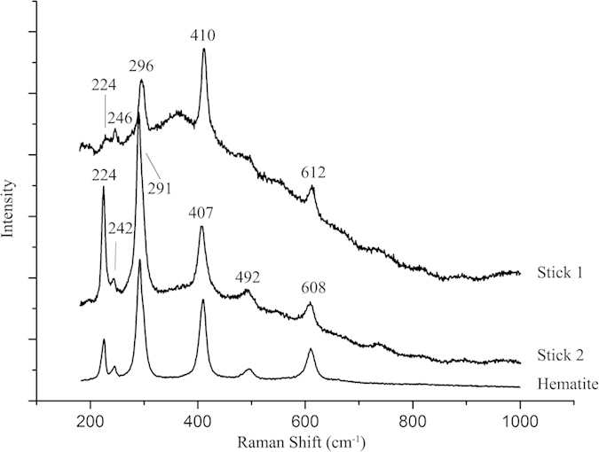
The Raman analysis was used to identify the red particles on the stick surface. The recognizable peaks of the red particles were showed in Fig. 5. The Raman spectra of two samples were compared with the Raman spectrum of hematite downloaded from the RRUFF database (Hematite R060190) and they had almost the same main peaks. The peaks at 224,246, 296, 410 and 612 cm−1 in the spectrum of stick1 and the peaks at 224, 242, 291,407, 492 and 608 cm−1 in the spectrum of stick2 were characteristic peaks of hematite (Fe2O3)34. The same characteristic peak would have small shifts in different spectra, which might be caused by complex reasons, such as the different instruments and parameters, influence of burial environment and tiny difference in the crystal structure. For example, Yang et al. pointed out that the longer the burial time, the weaker the main Raman peaks of chalcedony on the surface of stone axe might be35.
Figure 5. Raman Spectra of the hematite reference and the red particles on stick 1 and stick 2.
Organic residue analysis
The FTIR was used to analyse the inner material (yellow and translucent). The spectra of both sticks were presented in Supplementary Materials Fig. S1. Compared with literature data36,37, the spectra reveal the presence of protein, whose characteristic signals was the amide group (-N(H)-C = O-). More specifically, the peak at 3290–3300 cm−1 was assigned to N-H stretching vibration region; around 1650 cm−1 to C = O stretching vibration region; around 1540 cm−1 to N-H bending vibration region, and 1400–1420 cm−1 to C-N stretching region. These four functional groups belonged to the same molecule, e.g. an amino acid. The results suggested that the organic materials of the sticks were mainly made of protein.
Thus, proteomics was carried out to identify the nature and species of the protein materials of the sticks. Each sample had been analysed twice to gain enough information about the protein composition. Except for disregarded human background proteins, such as the keratin, all the identified peptides were BLAST searched against the NCBInr database to check the species-specificity of the sequences. The identified proteins and the specific peptides were listed in Table 1, Supplementary Materials Table S1 and S2, respectively.
Table 1. Identified protein and specific peptides in sticks.
| Sample | Identified proteins | Accession number | Seq. Cov. [%] | Matching Peptides | Unique Peptides* | Position | Score | Species |
|---|---|---|---|---|---|---|---|---|
| Stick 1 (from M17) | myosin-7 | gi|41386711 | 19 | 38 | – | – | – | – |
| Predicted: myosin-6 | gi|297479068 | 13 | 26 | – | – | – | – | |
| myosin light chain 3 | gi|270483786 | 36 | 6 | AAAAPAPAPAPPPAPEPSK | 19–37 | 49 | Bos taurus, Bison bison bison, Bubalus bubalis | |
| alpha-actinin-2 | gi|77736221 | 34 | 28 | – | – | – | – | |
| Predicted: Low Quality Protein: titin | gi|297465038 | 1 | 43 | – | – | – | – | |
| desmin | gi|2959452 | 37 | 16 | – | – | – | – | |
| myosin-binding protein C, cardiac-type | gi|115495853 | 15 | 19 | SIFTVEGAER | 728–737 | 45 | Bos taurus, Bison bison bison, Bos mutus | |
| AHNLAGAGPPVTTK | 941–954 | 89 | Bos taurus, Bos mutus | |||||
| Serum albumin | gi|1351907 | 33 | 21 | LVNELTEFAK | 66–75 | 74 | Bos taurus, Bison bison bison, Bubalus bubalis, Bos mutus | |
| TCVADESHAGCEK | 76–88 | 74 | Bos taurus, Bison bison bison, Bos mutus | |||||
| DAFLGSFLYEYSR | 347–359 | 42 | Bos indicus, Bos taurus, Bison bison bison, Bubalus bubalis, Bos mutus | |||||
| RPCFSALTPDETYVPK | 508–523 | 51 | Bos indicus, Bos taurus, Bison bison bison, Bubalus bubalis, Bos mutus | |||||
| myoglobin | gi|27806939 | 50 | 6 | HPSDFGADAQAAMSK | 120–134 | 47 | Bos taurus, Bubalus bubalis | |
| hemoglobin alpha chain | gi|6006425 | 30 | 4 | VGGHAAEYGAEALER | 18–32 | 82 | Bos taurus, Bison bison bison, Bubalus bubalis, Bos mutus, Bison bonasus, Bos grunniens | |
| collagen alpha-1(VI) chain precursor | gi|219804724 | 9 | 10 | AAEYDVVFGER | 990–1000 | 39 | Bos taurus, Bison bison bison, Bubalus bubalis, Bos mutus | |
| Predicted: collagen alpha-3(VI) chain isoform 4 | gi|297473452 | 9 | 24 | – | – | – | – | |
| Stick 2 (from M22) | myosin-7 | gi|41386711 | 15 | 35 | – | – | – | – |
| tropomyosin alpha-1 chain | gi|61888866 | 11 | 4 | – | – | – | – | |
| tropomyosin 4-like | gi|296486595 | 15 | 4 | IQLVEEELNRAQER | 56–69 | 39 | Bos taurus, Bison bison bison, Bubalus bubalis, Bos mutus | |
| Predicted: Low Quality Protein: titin | gi|297465038 | <1 | 31 | NGVVLESSDK | 2730–2739 | 19 | Bos taurus | |
| desmin | gi|2959452 | 11 | 5 | – | – | – | – | |
| myosin-binding protein C, cardiac-type | gi|115495853 | 4 | 5 | – | – | – | – | |
| Serum albumin | gi|1351907 | 31 | 21 | YICDNQDTISSK | 286–297 | 25 | Bos indicus, Bos taurus, Bison bison bison, Bubalus bubalis, Bos mutus | |
| DAIPENLPPLTADFAEDK | 319–336 | 64 | Bos indicus, Bos taurus | |||||
| Hemoglobin subunit alpha-2 | gi|122286 | 40 | 5 | VGGHAAEYGAEALER | 17–31 | 61 | Bos taurus, Bos grunniens, Bison bison bison, Bos mutus, Bison bonasus | |
| collagen, type VI, alpha 3-like isoform 3 | gi|296488813 | <1 | 8 | – | – | – | – |
The identified proteins contained myosin, actin, muscle auxiliary protein, collagen and others. Myosin is the main constituent units of myofibrils, which has two light chains and two long chains, and is also the components of the cytoskeleton38. Actin is a structural protein of microfilaments, which is the main protein of the cytoskeleton, and is widespread in the various types of muscle tissue and cell; especially the alpha-actin plays an important role in the regulation of the muscle fibers39. Titin, desmin and myomesin also belong to cytoskeletal proteins of muscle40. Besides, the collagen is the major component of connective tissue and serum albumin is the most abundant protein in animal plasma. According to the dataset of identified proteins, it was obvious that the inner material of sticks contained the muscle tissue. Notably, the myosin-binding protein C was identified as the cardiac-type. In addition, the type VI collagen was found and certified to exist in the heart tissue in previous work41,42. Thus, the sticks were made of animal heart.
According to the results of BLAST, the specific peptides equally present in cattle (Bos taurus), bison (Bison bison bison), yak (Bos mutus), zebu (Bos indicus), domestic yak (Bos grunniens), European bison (Bison bonasus) and water buffalo (Bubalus bubalis). The MS/MS spectra of bovine-specific peptides, such as AHNLAGAGPPVTTK from myosin-binding protein C, cardiac-type in stick1 and IQLVEEELNRAQER from tropomyosin 4-like in stick2 were shown in Fig. 6. Although the complete zooarchaeological data of Xiaohe Cemetery are not published, other species of bovine bones except of cattle have been not found28. Furthermore, some DNA analyses identified bovine remains in Xiaohe Cemetery as Bos taurus27,29. And the desert environment in Xiaohe is not suitable for yak to live. Thus, the cattle is the most possible interpretation for the origin of these specific peptides and the organic part of both sticks is from cattle heart.
Figure 6.

MS/MS spectra confidently identified two specific peptide sequences: (a) AHNLAGAGPPVTTK from stick 1 and (b) IQLVEEELNRAQER form stick 2.
Discussion
The red cosmetic sticks analysed were made of cattle heart covered with hematite powders. Hematite was one of the most widely used red pigments with the development of human being. About 18000 years ago, the hematite powder was found around the bones at Zhoukoudian, Beijing1. It was a common phenomenon of using hematite to paint red on human skull and axe in Qijia Culture and Xindian Culture thousands of years ago43. During the historical period, besides widely used as a kind of pigment in painting, mural painting and coloured painting sculpture, hematite is also treated as a traditional mineral drug to cure disease in China44. However, it was firstly reported that the cattle heart was used as cosmetic stick up to our knowledge, and the myocardium contained a certain amount of fat and collagen45, which were traditional and natural adhesives so as to attach the pigments conveniently to paint. And these sticks were irregular, which might be residue after painting or caused by the dehydration of muscle. Besides as the cosmetic tool, the stick also might be a kind of early painting tool, like brush or crayon, supported by plenty of relics painted red in Xiaohe Cemetery. Up to now, the earliest unearthed brush made of bamboo and rabbit hair was dated to the 4th-2nd centuries BC in China46, so the sticks in Xiaohe Cemetery could provide information for the development of early painting tool.
The cosmetic sticks were found in several archaeological sites in Xinjiang. In tomb M11 of Suebeixi Cemeteries47 (5th -3rd centuries BC), a leather bag was unearthed with string, comb, stone stick and red, black and white pigments and all of these were considered as a set of cosmetic implements. The stone stick had smooth surface and conical shape. A conical stick of 6.5 cm in length and 0.9 cm in diameter was found with a leather bag having brown powder in Sangeqiao Cemetery48 (4th -2nd centuries BC). The similar sticks and pigments were found in Zagunluk Cemetery49 (7th centuries BC -1st centuries AD), Baileqier Cemetery50 (cal. 2500–2700BP), Chawuhu Valley Cemetery51 (cal. 3000–2500BP) and so on. Since these sticks were usually unearthed with pigments, they were generally considered to be used for cosmetic, and sticks, pigment and/or leather bag were treated as a set of implements which was helpful to judge the gender of tomb occupier due to they were always buried with the female. These sticks share the similar shape and materials23: rod-shaped or conical and made of stone or wood (Fig. S3), which were distinct from those of Xiaohe Cemetery. Although cosmetic sticks and leather bags in Xiaohe Cemetery and other sites had the similar usage, the materials, shape and production of cosmetic sticks change greatly: more exquisite, suitable shape like today crayon and more wear-resistant materials.
More importantly, the cattle heart sticks had obvious religious significance. According to the excavated report, many of the relics in Xiaohe Cemetery were red-painted notably, such as the horn of the ox, the end of the arrow and the stand wooden pillar in front of the coffin24,32. The red colour was usually considered as a symbol of the worship for the blood52. The heart is one of the most important organs in cattle, which is the centre of the blood circulation. Using cattle heart as the painting tools might show that the worship of the blood was the mainly signature of the social religious culture. In addition, the stand wooden pillar in front of the coffin was considered as the sign of the reproduction worship. So using cattle heart as tools to paint red on human face and objects such as stand wooden pillar and wooden phallus indicated that painting red was a sacred and significant religious behaviour for Xiaohe people. Because the cosmetic sticks were in the leather bags buried with the female mummy, it is deduced that the woman carried out painting or making up with cosmetic sticks, which implied that woman played a special role in religious activities.
Plenty of evidences provide the wide use of cattle in Xiaohe culture, including cattle skulls tied to the wooden grave markers, cattle hide wrapping the coffins, cattle hide boots, buried cattle ears etc24,32. Xiaohe people widely used cattle’s products: sacrifice in the religious practices; a source of food, besides the meat, proteomic analysis indicated that milk and kefir cheese played an important role of Xiaohe people’ diet30,53; a kind of cloth; even the cow dung was used as a source of fertilizer or fuel54; and to manufacture animals glue55. Moreover, as indicated in this study, the heart of cattle was used as a cosmetic stick–a special painting tool.
Furthermore, Xiaohe Cemetery located in the desert where the output of hematite was never reported, so the source of the hematite was a significant issue to consider. Clarifying the details of communication areas and routes was required to do more research such as the distribution of hematite in Xinjiang, the migration of population among different culture and so on. All of these are the focus of the further work.
In this study, we identify that the red cosmetic sticks unearthed in Xiaohe Cemetery were made of cattle heart and the red pigment was hematite. Painting red and utility of cattle heart was the reflection of the society culture and worship in Xiaohe culture. More importantly, the women acted as the painter involved in religious activities. This work reveals the characterization of the cosmetic sticks used in ancient Xinjiang in China almost 3500 years ago, and the results provided a new perspective to understand the utilizations of cattle of Xiaohe people. Furthermore, it could broaden our understanding of the applications of the animal heart in prehistoric times.
Methods
SR-μCT scanning
The sticks were scanned by SR-μCT at the Shanghai Synchrotron Radiation Facility (SSRF), 13W, Shanghai, China. The parallel SR X-ray with the width of 2 cm and height of 4 mm was directed at the object with a source energy setting of 18 keV. The space resolution of the CCD detector was 9 μm. The data were analyzed by Mimics 12 software.
FTIR analysis
FTIR analysis was employed to characterize major constituent of the painting and guide further analysis. The samples were analysed as KBr micro pellets with a Nicolet 6700 (Thermo Scientific) FTIR spectrometer working in a transmission mode. Spectra were acquired over the range of 4000–400 cm−1 using a resolution of 4 cm−1, with 32 scans per spectrum. The software OMNIC 8.0 was applied to deal with the data.
Raman analysis
Raman analysis was performed on Laser Micro-Raman Spectrometer (JY XploRA, France). In the dark room and at room temperature, after being calibrated with a single crystal Si wafer sample, the sample excitation was performed with a 785 nm laser and focused through a 50× objective lens. The diffraction grating was 1200 g/mm and the width of the entrance slit of spectrometer was 100 μm. Spectra of the samples were recorded with a resolution of 2 cm−1 and three accumulations of 20 s each in the range 100–1000 cm−1 using the NGS LabSpec software.
Proteomic analysis
For each archaeological sample a subsample of 20 mg was suspended in 100 μL of extracting solution (Tris/HCl, pH 8.0, 10% SDS, 10 mM DTT and 0.0025% bromphenol blue). After being subjected to ultrasonic baths (3 × 15 min), the sample was incubated for 1 h at 56 °C. Then the sample was subjected to ultrasonic baths again for 15 min and centrifuged for 15 min at 12,000 g.
45 μL of the supernatant of each sample was mixed with 5 μL of glycerol, heated at 95 °C for 5 min. The extracting solution was loaded onto the gel (SDS-PAGE, sodium dodecyl sulfate polyacrylamide gel electrophoresis) with 25 μL each well. The electrophoresis apparatus was connected to a 200 V power. The gel with sample was stained by the solution (0.25% Coomassie Blue w/v, 50% ethanol, 10% acetic acid) and destained by the solution (25% ethanol, 8% acetic acid) until protein bands visible.
The gel with protein bands was cut into small pieces and washed by distilled water three times. Then the gel pieces were destained with 50% acetonitrile/25 mM NH4HCO3 and alkylated in the dark with 50 mM iodoacetamide for 30 min. After being washed with 25 mM NH4HCO3 buffer twice, the gel pieces of each sample were immersed in 12.5 ng/μL trypsin solutions, approximate 50–80 μL.And put in the microwave oven at 850W for 1 min for the digestion.
0.1% formic acid was added to dissolve the digested samples. Then the samples were analysed by RP C18 capillary LC column from Michrom Bioresources (100 μm × 150 mm, 3 μm). The parameters of LTQ Orbitrap Velos mass spectrometer was: 10 data-dependent HCD MS/MS scans per every full scan; a resolution of 30,000 and 30% normalized collision energy; charge state screening (excluding precursors with unknown charge state or +1 charge state); internal mass calibration (445.120025 ion as lock mass with a target lock mass abundance of 0%) and dynamic exclusion (exclusion size list 500, exclusion duration 30 s).
The MS/MS spectra were searched against the NCBInr (20093899 sequences; 6882348701 residues) by Mascot software version 2.4.1 (Matrix Science, UK). Trypsin was selected as the proteolytic enzyme and two missed cleavages were allowed. Carbamidomethylation (C) was selected as fixed modification. Deamidation (N and Q) and Dioxidation (M) were selected as variable amino-acid modifications. A peptide tolerance of 5 ppm and a product ion tolerance of 0.05 Da were used in the searches and the peptides were filtered with significance threshold p < 0.05 and ions score cut-off 13.
Additional Information
How to cite this article: Mai, H. et al. Characterization of cosmetic sticks at Xiaohe Cemetery in early Bronze Age Xinjiang, China. Sci. Rep. 6, 18939; doi: 10.1038/srep18939 (2016).
Supplementary Material
Acknowledgments
This study was supported by the grants from the National Science Foundation in China (41172164), the CAS Strategic Priority Research Program (XDA05130303) and Youth Innovation Promotion Association of CAS (2013281).
Footnotes
Author Contributions H.M. and Y.Y. designed the research, performed experiments and analysed data. I.A., W.L. and X.H. provided the sticks samples materials and resources. C.W. supervised the research and H.M. and Y.Y. wrote the paper, with input from the other co-authors.
References
- Yu Jinxiu Y. S. Archaeology in Chinese Vol. 3–10 (China Social Sciences Publishing House, 1996). [Google Scholar]
- Province I. o. C. R. i. L. The excavation of the “NüShen Temple” and Stone Barrows of Hongshan culture in Niuheliang, Liaoning province (in Chinese). Cultural Relics 8, 6 (1986). [Google Scholar]
- Cotte M., Dumas P., Richard G., Breniaux R. & Walter P. New insight on ancient cosmetic preparation by synchrotron-based infrared microscopy. Analytica Chimica Acta 553, 105–110, doi: 10.1016/j.aca.2005.07.067 (2005). [DOI] [Google Scholar]
- Ribechini E., Modugno F., Perez-Arantegui J. & Colombini M. P. Discovering the composition of ancient cosmetics and remedies: analytical techniques and materials. Analytical and bioanalytical chemistry 401, 1727–1738, doi: 10.1007/s00216-011-5112-2 (2011). [DOI] [PubMed] [Google Scholar]
- Ya L. The Chinese Dressing through the years (in Chinese). 3–6 (China Textile & Apparel Press, 2011). [Google Scholar]
- Xingliang H. Totem Culture of China (in Chinese). 33–54 (China Social Sciences Publishing House, 1992). [Google Scholar]
- Jiao Yongqin S. H. The Relationship between Totem Worship and the Facial Tattoos of Li Women (in Chinese). Literature Contend 3, 149–151 (2006). [Google Scholar]
- Zhou Xun G. C. The Chinese Women Dressing through the Years (in Chinese). 11–290 (Academia Press and Joint Publishing (Hong Kong) Company, 1991). [Google Scholar]
- Huafeng L. The Study of Face Makeup and Beauty Supplies and Its’ Production in Ancient China (in Chinese) Master thesis, Zhengzhou University, (2007).
- Colombini M. P., Giachi G., Iozzo M. & Ribechini E. An Etruscan ointment from Chiusi (Tuscany, Italy): its chemical characterization. Journal of Archaeological Science 36, 1488–1495, doi: 10.1016/j.jas.2009.02.011 (2009). [DOI] [Google Scholar]
- Van Elslande E. et al. Analysis of ancient Greco-Roman cosmetic materials using laser desorption ionization and electrospray ionization mass spectrometry. Analytical and bioanalytical chemistry 390, 1873–1879, doi: 10.1007/s00216-008-1924-0 (2008). [DOI] [PubMed] [Google Scholar]
- Gamberini M. C., Baraldi C., Palazzoli F., Ribechini E. & Baraldi P. MicroRaman and infrared spectroscopic characterization of ancient cosmetics. Vibrational Spectroscopy 47, 82–90, doi: 10.1016/j.vibspec.2008.02.005 (2008). [DOI] [Google Scholar]
- Dooryhée E., Martinetto P., Walter P. & Anne M. Synchrotron X-ray analyses in art and archaeology. Radiation Physics and Chemistry 71, 863–868, doi: 10.1016/j.radphyschem.2004.04.129 (2004). [DOI] [Google Scholar]
- Deeb C., Walter P., Castaing J., Penhoud P. & Veyssire P. Transmission Electron Microscopy (TEM) investigations of ancient Egyptian cosmetic powders. Applied Physics A: Materials Science & Processing 79, 393–396, doi: 10.1007/s00339-004-2543-z (2004). [DOI] [Google Scholar]
- Wang Zhendong. et al. Primary Study on Cosmetics Excavated from Yuan Dynasty Tomb in Huashizui (in Chinese). Rock and Miner Alanalysis 27, 255–258 (2008). [Google Scholar]
- Li Xiaoli Q. Y. & Jingsong X. Analysis of a “red powder” unearthed in Wulidun Ezhou city ,Hubei province (in Chinese). Sciences of Conservation and Archaeology 22, 46–48 (2010). [Google Scholar]
- Lucas A. Cosmetics, Perfumes and Incense in Ancient Egypy. The Journal of Egyptian Archaeology 16, 41–53 (1930). [Google Scholar]
- Martinetto P. et al. Cosmetic recipes and make-up manufacturing in ancient Egypt. Newsletter ESRFApril, 10–11 (1999). [Google Scholar]
- Walter. P. et al. Making make-up in ancient Egypt. Nature 397, 483–484 (1999). [Google Scholar]
- Patkar K. B. Herbal Cosmetic in ancient India. Indian J. Plast. Surg. Supplement. 41, 134–138 (2008). [PMC free article] [PubMed] [Google Scholar]
- Yamada M.-o. et al. Different element ratios of red cosmetics excavated from ancient burials of Japan. Science of The Total Environment 199, 293–298, doi: 10.1016/S0048-9697(97)05474-0 (1997). [DOI] [PubMed] [Google Scholar]
- Maier M. S. et al. Combined use of vibrational spectroscopy and GC–MS methods in the characterization of archaeological pastes from Patagonia. Vibrational Spectroscopy 44, 182–186, doi: 10.1016/j.vibspec.2006.09.003 (2007). [DOI] [Google Scholar]
- Penghui W. A Study on the Gender of the Sets of “the Cosmetic Stick” Implements io Xinjiang Prehistoric Archaeology (in Chinese). Research of China’s Frontier Archaeology 11, 329–341 (2012). [Google Scholar]
- Region C. R. a. A. I. o. X. A. The excavation report of xiaohe Cemetery (in Chinese). Research of China,s Frontier Archaeology 3, 338–398 (2004). [Google Scholar]
- Region C. R. a. A. I. o. X. A. A brief excavation report on Xiaohe graveyard located in Luobupo, Xinjiang Autonomous Region (in Chinese). Cultural Relics 10, 4–42 (2007). [Google Scholar]
- Mair V. H. The rediscovery and complete excavation of Ordek’s Necropolis. Journal of Indoeuropean Studies 34, 273 (2006). [Google Scholar]
- Chunxiang L. Molecular Genetic Analysis of Ancient Remains from Xiaohe Cemetery (in Chinese) PhD thesis, Jilin University, (2010).
- Wenying L. The Application of Scientific Archaeologly in Rsearch of the Xiaohe Culture (in Chinese) in China Cultural Relics News Vol. 2013-11-08 1–4 (Beijing, 2013).
- Cai Dawei S. Y., Zhuowei T. & Hui Z. Molecular Archaeological Research on the Origins of Cattle in Northern China (in Chinese). Quaternary International 34, 166–172 (2014). [Google Scholar]
- Yang Y. et al. Proteomics evidence for kefir dairy in Early Bronze Age China. Journal of Archaeological Science 45, 178–186, doi: 10.1016/j.jas.2014.02.005 (2014). [DOI] [Google Scholar]
- Binghua W. Reproductive Worship: spiritual and cultural core of early humans—the soul of the No. 5 Xiaohe Cemetery in Xinjiang (in Chinese). Root Exploration 04, 4–14 (2004). [Google Scholar]
- Region C. R. a. A. I. o. X. A. Important gains of Lop river cemetery archaeological excavations (in Chinese). Turfanological Research 1, 114–119 (2005). [Google Scholar]
- Yin Zongjun Z. M. & Tiqiao X. Application of synchrotron X-ray microtomography in paleontology for nondestructive 3-D imaging of fossil specimens (in Chinese). Physics 38, 504–509 (2009). [Google Scholar]
- Wang Jiying W. L. & Zhaojun L. Raman Spectra of Some MIneral Pigments Used in Anicent Chinese Artworks (in Chinese). The Journal of Egyptian Archaeology 24, 86–91 (2012). [Google Scholar]
- Yang Qun W. Y., Pengxiang Z. & Raman L. C. Micro-spectroscopy as a Technique Studying a Stone Ax of New Stone Age (in Chinese). Chinese Journal of Light Scattering 13, 119–123 (2001). [Google Scholar]
- Michael Jackson H. H. M. The Use and Misuse of FTIR Spectroscopy in the Determination of Protein Structure. Biochemistry and Molecular Biology 30, 95–120 (1995). [DOI] [PubMed] [Google Scholar]
- Zhou Ruiming S. Y. Study on Secondary Structures of a Prote in by Infrared Spectra (in Chinese). Journal of East China University of Science and Technology 23, 422–425 (1997). [Google Scholar]
- Li Dongying R. W. Influence of exer cise on the structur e and function of myosin (in Chinese). Journal of Clinical Rehabilitative Tissue Engineering Research 11, 6458–6464 (2007). [Google Scholar]
- Chen Ming L. A. Research Progress of the Actin, Actin-binding Protein and Cell Movement (in Chinese). Life Sciences 9, 1–5 (1997). [Google Scholar]
- Kong Baohua X. Y. Properties and roles of regulatory and cytoskeletal proteins in muscle (in Chinese). Science and Technology of Food Industry 32, 439–442 (2011). [Google Scholar]
- Kitten G., Kolker S., Krob S. & Klewer S. Type VI collagen in the cardiac valves and connective tissue septa during heart development. Brazilian journal of medical and biological research = Revista brasileira de pesquisas medicas e biologicas/Sociedade Brasileira de Biofisica…[et al.] 29, 1189–1193 (1996). [PubMed] [Google Scholar]
- Duff K., Williamson R. & Richards S.-J. Expression of genes encoding two chains of the collagen type VI molecule during human fetal heart development. International journal of cardiology 27, 128–129 (1990). [DOI] [PubMed] [Google Scholar]
- Zhiwei G. On the Ancient Sources of Ocher, Vermilion, Red Lead and its Application (in Chinese). Journal of Qinghai Nationalities University (social science) 37, 102–109 (2011). [Google Scholar]
- Chen Bingchun. et al. Research Progress of Traditional Mineral Chinese Medicine (in Chinese). China Journal of Chinese Materia Medica 39, 181–184 (2014). [PubMed] [Google Scholar]
- Sun Xiaoming L. L., Jiacheng Z., Songshan Z. & Baozhong S. Research on Prediction Chemical Composition of Beef by Near lnfrared Reflectance Spectroscopy (in Chinese). Spectroscopy and Spectral Analysis 31, 379–383 (2011). [PubMed] [Google Scholar]
- Tao C. The Manufacturing of Writing Brush during the Qin, Han, Wei, Jin, Northern and Southern Dynasties (in Chinese). Academic Forum of Nandu (Journal of the Humanities and Social Sciences) 32, 24–32 (2012). [Google Scholar]
- Cultural Relics and Archaeology Institute of Xinjiang Autonomous Region & Museum T. D. The Site and Cemetery at Subeixi in Shanshan County, Xinjiang (in Chinese). Archaeology 6, 42–57 (2002). [Google Scholar]
- Zhang Yongbing X. K. & Zhan L. A brief excavation report on Sangeqiao Cemetery in Shanshan, Xinjiang (in Chinese). Cultural Relics 6, 46–56 (2002). [Google Scholar]
- The Museum of Xinjiang Uygur Autonomous Region, C. o. B. M. A. P. a. C. o. C. C. Excavation of Graveyard No.1 at Zagunluk in Charchan, Xinjiang (in Chinese). Acta Archaeologica Sinica 1, 89–136 (2003). [Google Scholar]
- Cultural Relics and Archaeology Institute of Xinjiang Autonomous Region, H. N. M. A brief excavation report on Baileqier Cemetery in Hejing, Xinjiang (in Chinese) Cultural Relics of Xinjiang 3,4, 30–61 (1999). [Google Scholar]
- Archaeology, X. I. o. C. R. a. Xinjiang Chawuhu—a Large-scale Clan Cemetery Excavation Report (in Chinese). 1–328 (Oriental Press, 1999). [Google Scholar]
- Jinyuan C. Worship and taboos about the colour RED— a study of RED in Chinese belief systems (in Chinese) Master thesis, Sichuan University, (2007).
- Liang Yiming. et al. Proteomic analysis of residues in g rass basket excavated from Xiao_He graveyard (in Chinese). Sciences of Conservation and Archaeology 24, 81–85 (2012). [Google Scholar]
- Qiu Z. et al. Paleo-environment and paleo-diet inferred from Early Bronze Age cow dung at Xiaohe Cemetery, Xinjiang, NW China. Quaternary International 349, 167–177, doi: 10.1016/j.quaint.2014.03.029 (2014). [DOI] [Google Scholar]
- Rao H. et al. Proteomic identification of adhesive on a bone sculpture-inlaid wooden artifact from the Xiaohe Cemetery, Xinjiang, China. Journal of Archaeological Science 53, 148–155, doi: 10.1016/j.jas.2014.10.010 (2015). [DOI] [Google Scholar]
- Li J. F. et al. Buried in Sands: Environmental Analysis at the Archaeological Site of Xiaohe Cemetery, Xinjiang, China. PloS one 8, e68957 (2013). [DOI] [PMC free article] [PubMed] [Google Scholar]
Associated Data
This section collects any data citations, data availability statements, or supplementary materials included in this article.