Abstract
Objective:
To investigate whether reduced radiation dose abdominal CT images reconstructed with adaptive statistical iterative reconstruction V (ASIR-V) compromise the depiction of clinically competent features when compared with the currently used routine radiation dose CT images reconstructed with ASIR.
Methods:
27 consecutive patients (mean body mass index: 23.55 kg m−2 underwent CT of the abdomen at two time points. At the first time point, abdominal CT was scanned at 21.45 noise index levels of automatic current modulation at 120 kV. Images were reconstructed with 40% ASIR, the routine protocol of Dong-A University Hospital. At the second time point, follow-up scans were performed at 30 noise index levels. Images were reconstructed with filtered back projection (FBP), 40% ASIR, 30% ASIR-V, 50% ASIR-V and 70% ASIR-V for the reduced radiation dose. Both quantitative and qualitative analyses of image quality were conducted. The CT dose index was also recorded.
Results:
At the follow-up study, the mean dose reduction relative to the currently used common radiation dose was 35.37% (range: 19–49%). The overall subjective image quality and diagnostic acceptability of the 50% ASIR-V scores at the reduced radiation dose were nearly identical to those recorded when using the initial routine-dose CT with 40% ASIR. Subjective ratings of the qualitative analysis revealed that of all reduced radiation dose CT series reconstructed, 30% ASIR-V and 50% ASIR-V were associated with higher image quality with lower noise and artefacts as well as good sharpness when compared with 40% ASIR and FBP. However, the sharpness score at 70% ASIR-V was considered to be worse than that at 40% ASIR. Objective image noise for 50% ASIR-V was 34.24% and 46.34% which was lower than 40% ASIR and FBP.
Conclusion:
Abdominal CT images reconstructed with ASIR-V facilitate radiation dose reductions of to 35% when compared with the ASIR.
Advances in knowledge:
This study represents the first clinical research experiment to use ASIR-V, the newest version of iterative reconstruction. Use of the ASIR-V algorithm decreased image noise and increased image quality when compared with the ASIR and FBP methods. These results suggest that high-quality low-dose CT may represent a new clinical option.
INTRODUCTION
Dose reduction in body CT has become a top priority owing to concerns regarding the risks associated with ionizing radiation.1,2 However, dose reduction must be balanced by an acceptable level of image quality, and above all, diagnostic accuracy must be adequately maintained.
Although dose-reduction techniques, such as tube current modulation, low tube voltage and noise-reduction filters, have been implemented successfully, the most promising are the iterative reconstruction algorithms that have evolved beyond the traditional reconstruction method of filtered back projection (FBP).3–5 Adaptive statistical iterative reconstruction (ASIR; GE Healthcare, Waukesha, WI) is one of the most widely studied iterative reconstruction techniques and provides for clinically acceptable image quality with an estimated dose reduction in the range of 25–40%.6 However, the results from several studies have indicated that the use of ASIR was associated with quality problems, such as an artificial texture or a blotchy appearance, particularly when high-strength iterative reconstruction was used or large patients were scanned.3 Because many radiologists perceive this image alteration as unacceptable, the manufacturer (GE Healthcare) began offering this technology as a combination of the traditional FBP and 20–40% ASIR.7,8 Model-based iterative reconstruction (MBIR; Veo™; GE Healthcare, Waukesha, WI) has also become available as a fully iterative method. MBIR uses a more complex system of optical factor prediction such as X-ray tube response, detector response and many other aspects of X-ray physics such as scatter and crosstalk as well as exact geometric features of the cone beam and the absorbing voxels.9,10 It offers considerably better image quality than FBP and ASIR, even at ultralow doses.11 However, because MBIR requires a long processing time, it has not yet been widely applied in routine clinical practice.12
Most recently, a novel iterative reconstruction technique, ASIR-V was developed by GE Healthcare. This technique uses a nearly full iterative reconstruction process in-between ASIR and MBIR. It has the potential for clinically feasible dose reduction with better image quality than conventional ASIR, as well as a shorter imaging processing time than MBIR. Therefore, it can be considered conceptually as “augmented ASIR based on MBIR” or “modified MBIR”.
The purpose of this study was to compare objective and subjective image quality parameters of image reconstruction algorithms using ASIR and ASIR-V from reduced radiation dose abdominal CT examinations. The ultimate study goal was to investigate whether reduced radiation dose CT image reconstruction using ASIR-V was competitive when compared with our standard clinical radiation dose CT images reconstructed with ASIR, and whether the use of ASIR-V would result in a decrease in image quality.
METHODS AND MATERIALS
This prospective single institution study was Health Insurance Portability and Accountability Act compliant and was approved by our institutional review board. Written informed consent was obtained from all patients.
GE Healthcare provided the hardware and software support for the reconstruction of the ASIR-V images.
Patient population
The inclusion criterion was that a patient had undergone contrast-enhanced portal venous phase abdominal routine CT and was scheduled for a clinically indicated follow-up multiphase CT. To recruit patients for the study, we checked our radiology information system to identify patients scheduled for a follow-up clinical CT examination with the same scanner used in our study less than 3 months prior. Clinical exclusion criteria were severe allergy to iodinated contrast materials, compromised renal function (glomerular filtration rate of <40 ml min−1 1.73 m−2) and pregnancy. These inclusion and exclusion criteria were applied to find eligible patients from August to November 2014. Among the 35 total patients, 8 refused to undergo this research study. Thus, we enrolled a sequential series of 27 patients (mean age, 52.4 ± 12.8 years; body mass index, 23.5 ± 1.97 kg m−2; 10 females; 17 males). The volume CT dose indexes were recorded for both the matching routine and reduced radiation dose series to establish the level of dose reduction (Table 1).
Table 1.
Patient-specific demographic data and abdominal CT doses
| Patient number | Age (years) | Sex (M, F) | Body mass index (kg m−2) | CT dose index volume (mGy) |
Dose reduction from standard (%) | |
|---|---|---|---|---|---|---|
| Regular-dose series | Reduced-dose series | |||||
| 1 | 46 | M | 25.99 | 7.89 | 5.30 | 33 |
| 2 | 50 | F | 23.53 | 8.42 | 4.71 | 44 |
| 3 | 54 | M | 24.20 | 10.01 | 5.10 | 49 |
| 4 | 49 | F | 20.70 | 6.55 | 4.96 | 24 |
| 5 | 65 | M | 24.26 | 8.56 | 5.38 | 42 |
| 6 | 54 | M | 26.96 | 9.63 | 5.91 | 38 |
| 7 | 56 | M | 28.20 | 9.49 | 5.22 | 45 |
| 8 | 75 | F | 24.65 | 8.11 | 4.91 | 40 |
| 9 | 72 | M | 17.50 | 5.56 | 4.50 | 19 |
| 10 | 48 | M | 28.01 | 9.96 | 5.81 | 42 |
| 11 | 65 | M | 26.80 | 10.79 | 5.89 | 45 |
| 12 | 44 | M | 23.17 | 8.81 | 5.45 | 38 |
| 13 | 23 | F | 18.60 | 5.59 | 4.25 | 24 |
| 14 | 59 | F | 28.30 | 7.73 | 6.25 | 19 |
| 15 | 39 | F | 25.21 | 8.21 | 4.98 | 39 |
| 16 | 65 | M | 24.41 | 6.60 | 4.97 | 25 |
| 17 | 67 | F | 24.60 | 7.97 | 5.07 | 36 |
| 18 | 55 | M | 24.22 | 8.10 | 5.04 | 38 |
| 19 | 55 | M | 21.31 | 7.96 | 5.25 | 34 |
| 20 | 21 | F | 17.60 | 5.61 | 4.01 | 28 |
| 21 | 57 | M | 19.62 | 6.22 | 3.65 | 41 |
| 22 | 55 | M | 19.20 | 5.86 | 4.06 | 30 |
| 23 | 34 | F | 18.40 | 5.82 | 3.95 | 32 |
| 24 | 55 | M | 26.91 | 10.75 | 5.45 | 49 |
| 25 | 55 | M | 26.02 | 11.70 | 5.98 | 49 |
| 26 | 50 | M | 22.81 | 6.50 | 5.06 | 22 |
| 27 | 46 | F | 23.20 | 7.31 | 5.10 | 30 |
| Mean | 52.4 | 17 M, 10 F | 23.50 | 7.99 | 5.04 | 35.4% |
F, female; M, male.
CT scanning technique
All patients underwent 128-slice multidetector CT (GE Optima™ CT660; GE Healthcare) using the following parameters: fixed noise index of 21.25, 1.25-mm slice collimation, 120 kVp; gantry rotation time of 0.5 s and 40% ASIR at baseline study. The following reduced radiation dose CT scans were performed on using the same CT scanner with the following parameters: fixed noise index of 30: 1.25-mm slice collimation: 120 kVp: gantry rotation time of 0.5 s.
We employed automatic exposure control techniques (SMART mA; GE Healthcare) based on a noise index of 21.25 for clinical standard radiation dose passes and a noise index of 30 for the reduced radiation dose pass. Two portal venous phases were compared; however, the arterial or delayed phases were not evaluated in this study.
Post-processing and image reconstruction
Because our standard dose protocols are based on a 40% ASIR approach as well as the ASIR series, a 40% blend was used to optimize subjective image quality.4,5,13 The images obtained with the clinical routine radiation dose at the first time point from the portal venous phase were reconstructed using only 40% ASIR (ASiR; GE Healthcare, Milwaukee, WI) blended with 60% FBP, with reconstruction typically requiring 1–2 min. The images obtained with the reduced radiation dose at the second time point from the portal venous phase were reconstructed using FBP, 40% ASIR, 30% ASIR-V, 50% ASIR-V and 70% ASIR-V with reconstruction software support provided by GE Healthcare. All images (clinical routine dose, reduced dose) were reconstructed with a 2.5-mm slice thickness at 1.25-mm intervals in the axial plane (AW Advantage Workstation; GE Healthcare). Six series image sets (40% ASIR regular dose; FBP, 40% ASIR, 30% ASIR-V, 50% ASIR-V and 70% ASIR-V reduced radiation dose) were created. All image sets were then pushed to a research folder on a picture archiving and communication system (PACS). Display was calibrated, and all viewing conditions were held constant according to the applicable recommendations. All image analyses were performed on PACS software (Maroview 5.4; Infinitt, Seoul, Republic of Korea) with 5-MP light-emitting diode monitors (2048 × 2560, MDCG-5121; Barco, Kortrijk, Belgium).
Analysis of image quality
The 162 data sets (27 routine-dose CT scans with 40% ASIR, V series of 27 reduced-dose CT scans with FBP, 40% ASIR, 30% ASIR-V, 50% ASIR-V and 70% ASIR-V) were randomized and rendered anonymous such that the readers were unaware of which scanning technique had been used. All data sets were displayed using the soft-tissue window setting [window/level, 400/40 Hounsfield units (HUs)].
Qualitative analysis
For assessment of the qualitative analysis, all reduced radiation dose CT series reconstructed with FBP, 40% ASIR, 30% ASIR-V, 50% ASIR-V and 70% ASIR-V for each patient (identifying information removed) were randomized. Selected representative images were chosen by a radiologist who was not involved in grading the examinations. After all five randomized reduced radiation dose series were interpreted for each patient; the clinical routine-dose 40% ASIR series for each patient was evaluated for subjective quality in order to serve as the reference standard. Two sets of images were analysed; the first for image noise, diagnostic acceptability and artefacts; and the second matched to the same level of the aorta below the superior mesenteric artery in order to assess the image sharpness of the aortic wall.
Independent qualitative image analysis was then performed by two board-certified and fellowship-trained abdominal radiologists with 10 and 15 years' experience. On side-by-side monitors in a one-to-one format, the images were compared. In each case, the two readers were blinded to the reconstruction types for the reduced radiation dose series.
Image sharpness, image noise and diagnostic acceptability were all graded on a scale from 1 (worst) to 5 (best). Image sharpness was assessed by evaluating aortic wall sharpness. Artefacts were quantified on a scale from one to four (Table 2). To improve discrimination, 0.5-interval scores were allowed. Scores were derived for axial images using the soft-tissue window setting in a standard PACS system. The five pre-determined levels for scoring were through the portal vein bifurcation and sacroiliac joints. Mean image quality scores were calculated individually for each of the two radiologists. Based on the scores from all values, an overall image quality score was calculated for each aspect.
Table 2.
Grading score for qualitative image analysis
| Grading score | Qualitative analysis |
|||
|---|---|---|---|---|
| Subjective noise | Sharpness | Artefacts | Diagnostic acceptability | |
| 1 | Unacceptably noisy | Blurry | Affecting diagnostic information | Unacceptable |
| 2 | Better than one, poorer than three | Poorer than three | Major artefacts affecting the visualization of normal structure | Better than one, poorer than three |
| 3 | Some noise in an acceptable image | Minor burring in an acceptable image | Minor artefacts not affecting the visualization of any structure | Probably confident |
| 4 | Better than three, poorer than five | Better than three, poorer than five | Absent | Better than three, poorer than five |
| 5 | Minimal or no noise | Sharpest | Not applicable | Best |
Quantitative analysis
For all series, image noise, as reflected by the standard deviation and CT numbers (HU), was measured in a 250-mm2 circular region of interest (ROI). The ROI was placed in three standard locations: liver parenchyma in the mid liver (excluding large hepatic vessels), gluteal subcutaneous fat and the bladder. To ensure consistency, ROI placement per patient was matched as closely as possible to that used on the reduced radiation dose series.
Statistical analyses
The quantitative data were analysed by comparing the image noise and HUs among the six reconstructed image scan settings using one-way repeated measures analysis of variance and Kruskal–Wallis correction for multiple comparisons. The qualitative image analysis between the two radiologists was compared using Friedman test with Dunn post-test. The interobserver agreement was also statistically analysed using the intraclass correlation coefficient. All calculations and statistical analyses were performed using Microsoft® Excel® 2007 (Microsoft Office Excel 2007; Microsoft, Richmond, VA) and SPSS® software v. 21 (IBM Corporation, Armonk, NY; formerly SPSS Inc. Chicago, IL). p < 0.05 was considered to represent a statistically significant difference.
RESULTS
In the follow-up study, the mean dose reduction relative to the currently used common radiation dose was 35.37% (range: 19–49%).
Qualitative analysis
There was moderate to substantial interobserver agreement between the two radiologists regarding the subjective image quality criteria assessed in all 27 abdominal CT examinations (K = 0.651–0.830). Subjective image noise, sharpness, artefact, and diagnostic acceptability of the CT images reconstructed with routine-dose 40% ASIR and various lower dose reconstruction techniques are summarized in Tables 3 and 4.
Table 3.
Subjective image quality (IQ) and artefacts
| IQ criteria | 40% ASIR at routine dose |
Filtered back projection at lower dose |
40% ASIR at lower dose |
30% ASIR-V at lower dose |
50% ASIR-V at lower dose |
70% ASIR-V at lower dose |
Intraclass correlation coefficient | ||||||
|---|---|---|---|---|---|---|---|---|---|---|---|---|---|
| R1 | R2 | R1 | R2 | R1 | R2 | R1 | R2 | R1 | R2 | R1 | R2 | ||
| Sharpness | 4 | 4 | 2.31 | 2.4 | 3.08 | 3.19 | 3.71 | 3.85 | 3.67 | 3.79 | 2.31 | 2.42 | 0.692 |
| Subjective noise | 5 | 5 | 2.75 | 2.81 | 3.58 | 3.63 | 4.04 | 4.08 | 4.73 | 4.73 | 5 | 5 | 0.651 |
| Artefact | 4 | 4 | 2.98 | 3.04 | 3.56 | 3.62 | 3.98 | 3.98 | 4 | 4 | 4 | 4 | 0.682 |
| Diagnostic acceptability | 5 | 5 | 4.27 | 4.29 | 4.87 | 4.85 | 5 | 5 | 5 | 5 | 4.6 | 4.58 | 0.830 |
ASIR, adaptive statistical iterative reconstruction; R, reader.
Data are subjective image quality rating regarding sharpness, subjective noise, artefacts and diagnostic acceptability in comparison with baseline 40% ASIR image at routine dose rating for two readers.
Table 4.
Post Kruskal–Wallis test at subjective image quality
| Subjective image quality | Routine-dose 40% ASIR vs lower dose protocol |
Reduced-dose 40% ASIR vs reduced-dose ASIR-V |
||||||
|---|---|---|---|---|---|---|---|---|
| Filtered back projection | 40% ASIR | 30% ASIR-V | 50% ASIR-V | 70% ASIR-V | 30% ASIR-V | 50% ASIR-V | 70% ASIR-V | |
| Subjective noise | <0.001 | <0.001 | <0.001 | 0.057 | Same | 0.001 | <0.001 | <0.001 |
| Sharpness | <0.001 | <0.002 | 0.996 | 0.980 | <0.001 | 0.068 | 0.134 | 0.004 |
| Artefact | <0.001 | <0.001 | 0.890 | Same | Same | 0.002 | <0.001 | <0.001 |
| Diagnostic acceptability | <0.001 | 0.328 | Same | Same | <0.001 | 0.326 | 0.326 | 0.199 |
ASIR, adaptive statistical iterative reconstruction.
In the same lower dose CT scan series, 30% ASIR-V and 50% ASIR-V showed significantly decreased subjective image noise and lower artefacts with no significant differences in image sharpness or diagnostic acceptability when compared with 40% ASIR at lower dose. In cases of 70% ASIR-V, subjective image noise and artefacts were also decreased; however, the sharpness of the aortic wall was clearly worse than that observed for 40% ASIR at lower dose. Therefore, a high percentage of ASIR-V is not suitable for image reconstruction.
Among the ASIR-V series, a minor, blotchy pixilated appearance of the images was seen in 1 CT study at 50% ASIR-V and in 16 CT studies at 70% ASIR-V. No such appearance was seen for 30% ASIR-V. On the other hand, a blotchy pixilated appearance was regarded as substantial in two CT image series reconstructed using 70% ASIR-V (Table 5). The pixilation scores were graded as one (none), two (minor), three (substantial), or four (severe). The pixilation score also appeared to be affected by changes in patient's weight. In a group >75 kg, the mean pixilation score was 2.1 at 70% ASIR-V, whereas the mean score for those <60 kg was 1.5 at 70% ASIR-V. However, there were no differences in 30% ASIR-V or 50% ASIR-V according to patient size. Based on these results, it would seem that ASIR-V shares problems with ASIR such as artificial texture or blotchy appearance when a high percentage of ASIR-V is used and or large patients are scanned.
Table 5.
Scores for blotchy, pixilated imaging appearance of 27 abdominal CT studies
| Reconstruction technique | Filtered back projection | 30% ASIR-V | 50% ASIR-V | 70% ASIR-V |
|---|---|---|---|---|
| Pixilation score (1/2/3/4) | 27/0/0/0 | 27/0/0/0 | 26/1/0/0 | 9/16/2/0 |
ASIR-V, adaptive statistical iterative reconstruction V.
Data are numbers of CT studies given pixilation scores of 1 (none), 2 (minor), 3 (substantial), and 4 (severe) respectively. Because discrete values cannot be averaged, the lower or similar scores for pixilation given by the radiologists are presented.
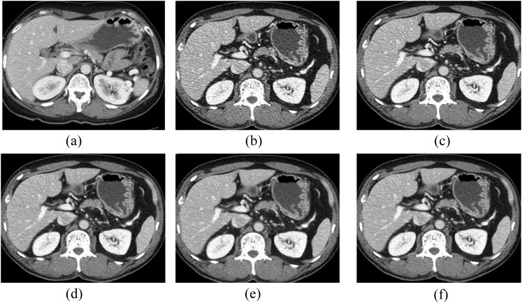
Compared with routine-dose CT scans reconstructed with 40% ASIR, the reduced radiation dose CT series reconstructed with 50% ASIR-V showed no significant differences in subjective image noise, sharpness, artefact or diagnostic acceptability (Table 4). Images reconstructed with 30% ASIR-V also did not show any significant differences in image quality pertaining to sharpness, artefact or diagnostic acceptability; however, subjective image noise was relatively high. In cases of images reconstructed with 70% ASIR-V, a relatively poor image quality was observed and attributed to worsening of image sharpness and diagnostic acceptability when compared with routine radiation dose CT reconstructed with 40% ASIR. The results from these analyses suggested that the overall subjective image quality and diagnostic acceptability scores of 50% ASIR-V at reduced radiation doses were nearly identical to the initial routine-dose CT with 40% ASIR (Figure 1a–f).
Figure 1.
Transverse abdominal CT images in a 50-year-old female with a body mass index of 23.53 kg m−2. Lower dose CT reconstruction included filtered back projection (b), adaptive statistical iterative reconstruction (ASIR) 40% (c), ASIR-V 30% (d), ASIR-V 50% (e) and ASIR-V 70% (f). The volume CT dose index for lower dose series was 4.7 mGy, representing 44% dose reduction relative to routine-dose ASIR 40% series (a). At the same reduced radiation dose CT series, all ASIR-V (d–f) images are less noisy without affecting the diagnostic acceptability. Compared with routine dose 40% ASIR series (a), both readers considered that, among the reduced radiation dose CT series, 50% ASIR-V series are nearly identical in image noise, sharpness and diagnostic acceptability without artefacts (e). The readers interpreted 70% ASIR-V series showing slightly blocky pixilated appearance at the tissue interfaces.
Quantitative analysis
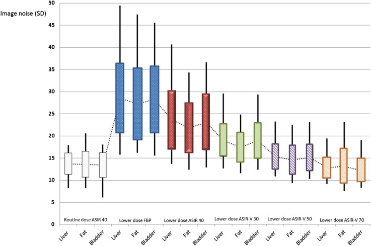
Table 6 shows the quantitative values for the image noise of the six different reconstruction algorithms. Overall, the results demonstrated that there was significant improvement in the objective image noise with the application of the reduced radiation dose CT series ASIR-V technique for abdominal CT examinations when compared with the reduced-dose 40% ASIR technique (p < 0.05; Table 7). Compared to the 40% ASIR in the routine-dose condition, objective image noise using ASIR-V at reduced radiation doses did not differ significantly at either the 50% or 70% settings (Figure 2).
Table 6.
Mean image noise according to dose and reconstruction method
| Site | Routine-dose 40% ASIR | Lower dose filtered back projection | Lower dose 40% ASIR | Lower dose 30% ASIR-V | Lower dose 50% ASIR-V | Lower dose 70% ASIR-V |
|---|---|---|---|---|---|---|
| Liver | 13.7 | 28.5 | 23.6 | 18.9 | 15.3 | 12.8 |
| Gluteal fat | 13.6 | 27.2 | 21.8 | 17.4 | 14.6 | 13.2 |
| Bladder | 13.4 | 28.2 | 23.1 | 19.0 | 15.1 | 12.2 |
| Total | 13.6 | 28.0 | 22.8 | 18.4 | 15.0 | 12.7 |
ASIR, adaptive statistical iterative reconstruction.
Table 7.
Post Kruskal–Wallis test at objective noise
| Soft tissue | Routine-dose 40% ASIR vs lower dose protocol |
Reduced-dose 40% ASIR vs Reduced-dose ASIR-V |
||||||
|---|---|---|---|---|---|---|---|---|
| Filtered back projection | 40% ASIR | 30% ASIR-V | 50% ASIR-V | 70% ASIR-V | 30% ASIR-V | 50% ASIR-V | 70% ASIR-V | |
| Liver | <0.001 | <0.001 | <0.001 | 0.324 | 0.916 | 0.037 | <0.001 | <0.001 |
| Gluteal fat | <0.001 | <0.001 | 0.001 | 0.976 | 1.000 | 0.018 | <0.001 | <0.001 |
| Bladder | <0.001 | <0.001 | <0.001 | 0.404 | 0.756 | 0.096 | <0.001 | <0.001 |
ASIR, adaptive statistical iterative reconstruction.
Figure 2.
Comparison of objective image noise values among routine dose 40% adaptive statistical iterative reconstruction (ASIR) and reduced dose with five reconstruction algorithms for liver, gluteral fat tissue and bladder are represented. This bar graph shows significant improvement in objective image noise with the application of the ASIR-V technique. Furthermore, 50% and 70% ASIR-V settings at reduced radiation doses are not significantly different compared with 40% ASIR in the routine-dose condition. Error bars indicate 95% confidence intervals. FBP, filtered back projection; SD, standard deviation.
DISCUSSION
Among the first generation of iterative reconstruction, ASIR has been the most widely studied iterative reconstruction (IR) technique and has been shown to effectively reduce image noise while maintaining image resolution by modelling the noise in every scanned projection and assuming the noise differences between neighbouring projections during the reconstruction process.14–18 This method provided for improved image quality at even lower doses than those used for FBP.
The second generation of IR, MBIR (Veo) significantly improved low-contrast resolution and decreased image noise compared with ASIR, even at ultralow doses. However, the main drawback of MBIR is its relatively long image creation processing time. For example, the creation of MBIR images takes about 1 h per case (0.09 frame per second), whereas ASIR images can be created in less than 1 min.19
ASIR-V is a new applicable method that has the potential to provide improved image quality at lower doses than ASIR and is more time effective than MBIR. ASIR-V differs from MBIR in that it excludes system optics in the modelling process, which is mainly responsible for the improvement in spatial resolution but is the most time-consuming portion of the IR process. Instead, ASIR-V focuses primarily on modelling the system noise statistics, objects and physics which are the main contributors to the reduction of noise, improvement of low-contrast detectability and reduction of artefacts in the reconstructed images. By omitting the most time-consuming component and focusing on the other factors during the IR process, ASIR-V significantly improves image quality while enabling a reconstruction speed similar to that of FBP. For this study, the reconstruction of CT images using ASIR-V at the system was not yet available. This was a preliminary study. However, the source image data were acquired on a commercially cleared Optima CT660 in routine clinical practice, with the data having additional off-line processing ASIR-V. Currently, the ASIR-V is US Food and Drug Administration (FDA) approved, with reconstruction speeds of up to 25 frames s−1.20
To our knowledge, this study is the first to use the FDA-approved commercial version of ASIR-V in a patient. The purpose of this study was to compare the image quality between ASIR and ASIR-V. Additionally, we sought to determine whether reduced radiation dose abdominal CT reconstructed with ASIR-V was a viable option when compared with regular-dose CT images reconstructed with ASIR. It should be noted that the regular-dose ASIR series already represents a reduced radiation dose CT when compared with standard dose FBP.
As with ASIR, images reconstructed with 70% ASIR-V showed low noise and a high degree of spatial correlation which may appear to have a “waxy” texture, unfamiliar to radiologists accustomed to images reconstructed with FBP or 20–40% ASIR. In our study, the overall image quality of images reconstructed with 70% ASIR-V was poor when compared with the routine-dose and lower dose CT images reconstructed with 40% ASIR.
When comparing image quality between routine-dose CT images with 40% ASIR and lower dose CT images with 50% ASIR-V, a tendency for the subjective image quality scores to be slightly lower than routine-dose 40% ASIR was observed and attributed to a mild level of increased image noise and a mild yet unfamiliar appearance of smoothing. However, this finding did not reach the level of statistical significance. Additionally, these conditions showed an equivalent highest score achieved for artefact and diagnostic acceptability. In the comparison of image quality between routine-dose CT images with 40% ASIR and lower dose CT images with 30% ASIR-V, subjective image noise was higher in 30% ASIR-V, whereas other subjective scores showed no significant differences.
This study had several limitations. For comparison purposes, we selected only a few of the possible percentage of blending techniques, such as 40% ASIR, and 30%, 50% and 70% ASIR-V. Additionally, we did not assess whether the same dose reduction with ASIR would also apply to other abdominal CT protocols that use thinner sections, such as multiphase liver, pancreatic and renal mass protocols in which 1.25- to 2.5-mm section thickness is used. Furthermore, the actual effect of ASIR-V on lesion detection and morphology was not assessed. Another limitation of our study was that the results may not be generalizable to other vendor scanners and perhaps not to other body regions using the ASIR-V technique for dose reduction. We also used a binary scoring system for the evaluation of image sharpness and noise of abdominal structures on ASIR, FBP and ASIR-V images, which may have masked subtle difference.
In conclusion, our data derived from the same patients suggest that the partial ASIR-V algorithm considerably improved image quality in comparison with both the partially iterative ASIR algorithm and traditionally applied FBP, which is not iterative. At the same time, portal venous phase CT images processed with a low-dose protocol reconstructed with partially iterative ASIR-V (50% ASIR-V) may have similar image noise, subjective image quality and a 35% lower patient radiation dose than CT images processed with a routine-dose protocol reconstructed with 40% ASIR. These results suggest that low-dose CT with ASIR-V may be a viable technique and is worthy of further study.
FUNDING
This study was supported by Dong-A University Hospital research funds, Busan, Republic of Korea and Heejin Kwon.
Contributor Information
Heejin Kwon, Email: risual@dau.ac.kr.
Jinhan Cho, Email: jojin@dau.ac.kr.
Jongyeong Oh, Email: jyoh@dau.ac.kr.
Dongwon Kim, Email: dwkim@dau.ac.kr.
Junghyun Cho, Email: jhcho@dau.ac.kr.
Sanghyun Kim, Email: shkim@dau.ac.kr.
Sangyun Lee, Email: shkim@dau.ac.kr.
Jihyun Lee, Email: risual@naver.com.
REFERENCES
- 1.Brenner DJ, Hall EJ. Computed tomography—an increasing source of radiation exposure. N Engl J Med 2007; 357: 2277–84. doi: 10.1056/NEJMra072149 [DOI] [PubMed] [Google Scholar]
- 2.Pearce MS, Salotti JA, Little MP, McHugh K, Lee C, Kim KP, et al. Radiation exposure from CT scans in childhood and subsequent risk of leukemia and brain tumors: a retrospective cohort study. Lancet 2012; 380: 499–505. doi: 10.1016/S0140-6736(12)60815-0 [DOI] [PMC free article] [PubMed] [Google Scholar]
- 3.Singh S, Kalra MK, Hsieh J, Licato PE, Do S, Pien HH, et al. Abdominal CT: comparison of adaptive statistical iterative and filtered back projection reconstruction techniques. Radiology 2010; 257: 373–83. doi: 10.1148/radiol.10092212 [DOI] [PubMed] [Google Scholar]
- 4.Sagara Y, Hara AK, Pavlicek W, Silva AC, Paden RG, Wu Q. Abdominal CT: comparison of low-dose CT with adaptive statistical iterative reconstruction and routine-dose CT with filtered back projection in 53 patients. AJR Am J Roentgenol 2010; 195: 713–19. doi: 10.2214/AJR.09.2989 [DOI] [PubMed] [Google Scholar]
- 5.Flicek KT, Hara AK, Silva AC, Wu Q, Peter MB, Johnson CD. Reducing the radiation dose for CT colonography using adaptive statistical iterative reconstruction: a pilot study. AJR Am J Roengenol 2010; 195: 126–1. doi: 10.2214/AJR.09.3855 [DOI] [PubMed] [Google Scholar]
- 6.Pickhardt PJ, Lubner MG, Kim DH, Tang J, Ruma JA, del Rio AM, et al. Abdominal CT with model-based iterative reconstruction (MBIR): initial results of a prospective trial comparing ultralow-dose with standard-dose imaging. AJR Am J Roentgenol 2012; 199: 1266–74. doi: 10.2214/AJR.12.9382 [DOI] [PMC free article] [PubMed] [Google Scholar]
- 7.Cornfeld D, Israel G, Detroy E, Bokhari J, Mojibian H. Impact of adaptive statistical iterative reconstruction (ASIR) on radiation dose and image quality in aortic dissection studies: a qualitative and quantitative analysis. AJR Am J Roentgenol 2011; 196: W336–40. doi: 10.2214/AJR.10.4573 [DOI] [PubMed] [Google Scholar]
- 8.Singh S, Kalra MK, Gilman MD, Hsieh J, Pien HH, Digumarthy SR, et al. Adaptive statistical iterative reconstruction technique for radiation dose reduction in chest CT: a pilot study. Radiology 2011; 259: 565–73. doi: 10.1148/radiol.11101450 [DOI] [PubMed] [Google Scholar]
- 9.Fleischmann D, Bose FE. Computed tomography—old ideas and new technology. Eur Radiol 2011; 21: 510–17. doi: 10.1007/s00330-011-2056-z [DOI] [PubMed] [Google Scholar]
- 10.Yu Z, Thibault JB, Bouman CA, Sauer KD, Hsich J. Fast model-based X-ray CT reconstruction using spatially nonhomogeneous ICD optimization. IEEE Trans Image Process 2011; 20: 161–75. doi: 10.1109/TIP.2010.2058811 [DOI] [PubMed] [Google Scholar]
- 11.Deák Z, Grimm JM, Treitl M, Geyer LL, Linsenmaier U, Körner M, et al. Filtered back projection, adaptive statistical iterative reconstruction, and a model-based iterative reconstruction in abdominal CT: an experimental clinical study. Radiology 2013; 266: 197–206. doi: 10.1148/radiol.12112707 [DOI] [PubMed] [Google Scholar]
- 12.Ning P, Zhu S, Shi D, Guo Y, Sun M. X-ray dose reduction in abdominal computed tomography using advanced iterative reconstruction algorithms. PLoS One 2014; 9: e92568. doi: 10.1371/journal.pone.0092568 [DOI] [PMC free article] [PubMed] [Google Scholar]
- 13.Prakash P, Kalra MK, Kambadakone AK, Pien H, Hsieh J, Blake MA, et al. Reducing abdominal CT radiation dose with adaptive statistical iterative reconstruction technique. Invest Radiol 2010; 45: 202–10. doi: 10.1097/RLI.ob013e3181dzfeec [DOI] [PubMed] [Google Scholar]
- 14.Leipsic J, Nguyen G, Brown J, Sin D, Mayo JR. A prospective evaluation of dose reduction and image quality in chest CT using adaptive statistical iterative reconstruction. AJR Am J Roentgenol 2010; 195: 1095–9. doi: 10.2214/AJR.09.4050 [DOI] [PubMed] [Google Scholar]
- 15.Pontana F, Duhamel A, Pagniez J, Flohr T, Faivre JB, Hachulla AL, et al. Chest computed tomography using iterative reconstruction vs filtered back projection (part 2): image quality of low-dose CT examinations in 80 patients. Eur Radiol 2011; 21: 636–43. doi: 10.1007/s00330-010-1991-4 [DOI] [PubMed] [Google Scholar]
- 16.May MS, Wüst W, Brand M, Stahl C, Allmendinger T, Schmidt B, et al. Dose reduction in abdominal computed tomography: intraindividual comparison of image quality of full-dose standard and half-dose iterative reconstructions with dual-source computed tomography. Invest Radiol 2011; 46: 465–70. doi: 10.1097/RLI.0b013e31821690a1 [DOI] [PubMed] [Google Scholar]
- 17.Hu XH, Ding XF, Wu RZ, Zhang MM. Radiation dose of non-enhanced chest CT can be reduced 40% by using iterative reconstruction in image space. Clin Radiol 2011; 66: 1023–9. doi: 10.1016/j.crad.2011.04.008 [DOI] [PubMed] [Google Scholar]
- 18.Vorona GA, Ceschin RC, Clayton BL, Sutcavage T, Tadros SS, Panigrahy A. Reducing abdominal CT radiation dose with the adaptive statistical iterative reconstruction technique in children: a feasibility study. Pediatr Radiol 2011; 41: 1174–82. doi: 10.1007/s00247-011-2063-x [DOI] [PubMed] [Google Scholar]
- 19.Koc G, Courtier JL, Phelps A, Marcovici PA, MacKenzie JD. Computed tomography depiction of small pediatric vessels with model-based iterative reconstruction. Pediatr Radiol 2014; 44: 787–94. doi: 10.1007/s00247-014-2899-y [DOI] [PubMed] [Google Scholar]
- 20.Lim K, Kwon H, Cho J, Oh J, Yoon S, Kang M, et al. Initial phantom study comparing image quality in computed tomography using adaptive statistical iterative reconstruction and new adaptive statistical iterative reconstruction V. J Comput Assist Tomogr 2015; 39: 443–8. doi: 10.1097/RCT.0000000000000216 [DOI] [PubMed] [Google Scholar]


