Abstract
Previously, we identified an adolescent idiopathic scoliosis susceptibility locus near human ladybird homeobox 1 (LBX1) and FLJ41350 by a genome-wide association study. Here, we characterized the associated non-coding variant and investigated the function of these genes. A chromosome conformation capture assay revealed that the genome region with the most significantly associated single nucleotide polymorphism (rs11190870) physically interacted with the promoter region of LBX1-FLJ41350. The promoter in the direction of LBX1, combined with a 590-bp region including rs11190870, had higher transcriptional activity with the risk allele than that with the non-risk allele in HEK 293T cells. The ubiquitous overexpression of human LBX1 or either of the zebrafish lbx genes (lbx1a, lbx1b, and lbx2), but not FLJ41350, in zebrafish embryos caused body curvature followed by death prior to vertebral column formation. Such body axis deformation was not observed in transcription activator-like effector nucleases mediated knockout zebrafish of lbx1b or lbx2. Mosaic expression of lbx1b driven by the GATA2 minimal promoter and the lbx1b enhancer in zebrafish significantly alleviated the embryonic lethal phenotype to allow observation of the later onset of the spinal curvature with or without vertebral malformation. Deformation of the embryonic body axis by lbx1b overexpression was associated with defects in convergent extension, which is a component of the main axis-elongation machinery in gastrulating embryos. In embryos overexpressing lbx1b, wnt5b, a ligand of the non-canonical Wnt/planar cell polarity (PCP) pathway, was significantly downregulated. Injection of mRNA for wnt5b or RhoA, a key downstream effector of Wnt/PCP signaling, rescued the defective convergent extension phenotype and attenuated the lbx1b-induced curvature of the body axis. Thus, our study presents a novel pathological feature of LBX1 and its zebrafish homologs in body axis deformation at various stages of embryonic and subsequent growth in zebrafish.
Author Summary
Scoliosis is the most common type of spinal deformity with a lateral spinal curvature of at least 10 degrees, affecting 2–4% of the population. Scoliosis caused by a primary problem related to the spine itself is classified into congenital scoliosis (CS) and idiopathic scoliosis (IS). Among these, adolescent idiopathic scoliosis (AIS), the most common form of scoliosis, is known as a common polygenic disease. Severe curving of the spine in scoliosis leads to profound psychological and social impacts, but etiology-based therapies have not been established since the precise pathological mechanisms of both IS and CS remain undefined. Previously, we identified an AIS susceptibility locus near human ladybird homeobox 1 (LBX1) by a genome-wide association study. Here, we report the functional characterization of the most significantly associated single nucleotide polymorphism (SNP), rs11190870 and LBX1 as well as its zebrafish homologues. Overexpression of LBX1 and zebrafish lbx genes caused lateral body curvature in association with the impairment of non-canonical Wnt/planar cell polarity signaling. Thus, our study presents a novel pathological feature of LBX1 in body axis deformation.
Introduction
Scoliosis is defined as lateral curvature of the spine with a Cobb angle greater than 10 degrees [1]. It is categorized into congenital, idiopathic, and secondary scoliosis [2]. Congenital scoliosis (CS) is caused by embryonic vertebral malformation that results in deviation of the normal spinal alignment [3]. Idiopathic scoliosis (IS) is a twisting condition of the spine characterized by rotation of the vertebrae without their malformation and is further categorized into infantile, juvenile, and adolescent type by age of onset. Among these forms, adolescent IS (AIS) accounts for 80% of all human scoliosis and develops in 2–4% of children aged between 10 and 16 years across all racial groups [1, 4]. Secondary scoliosis is attributed to a wide variety of causes such as cerebral palsy, paralysis, Duchenne muscular dystrophy, Marfan syndrome, and Ehlers-Danlos syndrome [5–7]. In contrast, the precise disease mechanisms of both IS and CS are understood poorly [8].
Axial skeletal development occurs through a sequential and coordinated series of events regulated by various growth/differentiation factors [2, 9]. During gastrulation, the vertebrate embryo elongates along the anterior-posterior axis through a process called convergent extension [10]. The notochord is then formed ventral to the neural tube as the transient embryonic backbone prior to vertebral bone formation in vertebrates [11]. Following somite segmentation in the paraxial mesoderm, which is formed in a well-defined order along the head to tail axis, the sclerotome derived from the ventral part of the somite eventually gives rise to the vertebrae, the annulus fibrosus of the intervertebral discs, and the rib cage [2]. Any anomalies in these processes are considered to result in the development of both CS and IS.
The role of hereditary or genetic factors especially in the development of AIS has been widely accepted [8]. AIS is a complex polygenic disease influenced by more than one allele at different loci [12]. Indeed, genome-wide association studies identified several novel susceptibility loci including ladybird homeobox 1 (LBX1), G protein-coupled receptor 126, zinc finger protein basonuclin 2, and paired box 1 (PAX1) [13–16]. Among them, a single nucleotide polymorphism (SNP), rs11190870 in the 3′-flanking region of LBX1, has been replicated consistently in independent studies using Chinese [17–19] and Caucasian populations [20].
Human LBX1 was first identified as a gene with homology to the ladybird late (lbl) gene in Drosophila [21]. The ladybird protein is a member of the homeobox transcription factor family with an engrailed repressor domain [22]. In vertebrates, Lbx genes are expressed in the dorsal spinal cord and hindbrain [23], a subpopulation of cardiac neural crest cells [24], muscle precursor cells, and satellite cells of regenerating adult skeletal muscle [25, 26]. Ectopic expression of LBX1 in chicken somites and limbs activates myogenic markers such as myogenin and myod, owing to the expansion of the myoblastic cell population [27]. Previous in vivo studies using Lbx1 knockout mice and lbx gene knockdown morphants in zebrafish or Xenopus did not reveal phenotypes associated with scoliosis [25, 28–30]. To our knowledge, the pathological features of Lbx1 and lbx genes in body axis deformation have not been explored.
Scoliosis has long been considered to be exclusive to bipedal vertebrates [31]. It has been proposed that the unique human upright posture alters spinal conditions toward the eventual development of scoliosis [32]. Naturally occurring scoliosis is quite rare in quadrupedal vertebrates such as rats and mice [31]. The lack of good animal models in vivo has been a major challenge for studying the etiology of scoliosis. Previously, the experimental animal model available for scoliosis research was the young melatonin-deficient chicken, which develops a three-dimensional spinal deformity consisting of lateral curvature after pinealectomy [33]. Recently, it has become clear that several types of fish including zebrafish are suitable for exploring human scoliosis [14, 34–38]. AIS-like scoliosis develops in loss-of-function mutants of protein tyrosine kinase 7 (ptk7) [37] and kinesin family member 6 in zebrafish [34]. Sharma et al. reported that the PAX1 enhancer locus in humans is associated with susceptibility to IS in females and its enhancer activity is disrupted by IS-associated SNPs [14]. Loss of collagen type VIII alpha 1 function also reportedly causes CS-like vertebral malformations [38].
In this study, we characterized the most significantly associated SNP, rs11190870, using chromosome conformation capture (3C), electrophoretic mobility shift assays (EMSAs), and dual luciferase assays, and then examined the effects of the misregulated expression of LBX1 on axial skeletal development using zebrafish as an animal model by both gain-of-function and loss-of-function approaches. We demonstrate that the elevated expression of human LBX1 or zebrafish lbx1 homologs in zebrafish causes axial developmental defects including defective convergent extension movement and body curvature, which could be attributed to the impairment of non-canonical Wnt/planar cell polarity (PCP) signaling. Some zebrafish transiently overexpressing lbx1b survived to develop mild body axis deformation including spinal curvature during larval or juvenile stage. Taken together, our study demonstrated the pathological contribution of lbx genes to body axis deformation in zebrafish.
Results
Characterization of the AIS-associated region
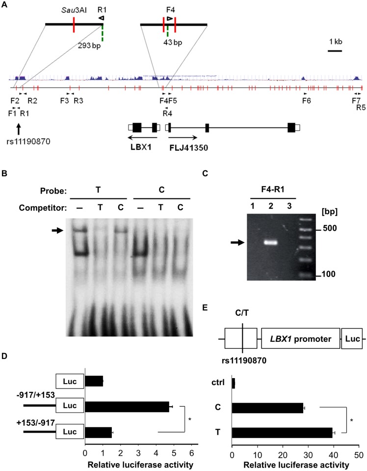
Human LBX1 and FLJ41350 are located approximately 0.6 kb apart in a head-to-head arrangement on human chromosome 10, and rs11190870 lies 7.5 kb downstream of LBX1 (Fig 1A). FLJ41350 is a hypothetical gene that is found only in the human genome, and its function is uncharacterized [15]. We characterized FLJ41350 through exon connection and 5′-rapid amplification of cDNA ends. We confirmed that FLJ41350 is composed of 3 exons, with the predicted translational start site located at exon 1 followed by an open reading frame of 120 amino acids with no known motif (S1 Fig). No orthologs of FLJ41350 are found in any other species except humans.
Fig 1. Characterization of rs11190870.
(A) A schematic representation of the most significantly associated SNP, rs11190870, and the surrounding genome region with the chromatin fragments obtained by the 3C assay. Cross-linked chromatin was digested with Sau3AI (vertical red lines). The green broken lines indicate primer positions. The blue lines indicate vertebrate conservation. The arrowheads indicate PCR primers. (B) Electrophoretic mobility shift assay of rs11190870 using 17-bp DIG-labeled probes for risk (T) and non-risk (C) alleles. Excess unlabeled probes were used as competitors. The arrow indicates that some nuclear factor(s) bound with higher affinity to the risk allele. (C) Physical interaction of rs11190870 and the nearby genomic region with the promoter region of LBX1-FLJ41350 in the 3C assay (lane 1: rhabdomyosarcoma cells, lane 2: A172 cells, lane 3: HeLa cells). The arrow indicates specific PCR products of the expected sizes derived from the indicated primer sets in (A). (D) Transcriptional activity of the LBX1-FLJ41350 promoter region. Luciferase activity of the promoter fragment (-917 to +153; direction of LBX1 transcription) and its reverse fragment (direction of FLJ41350 transcription) was measured in A172 cells. (E) The effect of the 590-bp conserved region containing rs11190870 on the LBX1 promoter in HEK 293T cells. Higher activity was observed for the risk allele (T) construct than for the non-risk allele construct (C). Luciferase activity was normalized to the internal control and expressed as a ratio relative to the promoter-less construct (ctrl). The data in (D) and (E) represent the mean ± standard error of two independent experiments. *p < 0.05.
To investigate the functional impact of rs11190870, we performed EMSAs and found that some nuclear proteins bound specifically to the genome sequences around rs11190870 with higher affinity to the risk allele than the non-risk allele (Fig 1B). We also analyzed the physical interaction between the genome sequence surrounding rs11190870 and its nearby genome regions using the 3C assay [39] with A172 human glioblastoma cells (A172 cells) (Fig 1C). The 3C assay is a powerful technique for analyzing chromatin organization to reveal the physical interaction between two distal genomic elements [40]. Digestion of cross-linked chromatin with a restriction enzyme and subsequent intra-molecular ligation produces novel junctions between restriction fragments in proximity in the nucleus, which can be detected by PCR. We confirmed that the specific band with primers F4 and R1 was of the expected length (Fig 1C) and corresponded to each primer region by sequencing. This result indicates that the F4 and R1 primer regions are adjacent to each other and that the genome sequence surrounding rs11190870 physically interacts with the promoter region of LBX1-FLJ41350.
We then cloned approximately 1 kb of the LBX1 promoter region (-917 to +153) and evaluated its promoter activity by luciferase assay. In A172 cells, the region had relatively high promoter activity in the direction of LBX1, but not in that of FLJ41350 (Fig 1D). Moreover, the LBX1 promoter, combined with a 590-bp sequence around rs11190870 that is highly conserved across species, had higher transcriptional activity with the risk allele than with the non-risk allele in HEK 293T cells (Fig 1E). These results suggest that rs11190870 confers AIS susceptibility by upregulating LBX1 transcription.
Induction of body curvature by ubiquitous overexpression of Lbx genes in zebrafish embryos
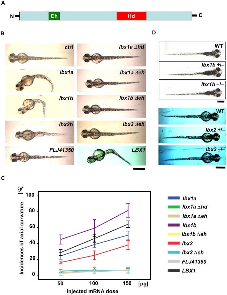
To investigate the effect of the elevated expression of LBX1 on body axis formation, we performed a series of gain-of-function experiments using zebrafish. We overexpressed zebrafish lbx1a, lbx1b, lbx2, and their mutated genes without the homeodomain or the engrailed homology domain by mRNA injection (Fig 2A and 2B). By 48 hours post-fertilization (hpf), the larvae developed body curvature by the ubiquitous overexpression of any one of these lbx genes, but not by that of the mutated genes without the functional domains (Fig 2B). The incidence of body curvature was highest with lbx1b overexpression and increased in a dose-dependent manner (Fig 2B and 2C). Notably, some lbx1b-overexpressing larvae exhibited notochord deformity and a displaced dorsal melanophore stripe (S2 Fig). In addition, a reduction or complete deletion of the forebrain and eyes was observed in many larvae (S2 Fig). Injection of human LBX1 mRNA caused body curvature in embryos, but injection of human FLJ41350 mRNA failed to induce any obvious phenotype related to body axis morphology (Fig 2B).
Fig 2. Body curvature induced by injection of lbx mRNAs.
(A) Protein structure of Lbx family members with an engrailed homology domain (Eh) and homeobox domain (Hd). (B) Dorsal views of live embryos (48 hpf) injected with zebrafish lbx1a, lbx1b, or lbx2b mRNA, as well as human LBX1 or FLJ41350 mRNA, in comparison with those injected with lbx1aΔhd mRNA which lacks the homeodomain, and lbx1aΔeh, lbx1bΔeh, and lbx2Δeh mRNA which lacks the engrailed domain. Buffer was injected as a control (ctrl). Severe curvature was observed in embryos injected with lbx1a, lbx1b, and LBX1 mRNA, while mild body curvature was observed in those injected with lbx2 mRNA, but no curvature was observed in those injected with lbx genes lacking the functional domains. Bending of the body axis was not observed in embryos injected with FLJ41350. (C) Quantitative analysis of the phenotype of embryos injected with different doses of mRNA. Body curvature occurred in lbx1b, LBX1, lbx1a, and lbx2 embryos in a dose-dependent manner. lbx1b embryos presented with the highest incidence of curvature (45% for 50 pg, 59% for 100 pg, and 82% for 150 pg), and then LBX1 (29% for 50 pg, 46% for 100 pg, and 64% for 150 pg), lbx1a (24% for 50 pg, 39% for 100 pg, and 51% for 150 pg), and lbx2 (16% for 50 pg, 25% for 100 pg, and 38% for 150 pg). The incidence of curvature was less than 10% in lbx1aΔhd, lbx1aΔeh, lbx1bΔeh, lbx2Δeh, and FLJ41350 embryos regardless of dose. The data represent the mean ± standard error of three independent injections (n = 27–37). (D) Dorsal views of live larva are shown. lbx1b+/- and lbx1b-/- mutants as well as lbx2+/- and lbx2-/- mutants are comparable to WT with a straight body axis. The scale bar represents 500 μm.
We also examined the loss-of-function effect on axial development in transcription activator-like effector nuclease (TALEN)-mediated knockout zebrafish (S3 and S4 Figs). Unlike overexpression of lbx genes, lbx1b-/- and lbx2-/- mutant larvae displayed a straight trunk comparable to wild-type larvae (Fig 2D), suggesting the involvement of gain-of-function but not loss-of-function of lbx1b in the body curvature phenotype that might be related to scoliosis susceptibility.
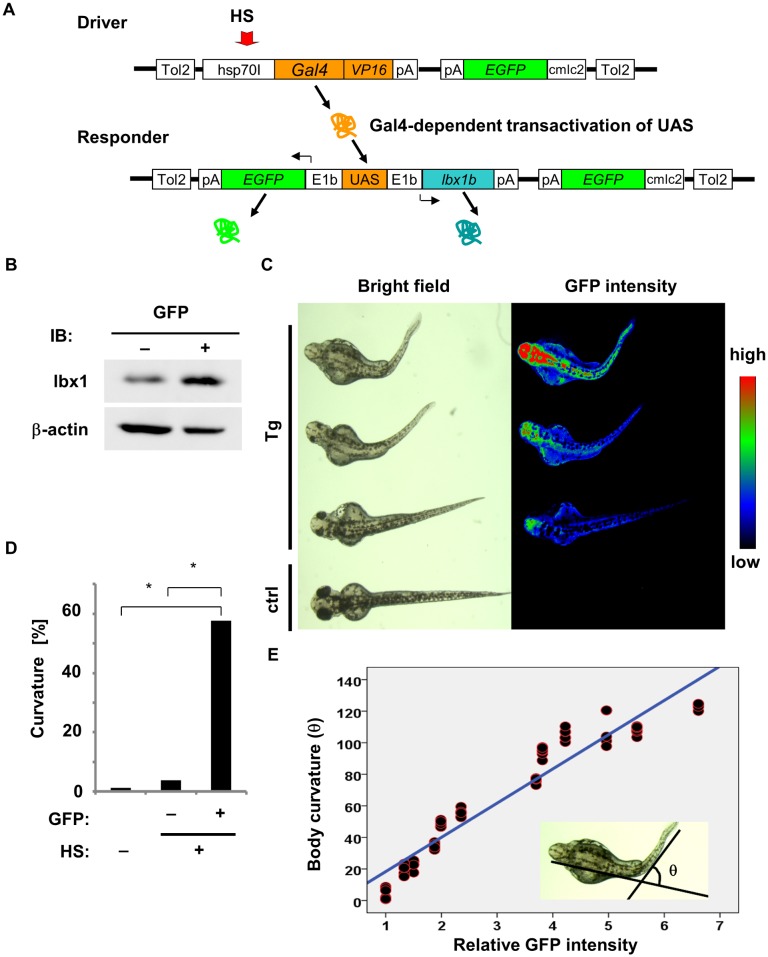
To confirm defective axial development in the established line with uniform expression of lbx1 in an inducible manner, we employed a Gal4/UAS-based bidirectional expression system for the stable overexpression of lbx1b in zebrafish [41]. The F0 driver transgenic carriers were crossed with the F0 responder transgenic carriers to produce Tg(hsp:Gal-VP;EGFP:UAS:lbx1b) F1 progeny with different copies of the Tol2 insertion. Responding to heat shock, embryos with both driver and responder transgenes expressed EGFP and lbx1b driven by the E1b promoter (Fig 3A). The positive correlation between the levels of EGFP and lbx1b expression was examined in Tg(hsp:Gal-VP;EGFP:UAS:mcherry) (S5 Fig). Overexpression of lbx1b in Tg(hsp:Gal-VP;EGFP:UAS:lbx1b) after heat shock was confirmed by western blotting (Fig 3B). By 48 hpf, body curvature became evident in Tg(hsp:Gal-VP;EGFP:UAS:lbx1b) embryos exposed to heat shock at 4 hpf (Fig 3C and 3D). All larvae with lbx1b overexpression died within 7 days post-fertilization (dpf). The severity of body curvature was related to the fluorescence intensity of EGFP in a dose-dependent manner (Fig 3C and 3E). Taken together, we conclude that overexpression of lbx genes, especially lbx1b, induces body curvature in zebrafish embryos.
Fig 3. Body curvature induced by lbx1b overexpression under the control of the heat shock-inducible hsp70I promoter.
(A) The constructs in the Gal4/UAS—based bidirectional expression system. The F0 driver transgenic carriers were crossed with the F0 responder transgenic carriers to produce Tg(hsp:Gal-VP;EGFP:UAS:lbx1b) F1. Heat shock (HS) treatment activated the hsp70I promoter in the transgene driver (Driver), which expresses Gal4-VP16. Gal4-VP16 protein bound to UAS on the transgene responder (Responder) and activates the expression of lbx1b and EGFP via E1b minimal promoters. (B) Expression of lbx1 protein in 48 hpf transgenic zebrafish with (GFP+) or without HS (GFP-). (C) Body curvature of transgenic 48 hpf zebrafish that received HS at 4 hpf. The severity of body curvature was correlated with GFP intensity. (D) Incidence of body curvature at 48 hpf in embryos with (HS+EGFP+, n = 85) and without (HS+EGFP-, n = 129) EGFP expression and without HS (n = 79). The incidence of curvature was significantly increased in the HS+EGFP+ embryos (*p < 0.01). (E) lbx1b expression level and body curvature. A linear regression line was obtained between the relative fluorescence intensity of GFP and the severity of body curvature quantified by the angle θ: [θ/degree] = 21.70 × [relative fluorescence intensity]– 3.37, with the slope coefficient different from 0 (p < 0.01). This model accounted for 92.2% of the θ variance.
Defective convergence and extension movements caused by lbx1b overexpression in zebrafish embryos
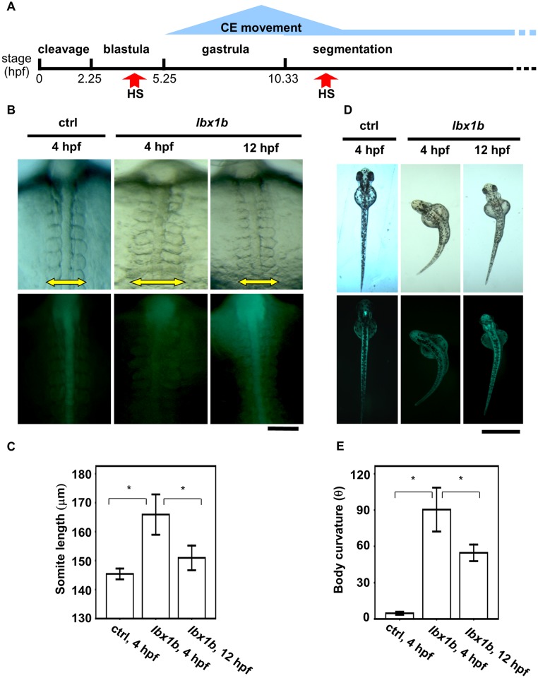
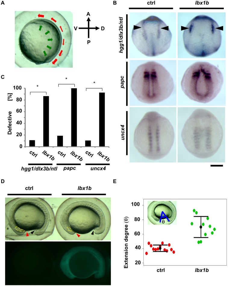
To elucidate the mechanism by which lbx1 overexpression causes embryonic body curvature, we traced back to the pregastrulation stages. Convergent extension movement during gastrulation (5.25–10.33 hpf) shapes the body axis, narrowing all germ layers in the mediolateral direction and extending them along the anterioposterior axis (Figs 4A and 5A). Embryos overexpressing lbx1b in the gastrulation stage showed mediolateral elongation of somites (Fig 4B and 4C), suggesting some perturbations occur in the formation of the body axis due to abnormal convergent extension. Embryos exposed to heat shock at 4 hpf exhibited a more profound convergent extension defect and more severe body curvature than those at 12 hpf (Fig 4B–4E), demonstrating a positive correlation between the extent of defective convergent extension with the severity of body curvature and the presence of a critical time window for lbx1b overexpression. In situ hybridization for the characteristic markers for the ectoderm or mesoderm revealed a marked delay of convergent movement in embryos overexpressing lbx1b (Fig 5B and 5C). Compared with sibling controls, lbx1b-overexpressing embryos showed a wider neural ectoderm border (dlx3b), broader paraxial mesoderm (papc), and mediolateral elongation (uncx4) of somites (Fig 5B). We also found a significant delay of extension movement (Fig 5D and 5E), which elongated the embryo from head to tail. By contrast, the expression pattern of a dorsal marker, chordin (chd), and a ventral marker, ventral homeobox (vox), in early gastrula was not significantly altered in lbx1b-overexpressing embryos (S6 Fig), indicating that dorsoventral patterning was not affected in these embryos. These results indicate that defective convergent extension resulting from elevated lbx1b expression during gastrulation provokes impaired body axis formation.
Fig 4. Time-dependent induction of somite mediolateral elongation and body curvature by lbx1b overexpression.
(A) A schematic diagram of zebrafish early developmental stages. Convergent extension (CE) is mainly involved in gastrulation. (B) Comparison of somite mediolateral length (16 hpf) between Tg(hsp:Gal-VP; UAS:EGFP) with heat shock (HS) treatment at 4 hpf (ctrl 4 hpf), Tg(hsp:Gal-VP; EGFP:UAS:lbx1b) with HS at 4 hpf (lbx1b 4 hpf), and Tg(hsp:Gal4-VP; EGFP:UAS:lbx1b) with HS at 12 hpf (lbx1b 12 hpf). A significant elongation of somite mediolateral length (yellow arrow in the upper panels) was observed in the lbx1b embryos at 4 hpf. The scale bar represents 100 μm. (C) Quantitative analysis of somite mediolateral length in embryos at 16 hpf. A significant difference was observed between ctrl at 4 hpf (n = 16) and lbx1b at 4 hpf (n = 13), and lbx1b at 4 hpf (n = 13) and lbx1b at 12 hpf (n = 14), *p < 0.01. (D) Comparison of body curvature (48 hpf) between Tg(hsp:Gal-VP; UAS:EGFP) with HS treatment at 4 hpf (ctrl 4 hpf), Tg(hsp:Gal-VP:EGFP:UAS:lbx1b) with HS at 4 hpf (lbx1b 4 hpf), and Tg(hsp:Gal-VP:EGFP:UAS:lbx1b) with HS at 12 hpf (lbx1b 12 hpf). More severe curvature of the body axis was induced in lbx1b embryos at 4 hpf than in lbx1b embryos at 12 hpf. The scale bar represents 1 mm (E) Quantitative analysis of body curvature at 48 hpf. A significant difference was observed between ctrl 4 hpf (n = 13) and lbx1b 4 hpf (n = 10), and between lbx1b 4 hpf (n = 10) and lbx1b 12 hpf (n = 11), *p < 0.05. The severity of body curvature was quantified by the angle described in the legend of Fig 3. Both driver transgenic and responder transgenic fish were F2 lines.
Fig 5. Defective convergent extension in lbx1b overexpressing zebrafish embryos.
(A) A diagram of convergent extension during zebrafish gastrulation. A, P, D, and V indicate anterior, posterior, dorsal, and ventral sides, respectively. The green and red arrows indicate the directions of convergence and extension movements, respectively. (B) In situ hybridization for germ layer markers at the early stages of development. Dorsal views of embryos injected with buffer as control (ctrl) or lbx1b mRNA (lbx1b). Upper panels show marker expression of hgg1 (rostral mesoderm), ntl (notochord), and dlx3b (border between neural and non-neural ectoderm, black arrowheads) at the tail bud stage. Middle panels show the expression of papc (paraxial mesoderm) at 11 hpf. Lower panels show the expression of uncx4 (somite) at 13 hpf. The scale bar represents 200 μm. (C) Incidence of defective expression patterns. Significant differences (*p < 0.01) were observed for the germ layer markers. The numbers of control and lbx1b zebrafish embryos were 36 and 30 for hgg1/ntl/dlx3b, 32 and 28 for papc, and 28 and 27 for uncx4, respectively. (D) Lateral views of 16 hpf normal sibling (ctrl) and Tg(hsp:Gal4-VP16:EGFP:UAS:lbx1b) (lbx1b) embryos upon heat shock at 4 hpf. The red and black arrowheads indicate the border between the yolk and the rostral part and the yolk and the caudal part, respectively. The yellow lines indicate the developing eyes. (E) Quantitative analysis of extension movement. Progression of the movement was quantified by the angle θ at 16 hpf for sibling control (ctrl; n = 13) and transgenic (lbx1b; n = 11) embryos. Convergent extension movement was significantly delayed in lbx1b embryos (p < 0.01).
Downregulation of wnt5b in lbx1b-overexpressing embryos
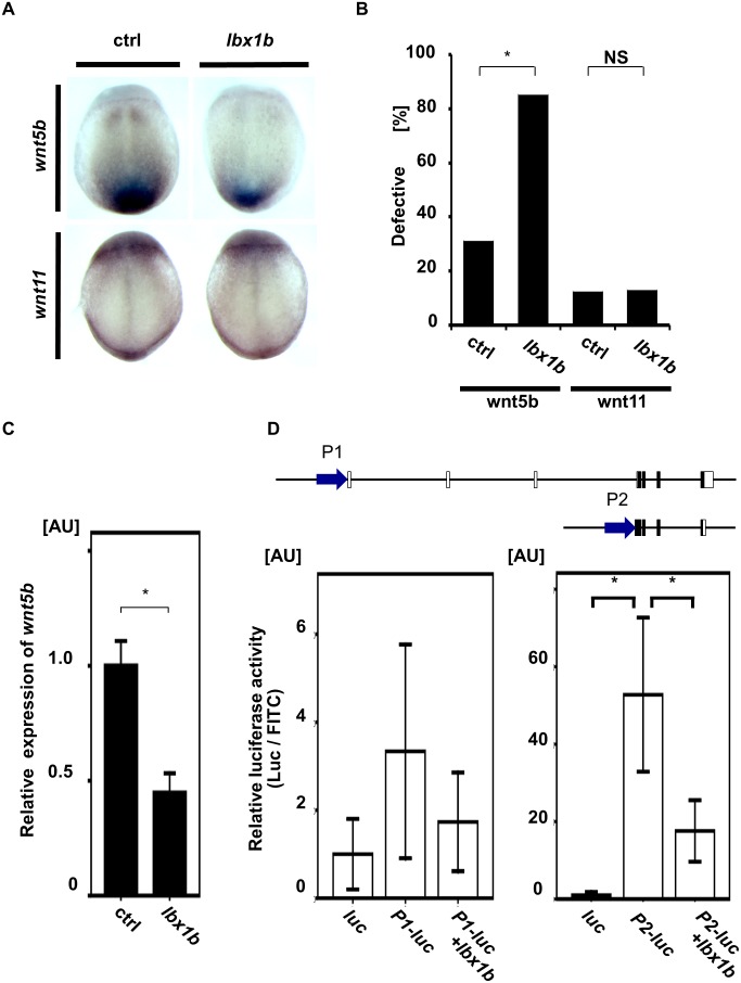
In vertebrates, non-canonical Wnt/PCP signals are mainly involved in the regulation of convergent extension [10]. Loss of function of wnt5b or wnt11, the ligand for the non-canonical Wnt/PCP signaling pathway, leads to severely defective convergent extension movement in zebrafish [42, 43]. Our in situ hybridization study revealed that wnt5b was downregulated in gastrulation embryos upon lbx1b overexpression (Fig 6A and 6B and S7 Fig). In contrast, no significant change was observed in wnt11 expression (Fig 6A and 6B and S7 Fig). We also confirmed by quantitative RT-PCR that wnt5b expression was significantly downregulated at the gastrulation stage in lbx1b-overexpressing embryos (Fig 6C).
Fig 6. Downregulation of wnt5b during gastrulation by lbx1b overexpression.
(A) In situ hybridization for non-canonical Wnt/PCP ligands (wnt5b and wnt11) in control and lbx1b-overexpressing embryos at 90% epiboly. Dorsal views (anterior to the top) for wnt5b and wnt11 of embryos injected with buffer (ctrl) or lbx1b mRNA (lbx1b). (B) Incidence of the defective expression patterns observed in the embryos shown in panel A. Significant differences (*p < 0.01) were observed for wnt5b. The numbers of control and lbx1b zebrafish embryos were 22 and 21 for wnt5b and 23 and 22 for wnt11, respectively. NS: not significant. (C) Decreased wnt5b expression in 90% epiboly embryos injected with lbx1b mRNA by quantitative RT-PCR assays. *p < 0.01. (D) In vivo luciferase assay in 90% epiboly embryos injected with control vector (luc) or the putative promoter regions (P1 and P2) of zebrafish wnt5b. P2 showed higher transcriptional activity than P1. Co-injection of lbx1b mRNA in P2 significantly repressed the transcriptional activity. *p < 0.01. AU, arbitrary unit.
We then performed an in vivo luciferase assay in zebrafish embryos to analyze the in vivo effects of lbx1b overexpression on the transcriptional activity of two potential promoter regions of wnt5b. Two transcripts encoding 363 (MN1309037) or 380 amino acids were found on the Ensembl website. We tested the sequences upstream of wnt5b including these promoters (P1 and P2). In 90% epiboly embryos, the P2 promoter had much stronger transcriptional activity (about 50-fold induction) than the P1 promoter. Co-injection of lbx1b mRNA repressed the transcriptional activity of P2 by 66.7% (Fig 6D). Thus, lbx1b overexpression during gastrulation downregulated the expression of wnt5b largely through repression of the P2 promoter. These results suggest that defective convergent extension caused by the overexpression of lbx1b in embryos could be attributed to impairment of non-canonical Wnt/PCP signaling.
Impairment of non-canonical Wnt/PCP signaling during gastrulation in embryos overexpressing lbx1b
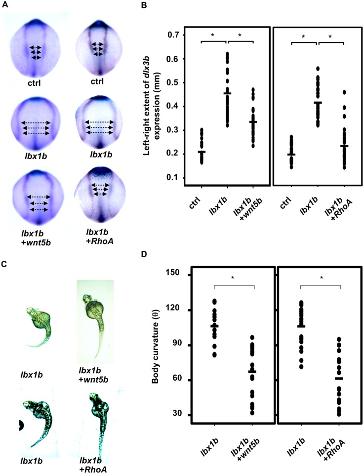
To evaluate further the effect of misregulation of non-canonical Wnt/PCP signaling in defective convergent extension caused by lbx1b overexpression, we performed a rescue experiment by overexpressing wnt5b, a ligand of the Wnt/PCP pathway. We optimized the amount of wnt5b mRNA injection to avoid defects caused by its overexpression in embryos. Defective migration of dlx3b-positive cells in embryos injected with lbx1b mRNA was rescued by co-injection of lbx1b and wnt5b mRNA (Fig 7A and 7B). Wnt5b mRNA injection mostly rescued the body curvature phenotype in Tg(hsp:Gal-VP;EGFP:UAS:lbx1b) with heat shock at 4hpf (Fig 7C and 7D). We further examined whether defects caused by lbx1b overexpression can be rescued by overexpressing RhoA or Rac1 small GTPases, both of which are downstream effectors of the Wnt/PCP pathway. RhoA rescued both the defective convergent extension and body curvature phenotype (Fig 7A and 7B), whereas Rac1 failed to rescue the convergent extension defects and body curvature (S8 Fig). Interestingly, RhoA overexpression was not effective in larvae with heat shock at 12 hpf (S9 Fig). These results demonstrate that impairment of non-canonical Wnt/PCP signaling, especially the wnt5b/RhoA pathway, caused by lbx1b overexpression, contributes to defective convergent extension and curvature of the body axis.
Fig 7. Rescue of defective convergent extension in lbx1b-overexpressing embryos by wnt5b and RhoA mRNA injection.
(A) Dorsal view of dlx3b/hgg1/ntl expression in tail bud embryos injected with buffer (ctrl), lbx1b mRNA (lbx1b), lbx1b plus zebrafish wnt5b mRNA (lbx1b+wnt5b), or lbx1b plus human RhoA mRNA (lbx1b+RhoA). (B) Quantitative analysis of convergent extension movement in the tail bud embryos in (A). (ctrl, n = 26 and 29; lbx1b, n = 27 and 33; lbx1b+wnt5b, n = 27; lbx1b+RhoA, n = 32). The extent of defective convergent extension was evaluated by measuring the distance between the inner edges of bilateral dlx3b expression at 3 regions as indicated by the arrows in panel A. Wnt5b or RhoA mRNA injection significantly rescued defective convergent extension (*p < 0.01). (C) Dorsal view of 48 hpf Tg(hsp:Gal-VP;EGFP:UAS:lbx1b) embryos upon heat shock at 4 hpf with buffer (lbx1b), wnt5b mRNA (lbx1b+wnt5b), or RhoA mRNA (lbx1b+RhoA) injection. (D) Quantitative analysis of body curvature in Tg(hsp:Gal4-VP;EGFP:UAS:lbx1b) embryos upon heat shock at 4 hpf with buffer injection (lbx1b, n = 16 and 21; lbx1b+wnt5b, n = 18; lbx1b+RhoA, n = 18).The severity of body curvature was significantly alleviated in Wnt5b or RhoA mRNA-injected embryos with heat shock at 4 hpf, *p < 0.01. Severity of body curvature was quantified by the angle as described in Fig 3. F2 lines of driver transgenic and responder transgenic fish were used. Injections were performed at doses of 50 pg/embryo for lbx1b mRNA, 40 pg/embryo for wnt5b mRNA, and 15 pg/embryo for RhoA mRNA.
Scoliosis in zebrafish expressing lbx1b under the control of the GATA2 minimal promoter and the lbx1b enhancer
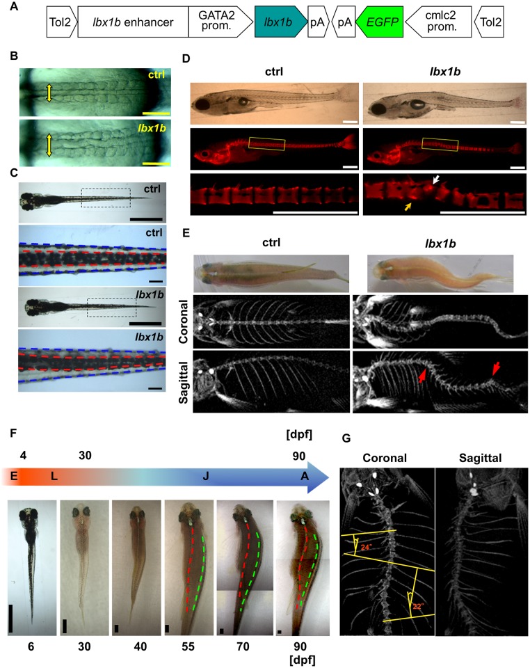
To investigate the effects of lbx1b overexpression on endogenous expression domains during axial development, we forced lbx1b expression under the control of the previously characterized lbx1b enhancer [44] and the GATA2 minimal promoter by microinjecting a GATA2-1b:lbx1b plasmid (Fig 8A). We confirmed that reporter expression driven by the regulatory elements in Tg(GATA2-1b:EGFP) generally recapitulated the endogenous expression of lbx1b, lbx1a, or lbx2 at different developmental stages (S10 Fig). Similarly to embryos injected with mRNAs, many 48 hpf embryos injected with GATA2-1b:lbx1b developed severe body curvature (S11 Fig), abnormalities in somite morphology (Fig 8B), notochord deformity (S12A Fig), and a displaced dorsal melanophore stripe (Fig 8C). Some of the larvae with a displaced dorsal melanophore stripe had no apparent notochord deformity (S12B Fig). The majority of Tg(GATA2-1b:lbx1b) F1 embryos presented with a severe malformation and died within 24 hpf (S13 Fig). Some were alive at 48 hpf, developing serious axial body curvature, but died within 72 hpf (S13 Fig).
Fig 8. Scoliosis in transgenic founder zebrafish expressing lbx1b using the GATA2 minimal promoter and the lbx1b enhancer.
(A) The transgene construct is shown. The GATA2 minimal promoter and lbx1b enhancer cooperatively drive lbx1b expression. (B) Dorsal views of live GATA2-1b:MCS (ctrl) or GATA2-1b:lbx1b (lbx1b) injected embryos with 10–13 somites. Somite arrangement is bilateral and symmetric in control embryos, but asymmetrical in lbx1b embryos. The yellow arrows indicate the mediolateral length of somites. (C) Dorsal views of 6 dpf larvae. The red and blue dotted lines indicate the boundaries of the dorsal melanophore stripes and the trunk, respectively. A displaced dorsal melanophore stripe was observed in lbx1b larvae. (D) Lateral views of alizarin red-stained larvae at 21 dpf. The white and yellow arrows indicate the hemivertebrae and block vertebra that developed from the deformed notochord in lbx1b larva, respectively. (E) Dorsal views and micro-computed tomography (μCT) analysis of adult zebrafish. Scoliosis was observed in adult fish grown from embryos with mild notochord deformities. The coronal and sagittal planes are reconstructed from μCT images of the fish in the upper panels, each showing a gross appearance. The red arrows indicate vertebral malformation. (F) Diagram of zebrafish growth stages. Continuous observation of one GATA2-1b:lbx1b-injected larva with a displaced dorsal melanophore stripe, but without notochord deformation, from 6 to 90 dpf. The red and green dotted lines indicate the dorsal middle lines and the right upper boundary of the lateral stripe, respectively. Progressive scoliosis with rotation of the longitudinal axis of the body was observed after 55 dpf. E, embryo; L, larva; J, juvenile; A, adult. (G) μCT analysis of the three-dimensional structure of the spine of the same zebrafish in (F) showing scoliosis. Cobb’s angle was measured in the coronal plane. Scale bars in (B): 200 μm; (C): 1 mm or 100 μm; (D): 500 μm; and (F): 1 mm.
Unlike the F1 generation of Tg(GATA2-1b:lbx1b), which is embryonic lethal, some founder Tg(GATA2-1b:lbx1b) with almost a straight trunk could survive to adulthood, thus allowing our observation of the later developmental stages in this model. We monitored embryos with a mild notochord deformity induced by injection of GATA2-1b:lbx1b (S12A Fig) (n = 41) until adulthood, together with wild-type siblings as controls (n = 45). Thirteen Tg(GATA2-1b:lbx1b) and two control zebrafish died within 21 days. The deformed notochord (S12A Fig red arrow) gradually ossified to form a spine, leading to vertebral malformations (n = 27, p < 0.01) such as hemivertebrae (Fig 8D, white arrow) and block vertebra (Fig 8D, yellow arrow) at the location of the notochord deformity. Eventually, these zebrafish showed scoliosis with vertebral malformations mimicking CS (Fig 8E and S1 video). No apparent spinal deformity was identified in the control (Fig 8B–8E). Thus, local notochord deformity in founder Tg(GATA2-1b:lbx1b) develops into CS-like scoliosis with vertebral malformations.
To investigate the possibility of AIS-mimicking scoliosis in Tg(GATA2-1b:lbx1b) during the period corresponding to human adolescence (Fig 8F), we kept transgenic larvae that had a displaced dorsal melanophore stripe without an apparent notochord deformity (n = 45), together with their wild-type siblings (n = 60). Eight Tg(GATA2-1b:lbx1b) and two control zebrafish died within 30 days. In 19 of the 37 surviving Tg(GATA2-1b:lbx1b) (p < 0.01), significant scoliosis, with rotation of the longitudinal body axis but without visible vertebral malformations, was observed by 55 dpf and then developed progressively until 90 dpf (Fig 8F, 8G and S2 video). Additionally, there was a significant female bias (16/19) for the prevalence of scoliosis (p < 0.01). No apparent spinal deformity was identified in the control group (0/58). These results indicate that mild body axis deformation resulting from the increased expression of lbx1b could cause irregular trunk development such as notochord deformity and a displaced dorsal melanophore stripe, further leading to the later development of CS- or AIS-like scoliosis.
Discussion
We demonstrate here that the most significantly associated SNP, rs11190870 [15] could confer AIS susceptibility by activating LBX1 transcription. Our gain-of-function approaches using the zebrafish model revealed that the elevated expression of human LBX1 or any of the zebrafish genes lbx1a, lbx1b, and lbx2 causes body axis deformation at various stages of embryonic and subsequent growth in zebrafish. Embryonic body curvature prior to vertebral column formation is associated with defective convergent extension involving the downregulation of wnt5b during gastrulation to disrupt axial development. Defective convergent extension and embryonic body curvature phenotypes were mostly rescued by the overexpression of wnt5b and RhoA, key molecules in the Wnt/PCP signaling pathway. An embryonic lethal phenotype could be alleviated by chimeric expression of lbx1b under the control of the GATA2 minimal promoter and the lbx1b enhancer in larvae, thus allowing observation of the later onset of the spinal curvature with or without vertebral malformation in zebrafish. Thus, as a step towards better understanding of the genetic pathophysiology of scoliosis, our study provide a new evidence for a pathological role of LBX1 and its zebrafish homologs in body axis deformation.
The most significant SNP associated with AIS (rs11190870) is located in the intergenic region [15]. The nearest gens are LBX1 and FLJ41350, which are 7.5 kb upstream and 8.1 kb downstream of rs11190870, respectively. Using 3C assays, we found that the genome sequence surrounding rs11190870 physically interacts with the LBX1 and FLJ41350 promoters. In luciferase assays, significantly higher promoter activity was detected in the direction toward LBX1, but not toward FLJ41350. EMSAs revealed that some nuclear proteins bound specifically to the genome sequences around rs11190870 with higher affinity to the risk allele. Given that risk variants could disrupt or create a binding site for a transcription factor, any change of LBX1 expression driven by the variants, including downregulation, upregulation, and alteration in temporospatial distribution, would be possible. Expression quantitative trait loci (eQTL) data are available only for peripheral blood cells, which showed no association between the LBX1 expression level and the rs11190870 genotype (Human genetic variation database. (http://www.genome.med.kyoto-u.ac.jp/SnpDB/index.html). However, further studies on eQTL are hampered by a lack of information on which types of tissues or cells are relevant to AIS pathogenesis.
So far, phenotypes associated with CS and AIS have not been reported in Lbx1 null mice and lbx gene knockdown morphants in zebrafish or Xenopus [25, 28–30]. Our lbx1b or lbx2 single knockout zebrafish mutants generated by targeting the first exon using Platinum TALENs [45] also did not exhibit embryonic axial body curvature or scoliosis. The database of Zebrafish Mutation Project also shows that normal development is observed in lbx1a or lbx2 nonsense mutants (https://www.sanger.ac.uk/sanger/Zebrafish_Zmpsearch/lbx1), although information on the associated phenotype of double or triple knockout zebrafish is not available. Previous studies demonstrated a dominant-negative effect by the removal of the engrailed domain from Xenopus Lbx1 that normally functions as a repressor [22, 46]. Injection of lbx1aΔeh, lbx1bΔeh, or lbx2Δeh mRNA did not cause any body curvature as shown in our study. Thus, the current data do not support the possibility that loss-of-function of LBX1 is involved in susceptibility to scoliosis. In contrast, we found a significant increase of promoter activity in the presence of the genomic region with rs11190870 found in the risk allele. Considering that rs11190870 could confer AIS susceptibility by activating LBX1 transcription, it would be reasonable to assume that upregulation of human LBX1 may contribute to some aspects of the pathogenic mechanism in scoliosis.
The ladybird protein is a member of the homeobox transcription factor family with an engrailed repressor domain at the N-terminus [22]. Overexpression of LBX1 and any one of lbx1a, lbx1b, or lbx2 caused defective convergent extension movements that led to curvature of the body axis. Upon overexpression of the lbx genes without the engrailed repressor domain, body curvature was not observed in the embryos, suggesting that Lbx genes negatively regulate their target genes as repressors. Indeed, our in vivo luciferase assays revealed that lbx1b significantly represses the promoter activity of wnt5b. Hence, lbx1b downregulates wnt5b expression during gastrulation at the transcriptional level, thereby causing defective convergent extension followed by deformation of the body axis.
Both canonical and non-canonical Wnt signaling pathways are involved in convergent extension movements during gastrulation. A shortened-curled tail was reported in a Wnt-5 mutant (ppt−/−) with defective convergent extension [47]. AIS- and CS-like scoliosis are also observed in zebrafish mutants of ptk7, which regulates both canonical and non-canonical Wnt signaling activity [37, 48]. The same group identified a novel sequence variant within a single IS patient that disrupted PTK7. In this study, we found that the elevated expression of lbx1 in zebrafish evokes wnt5b downregulation, suggesting that aberrant Wnt/PCP signaling causes defective convergent extension in our experimental model. Interestingly enough, our approach investigating the etiology of scoliosis from the opposite direction also led to the hypothesis that a dysregulated Wnt signaling pathway is involved in both CS and IS pathogenesis.
Non-canonical Wnt/PCP signaling is involved in a variety of events independently of β-catenin [49]. During axis formation in vertebrates, the Wnt/PCP pathway regulates cell polarity and cell motility by modulating the activity of Rho family small GTPases. Especially, RhoA-ROCK signaling mainly acts downstream of wnt5 and wnt11 in zebrafish embryos [50]. Co-injection of mRNA for wnt5b or RhoA mRNA with lbx1b mRNA rescued defective convergent extension leading to embryonic body curvature. These findings strongly support our hypothesis that misregulation of Wnt/PCP signaling induced by lbx1b overexpression is responsible for defective convergent extension followed by body axis deformation.
To date, CS and IS have been considered not to be etiologically relevant, but it has been reported previously that a family history of IS was observed in 17.3% of 237 families with CS [51]. Another study of 31 CS cases also reported that three (10%) had first-degree relatives with IS [52]. The overlapping familial aggregates of CS and IS suggested the possibility of a common cause for these clinically distinct diseases. The uniform overexpression of lbx1b either ubiquitously or in the endogenous expression domain results in severe defective convergent extension leading to morphological defects in both mesoderm and ectoderm patterning followed by early death prior to notochord mineralization to form the spine. In contrast, mosaic expression of lbx1b under the control of the lbx1b enhancer in larvae alleviated the embryonic lethal phenotype with body curvature and thereby allowed the later onset of scoliosis with or without vertebral malformation in zebrafish. The F1 embryos generated by the AIS- and CS-like mosaic transgenic founders presented with severe body axis deformation including convergent extension defects and body curvature. Thus, our observations that the elevated expression of lbx1b causes both AIS- and CS-like scoliosis may provide a new perspective for the shared genetic basis of AIS and CS.
Some of the founder Tg(GATA2-1b:lbx1b) with a displaced dorsal melanophore stripe without apparent notochord deformity developed scoliosis with rotation around the longitudinal axis of the body, but without visible vertebral malformations. These results suggest that subtle deformities in the early body axis may be later accentuated during the growth spurt. In fact, AIS patients appear to be quite normal until adolescence. It is reasonable to postulate that an early event such as defective axial development resulting from the upregulation of LBX1 may be too mild to be detected in potential AIS patients until the growth spurt. In a late-onset polygenic disease such as AIS, even such subtle abnormalities may be sufficient to accumulate growth irregularities and greatly aggravate biomechanical instability during adolescence in association with additional genetic or environmental factors. However, at present, considering that the GATA2 minimal promoter and an lbx1b enhancer could drive lbx1b expression in neural tissue later in development [44], we cannot exclude the possibility that lbx1b expression after convergent extension causes idiopathic scoliosis. Thus, we need to determine carefully the mechanism by which lbx1b causes the AIS-like phenotype in the mosaic transgenic founders.
Polygenic diseases including AIS are triggered by the combination of a number of susceptibility genes whose individual contribution may be relatively small. It is also considered that these diseases could occur when a threshold of quantitatively-varying risk or liability influenced genetically and environmentally is exceeded [53]. Unlike a monogenic disease caused by a mutation in one gene, it appears that the cumulative effects combined with additional factors for a relatively long time lead to the onset of clinical manifestations of AIS, even though the contribution of each individual gene is small. Our study provides a new evidence for the possible involvement of LBX1-induced mild defects during embryonic axial development in AIS susceptibility. As the faithful recapitulation of the late-onset polygenic disease in the animal model has not been generally established yet, our current experimental approaches are still fraught with limitations. Further studies are necessary for establishment of a genetic animal model recapitulating the expression of LBX1 in an analogous way to that in AIS patients.
Materials and Methods
Ethics statement
All of the animal experimental procedures used in this study were approved by the Animal Care Committee of the Institute for Frontier Medical Sciences, Kyoto University and conformed to institutional guidelines for the study of vertebrates
Cell culture and 3C assay
Rhabdomyosarcoma cells and A172 human glioblastoma cells (A172 cells) from ATCC and HeLa cells were obtained from the Japanese Collection of Research Bioresources Cell Bank (Osaka, Japan). The cells were maintained at 37°C under 5% CO2 in Dulbecco’s modified Eagle’s medium-high glucose supplemented with penicillin (50 U/mL), streptomycin (50 g/mL), and 10% fetal bovine serum. The cells were crosslinked with 37% formaldehyde solution at a final concentration of 1% in a 37°C dry incubator for 10 min, followed by an additional incubation at 4°C for 2 h. The crosslinked protein-chromatin material was purified by 8 M urea ultracentrifugation and digested with Sau3AI as described previously. A 2-g aliquot of chromatin was diluted in a ligation buffer and ligated with T4 DNA ligase (Fermentas) for 4 h. After reversing the crosslinks, the ligated DNA was amplified by PCR with various combinations of primers using GoTaq Hot Start Master Mix (Promega).
Electrophoretic mobility shift assay
We prepared nuclear extracts from rhabdomyosarcoma and A172 cells as described previously [54]. We prepared probes for the risk (R) and non-risk (N) alleles of rs11190870 by annealing 17-bp complementary oligonucleotides and labeling with digoxigenin (DIG)-11-ddUTP (Roche). For competition experiments, nuclear extracts were pre-incubated with excess unlabeled probes. We detected DNA-protein complexes using a DIG gel shift kit according to the manufacturer’s instructions (Roche).
Construction of plasmids, transfection, and luciferase reporter assay
We amplified the LBX1 promoter fragment (-917 to +153) in both directions by PCR and cloned them into the pGL4.10 promoter-less luciferase reporter vector (Promega). The constructs were co-transfected with the pGL4.73 Renilla luciferase vector (hRluc/SV40) as an internal control. Transfection of each construct was performed using TransIT-LT1 (Mirus Bio LLC). HEK 293T cells were maintained at 37°C under 5% CO2 in Dulbecco’s modified Eagle’s medium-high glucose supplemented with 10% fetal bovine serum. Transfection was performed with Lipofectamine LTX and PLUS reagent (Life Technologies). After 24 h of transfection, the cells were harvested and luciferase activity was measured using a Pick&gene dual luciferase detection kit (Toyo B-Net Co.).
Zebrafish
The RIKEN Wako (RW) strain and AB strain were obtained from the Zebrafish National BioResource Center of Japan (http://www.shigen.nig.ac.jp/zebra/) and Kondoh ERATO Laboratory, respectively. Adult fish were maintained under a 14 h light–10 h dark cycle at 28°C. Embryos were kept at 28°C and staged by hpf or dpf [55]. The RW strain was subjected to micro-injection and whole-mount in situ hybridization. The AB strain was used for the preparation of total RNA. The established line Tg(UAS:EGFP) [41] was generously provided by Dr. Koichi Kawakami (National Institute of Genetics).
In vitro synthesis and microinjection of mRNA
Specific primers for zebrafish lbx1a (NM_001025532), lbx1b (NM_001163312), and lbx2 (NM_001007134), and human LBX1 (NM_006562.4), FLJ41350 (NR_029380), and RhoA (NM_001664.2) were designed based on the nucleotide sequences from GenBank (S1 Table). The cDNAs were amplified by PCR from a cDNA library and cloned into the pCS2(+) vector. Deletion constructs of the engrailed domain and homeodomain of lbx1 were generated by inverse PCR. Capped mRNAs were synthesized using an SP6 RNA polymerase in vitro transcription kit (Life Technologies) and purified using a MEGAclear Kit (Life Technologies) according to the manufacturer’s instructions. A mixture containing 50/100/150 pg mRNA for lbx1a, lbx1b, lbx2, LBX1, and FLJ41350, 15 pg mRNA for RhoA, and 40 pg mRNA for wnt5b and RAC1 was injected into the cytoplasm of one-cell-stage embryos.
Construction of TALENs
Highly active Platinum TALENs were constructed using two-step Golden Gate assembly method as described previously with a slight modification [45]. DNA-binding modules were assembled with the two-step Golden Gate method using the Platinum Gate TALEN Kit (Addgene, Kit #1000000043). pCS2-based vectors were used as destination vectors. The target sequence was 5’-TAAACCCCCTGGACCACcttccaccacccgcgAGCTCCAACAA GCCCTTA-3’, where uppercase and lowercase letters indicate lbx1b TALEN recognition sequence and spacer sequence, respectively. We found polymorphism of RW WT in the left TALE-binding sequence; TAAACCCCCTGGACCAC and TGAATCCCCTGGACCAC, both of which are silent mutations. The target sequence was 5’-TTGCAGTCCAGCGGCGAG gagaggcggcggggtCCCTTGGACCAACTCCCA-3’, where uppercase and lowercase letters indicate lbx2 TALEN recognition sequence and spacer sequence, respectively.
Generation of lbx1b mutant
Genomic DNA was extracted from the caudal fins of lbx1b TALENs mRNA-injected zebrafish. For sequencing, PCR products were amplified from the genomic DNA and phosphorylated by T4PNK (TAKARA BIO INC.), and then subcloned into EcoRV site of pBluescript II SK(+) vector. We identified F0 fishes carrying multiple mutations in the target site, and then generated and screened a F1 fish with a nonsense mutation by crossing the F0 and wild type fishes. lbx1b+/- and lbx2+/- mutant were generated by crossing F1 and wild type fishes. lbx1b-/- and lbx2-/- were further generated by intercrossing lbx1b+/- and lbx2+/-, respectively.
Generation of transgenic zebrafish
The Tol2 transposon/transposase system [41, 56–59] was employed for the establishment of transgenic zebrafish. The coding sequence of lbx1b was cloned into pME-MCS to generate pME-lbx1b. The complementary sequence of the E1b promoter, EGFP, and polyA were cloned into p5E-UAS-E1b to generate p5E-polyA-EGFP-E1b-UAS-E1b. The driver construct (Fig 3A) was generated by recombining p5E-hsp70I, pME-Gal4VP16, p3E-polyA, and pDestTol2CG2 with Gateway LR Clonase II Enzyme mix (Life Technologies). Similarly, the responder construct (Fig 3A) was generated by recombining p5E-polyA-EGFP-E1b-UAS-E1b, pME-lbx1b, p3E-polyA, and pDestTol2CG2. Capped mRNA of medaka Tol2 transposase was prepared by in vitro translation as described above. A mixture containing 50 pg transposase mRNA and 40 pg Tol2 transgenic plasmid was injected into the cytoplasm of one-cell-stage embryos. F1 fish were acquired by outcrossing EGFP-positive F0 with RW fish, and screened by cardiac fluorescence. F2 lines were then generated by outcrossing F1 and RW fish. All kept F2 lines yielded about 50% EGFP-positive progeny when mated to RW fish, which suggested there was a single Tol2 insertion site. The lbx1b enhancer located from +1316 to +2383 bp downstream of the lbx1b transcription start site [44] was cloned into pME-MCS. The enhancer activity in vivo was confirmed using zebrafish enhancer detection (ZED) [60]. To generate p5E-lbx1b enhancer-GATA2, the 2.3-kb BamHI fragment from the ZED-lbx1b enhancer, was cloned into the BamHI site of p5E-MCS. The constructs GATA2-1b:lbx1b or GATA2-1b:MCS were generated by recombining p5E-lbx1b enhance-GATA2, pME-lbx1b or pME-MCS, p3E-polyA, and pDestTol2CG2.
Heat shock treatment
Embryos at 4 hpf or 12 hpf in E3 buffer were placed on block incubator and heated up to 38°C gradually, and then maintained at 38°C for 30 min. After heat shock treatment, they were gradually cooled to 28.5°C. The heat shock treatment causes neither an anomaly nor a decrease in viability.
Western blot analysis
Larvae were homogenized with a Dounce tissue grinder and lysed with lysis buffer (50 mM Tris-HCl [pH 7.5], 150 mM NaCl, 1% NP-40, 0.1% SDS) containing protease inhibitor cocktail (Roche). Lysates were mixed with 5× Laemmli sampling buffer containing 100 mM DTT and boiled at 95°C for 3 min. Proteins were separated by SDS-PAGE and transferred onto PVDF membranes (Merck Millipore). After blocking with BLOCKING ONE (Nacalai Tesque), the membranes were incubated with primary antibodies in phosphate-buffered saline containing 0.1% Tween 20 and 10% BLOCKING ONE, followed by incubation with horseradish peroxidase-conjugated secondary antibodies. Signals were detected with SuperSignal West Pico Chemiluminescent Substrate (Thermo Scientific) and images were captured by ImageQuanta LAS 4000 (GE Healthcare Bio-Sciences).
Whole-mount in situ hybridization
Whole-mount in situ hybridization was performed as described previously [61]. Sense and antisense riboprobes for hgg1, dlx3b, ntl, papc, uncx4, wnt5b, wnt11 [42], chd and vox [48] were generated by in vitro translation using a digoxigenin (DIG) RNA labeling kit with T7 or T3 RNA polymerase (Roche). Hybridization signals were detected with an alkaline phosphatase-conjugated anti-DIG antibody (Roche) according to the manufacturer’s instructions. For quantification, the image colors of in situ hybridization were inverted, and Area, Integrated Density, and Mean Gray Value were measured by ImageJ. The corrected Gray Value = Integrated Density − (Area of the selected embryos × Mean Gray Value of background readings).
RNA extraction and quantitative RT—PCR
Total RNA was extracted from zebrafish embryos injected with lbx1b mRNA using an RNeasy Plus Mini kit (QIAGEN). Two hundred nanograms of total RNA were used to synthesize cDNA with a PrimeScript RT reagent Kit (Takara Bio). Quantitative RT-PCR was performed using SYBR Premix Ex Taq II (Takara Bio) on a StepOne instrument (Life Technologies). Relative mRNA expression was normalized to ef-1α and calculated using the 2−ΔΔCT method. Specific primers for quantitative RT—PCR are listed in S1 Table.
In vivo luciferase assay
A 569-bp insulator of chicken β-globin (BGI) and firefly luciferase (luc) were amplified from the ZED vector and pGL3-basic vector (Promega), respectively. The resultant amplification products were cloned into pME-MCS to construct the promoter-less pME-BGI-luc plasmid. Two fragments of approximately 2 kb upstream of each transcription start site of wnt5b were amplified from zebrafish genome DNA with the primers listed in S1 Table. These were cloned into pME-BGI-luc, pME-BGI-P1-luc plasmid (P1), and pME-BGI-P2-luc (P2) by the In-Fusion technique (Clontech). A mixture containing fluorescein isothiocyanate (FITC)-dextran (SIGMA) and luciferase plasmids with or without lbx1b mRNA was injected into one-cell-stage embryos. FITC fluorescence intensity was quantified using a fluorescence microscope (Leica MZ 16 FA) and ImageJ software. Embryos were then lysed individually and luciferase activity was measured as described previously [62]. The measured activity was normalized by the FITC fluorescence intensity of an individual embryo.
Skeletal preparations
Bones in fixed larvae were stained with alizarin red (Wako).
Skeletal imaging by micro-computed tomography analysis
Vertebral bone morphology of adult zebrafish was analyzed by micro-computed tomography scans with inspeXio SMX-90CT (SHIMADZU). Three-dimensional reconstruction and videos were generated with ImageJ software.
Phenotype scoring and statistical analysis
Embryos were examined and scored for relevant phenotypes. Statistical analysis (SPSS 16.0) was performed by chi-square analysis for enumeration data and independent-samples t test or one-way ANOVA for measurement data to calculate p values under various conditions. Spearman’s correlation between relative fluorescence intensity and body curvature severity was calculated. A linear regression equation was calculated with SPSS.
Supporting Information
Nucleotide sequences were determined by Sanger sequencing of RT-PCR and rapid amplification of cDNA ends (RACE) products. Nucleotides are numbered on the right. The initiation codon is underlined. It conformed to the Kozak sequence. The stop codon is indicated by an asterisk and a putative poly-adenylation signal is enclosed in an open box. Multiple transcription start sites (TSSs) clustered in a region of a few dozen base pairs were identified by 5′-RACE, but only the most major TSS is shown.
(TIF)
Dorsal views of larvae at 6 dpf. The deformed notochord, displaced dorsal melanophore stripe, and anophthalmia in lbx1b mRNA-injected zebrafish are indicated by a green, yellow, and red arrow, respectively. Scale bar: 1 mm.
(TIF)
(A) Alignment of representative sequences of the PCR amplicons from lbx1b transcription activator-like effector nuclease (TALEN) mRNA-injected embryos showing insertions and deletions. TALEN-binding sites in exon 1 of lbx1b are indicated in red. Indels were detected in 32.7% (17/52) of clones from embryos injected with TALEN mRNA. A dash indicates a single nucleotide deletion. Inserted nucleotides are underlined. (B) DNA chromatographs for sequences of a wild-type (WT) and an established lbx1b nonsense mutant line. Three nucleotides (CAC) in WT were deleted and/or replaced with two nucleotides (TT) (indicated in red). (C) Schematic diagram showing a premature stop site caused by a TALEN-induced frameshift mutation in the first exon of lbx1b. Altered amino acids in the mutant are indicated in red. E1: exon 1; E2: exon 2; Eh, engrailed homology domain; Hd: homeobox domain.
(TIF)
(A) Alignment of representative sequences of the PCR amplicons from lbx2 transcription activator-like effector nuclease (TALEN) mRNA-injected embryos showing insertions and deletions. TALEN-binding sites in exon 1 of lbx2 are indicated in red. Indels were detected in 40.9% (56/137) of clones from embryos injected with TALEN mRNA. A dash indicates a single nucleotide deletion. Inserted nucleotides are underlined. (B) DNA chromatographs for sequences of a wild-type (WT) and an established lbx2 nonsense mutant line. Sixteen nucleotides were deleted and/or replaced with 32 nucleotides in the mutant (indicated in red). (C) Schematic diagram showing a premature stop site caused by a TALEN-induced frameshift mutation in the first exon of lbx2. Altered amino acids in the mutant are indicated in red. E1: exon 1; E2: exon 2; Eh, engrailed homology domain; Hd: homeobox domain.
(TIF)
(A) The constructs of the Gal4/UAS-based bidirectional expression system. Heat shock (HS) treatment activates the hsp70I promoter in the driver construct (Driver) to express Gal4-VP16. Gal4-VP16 protein binds to the UAS on the responder construct (Responder) and activates the expression of mCherry and EGFP via E1b minimal promoters. (B) A positive correlation of expression level between the two genes flanking the UAS in Tg(hsp:Gal4-VP: EGFP:UAS:mcherry). The scale bar represents 1 mm.
(TIF)
(A) Whole-mount in situ hybridization (WISH) for dorsal organizer gene chd and ventral gene vox in shield-stage embryos injected with buffer as control (ctrl) or lbx1b mRNA (lbx1b). Photos are taken from the animal pole side of embryos with dorsal to the left. (B) Quantitative analysis of the WISH signals shown in A. For quantification of the dorsoventral position, the angle θ [degree] shown in the panel A as measured. No significant change was observed for both chd (p = 0.261) and vox (p = 0.071) expression pattern. The numbers of control and lbx1b zebrafish embryos were 22 and 23 for chd, 23 and 23 for vox, respectively.
(TIF)
Quantitative analysis of the signal intensity of in situ hybridization for wnt5b or wnt11 by processing the images shown in Fig 6A. Significant differences (*p < 0.01) were observed in wnt5b.
(TIF)
(A) Dorsal view of dlx3b/hgg1/ntl expression in the tail bud of embryos injected with buffer (ctrl), lbx1b mRNA (lbx1b), or lbx1b and human RAC1 mRNA (lbx1b+Rac1). (B) Quantitative analysis of convergent extension (CE) movement with embryos in (A) (ctrl, n = 15; lbx1b, n = 15; lbx1b+Rac1, n = 13). The extent of defective CE was evaluated by measuring the distance between the inner edges of bilateral dlx3b expression at 3 regions as indicated by the arrows in panel A. RAC1 mRNA injection failed to rescue defective CE. *p < 0.01, NS: not significant. (C) Dorsal view of 48 hpf Tg(hsp:Gal-VP; EGFP:UAS:lbx1b) embryos upon heat shock at 4 hpf with buffer (lbx1b) or RAC1 mRNA (lbx1b+Rac1) injection. (D) Quantitative analysis of body curvature with the embryos in (C) (lbx1b, n = 11; lbx1b+Rac1, n = 11). No significant change of the severity of body curvature was observed in RAC1 mRNA-injected embryos. NS: not significant. Severity of body curvature was quantified by the angle as described in Fig 3. F2 lines of driver and responder transgenic fish were used. The dosages for embryo injections were 50 pg/embryo for lbx1b mRNA and 40 pg/embryo for RAC1 mRNA.
(TIF)
(A) Dorsal views of 48 hpf Tg(hsp:Gal-VP:EGFP:UAS:lbx1b) embryos upon heat shock (HS) at 12 hpf with buffer (HS12h) or RhoA mRNA (HS12h+RhoA) injection. (B) Quantitative analysis of body curvature in Tg(hsp:Gal-VP:EGFP:UAS:lbx1b) embryos upon HS at 12 hpf with buffer injection (lbx1b, n = 13) or RhoA mRNA injection (lbx1b+RhoA, n = 13). Significant change in axis development was not observed in RhoA-injected embryos with HS at 12 hpf. Severity of body curvature was quantified by the angle as shown in Fig 3E. F2 lines of driver and responder transgenic fish were used.
(TIF)
(A) Construction of the transgene. The GATA2 minimal promoter and an lbx1b enhancer cooperatively drive the expression of EGFP. The cardiac specific promoter cmlc2 drives EGFP expression in the heart as a transgenic marker. (B–D) Comparison of EGFP fluorescence with lbx1a, lbx1b, and lbx2 expression in lateral views at the 90% epiboly-bud stage (B), 13–15 somites stage (C), and 24–26 somites stage (D).
(TIF)
(A) Dorsal views of live embryos at 48 hpf. Body curvature was observed in zebrafish injected with GATA2-1b:lbx1b (lbx1b), but not in those injected with GATA2-1b:MCS (ctrl). The scale bar represents 500 μm. (B) Quantitative analysis of body curvature (cur) in 48 hpf embryos. The incidence of body curvature was significantly increased in lbx1b embryos (ctrl, 5%, n = 83; lbx1b, 46%, n = 94. p < 0.01). Scale bar in (A): 500 μm.
(TIF)
(A) Lateral views of embryos (48 hpf) injected with GATA2-1b:mCherry (ctrl) or GATA2-1b:lbx1b (lbx1b). Local notochord deformation (red arrow) was observed in lbx1b embryos. The lower panels show magnified views of the areas indicated by the dotted boxes in the corresponding middle panels. Scale bars represent 500 μm. (B) Quantitative analysis of the phenotypes of notochord deformation (nc+) and displaced dorsal melanophore stripe (str+) in 6 dpf zebrafish (ctrl, n = 224; lbx1b, n = 288).
(TIF)
(A) The constructs used for transgenesis in zebrafish with the tol2 transposon system are shown. The cardiac specific promoter cmlc2 drives EGFP expression in the heart as a transgenic marker. (B) RT-PCR for 11 hpf embryos of Tg(GATA2-1b:MCS) F1 (MCS) and Tg(GATA2-1b:lbx1b) F1 (lbx1b) shows the elevated expression of lbx1b in lbx1b embryos. Ef-1α was used as a constitutive control. (C) Dorsal views of In situ hybridization for papc (paraxial mesoderm marker) at 11 hpf. Severe convergent defects were found in lbx1b embryos. The scale bar represents 200 μm. (D) Dorsal views of embryos at 48 hpf. Severe body curvature was observed in lbx1b embryos. The scale bar represents 1 mm.
(TIF)
(DOC)
(AVI)
(AVI)
Acknowledgments
We thank Dr. Koichi Kawakami (National Institute of Genetics) for providing Tol2 kit plasmids and Tg(UAS:EGFP). We also thank Dr. José Luis Gómez-Skarmeta and Dr. José Bessa (Centro Andaluz de Biologia del Desarrollo) for providing ZED vector. We thank Ms. H. Sugiyama for valuable technical and secretarial help.
Data Availability
All relevant data are within the paper and its Supporting Information files.
Funding Statement
This research was supported by research funds from Frontier Medical Sciences, Kyoto University; Institute of Biomedical & Health Sciences, Hiroshima University; and RIKEN Center for Integrative Medical Sciences. The funders had no role in study design, data collection and analysis, decision to publish, or preparation of the manuscript.
References
- 1.Weinstein SL. Natural history. Spine. 1999;24(24):2592–600. . [DOI] [PubMed] [Google Scholar]
- 2.Pourquie O. Vertebrate segmentation: from cyclic gene networks to scoliosis. Cell. 2011;145(5):650–63. 10.1016/j.cell.2011.05.011 [DOI] [PMC free article] [PubMed] [Google Scholar]
- 3.Hedequist D, Emans J. Congenital scoliosis: a review and update. Journal of pediatric orthopedics. 2007;27(1):106–16. 10.1097/BPO.0b013e31802b4993 . [DOI] [PubMed] [Google Scholar]
- 4.Konieczny MR, Senyurt H, Krauspe R. Epidemiology of adolescent idiopathic scoliosis. Journal of children's orthopaedics. 2013;7(1):3–9. 10.1007/s11832-012-0457-4 [DOI] [PMC free article] [PubMed] [Google Scholar]
- 5.Avivi E, Arzi H, Paz L, Caspi I, Chechik A. Skeletal manifestations of Marfan syndrome. The Israel Medical Association journal: IMAJ. 2008;10(3):186–8. . [PubMed] [Google Scholar]
- 6.Shirley ED, Demaio M, Bodurtha J. Ehlers-danlos syndrome in orthopaedics: etiology, diagnosis, and treatment implications. Sports health. 2012;4(5):394–403. 10.1177/1941738112452385 [DOI] [PMC free article] [PubMed] [Google Scholar]
- 7.Vialle R, Thevenin-Lemoine C, Mary P. Neuromuscular scoliosis. Orthopaedics & traumatology, surgery & research: OTSR. 2013;99(1 Suppl):S124–39. 10.1016/j.otsr.2012.11.002 . [DOI] [PubMed] [Google Scholar]
- 8.Kouwenhoven JW, Castelein RM. The pathogenesis of adolescent idiopathic scoliosis: review of the literature. Spine. 2008;33(26):2898–908. 10.1097/BRS.0b013e3181891751 . [DOI] [PubMed] [Google Scholar]
- 9.Brent AE, Tabin CJ. Developmental regulation of somite derivatives: muscle, cartilage and tendon. Current opinion in genetics & development. 2002;12(5):548–57. . [DOI] [PubMed] [Google Scholar]
- 10.Wallingford JB, Fraser SE, Harland RM. Convergent extension: the molecular control of polarized cell movement during embryonic development. Developmental cell. 2002;2(6):695–706. . [DOI] [PubMed] [Google Scholar]
- 11.Stemple DL. Structure and function of the notochord: an essential organ for chordate development. Development. 2005;132(11):2503–12. 10.1242/dev.01812 . [DOI] [PubMed] [Google Scholar]
- 12.Ward K, Ogilvie J, Argyle V, Nelson L, Meade M, Braun J, et al. Polygenic inheritance of adolescent idiopathic scoliosis: a study of extended families in Utah. American Journal of Medical Genetics Part A. 2010;152A:1178–88. 10.1002/ajmg.a.33145 . [DOI] [PubMed] [Google Scholar]
- 13.Ogura Y, Kou I, Miura S, Takahashi A, Xu L, Takeda K, et al. A Functional SNP in BNC2 Is Associated with Adolescent Idiopathic Scoliosis. American journal of human genetics. 2015;97(2):337–42. 10.1016/j.ajhg.2015.06.012 . [DOI] [PMC free article] [PubMed] [Google Scholar]
- 14.Sharma S, Londono D, Eckalbar WL, Gao X, Zhang D, Mauldin K, et al. A PAX1 enhancer locus is associated with susceptibility to idiopathic scoliosis in females. Nature Communications. 2015;6:6452 10.1038/ncomms7452 . [DOI] [PMC free article] [PubMed] [Google Scholar]
- 15.Takahashi Y, Kou I, Takahashi A, Johnson Ta, Kono K, Kawakami N, et al. A genome-wide association study identifies common variants near LBX1 associated with adolescent idiopathic scoliosis. Nature Genetics. 2011;43:1237–40. 10.1038/ng.974 . [DOI] [PubMed] [Google Scholar]
- 16.Kou I, Takahashi Y, Johnson TA, Takahashi A, Guo L, Dai J, et al. Genetic variants in GPR126 are associated with adolescent idiopathic scoliosis. Nat Genet. 2013;45(6):676–9. 10.1038/ng.2639 . [DOI] [PubMed] [Google Scholar]
- 17.Fan Y-H, Song Y-Q, Chan D, Takahashi Y, Ikegawa S, Matsumoto M, et al. SNP rs11190870 near LBX1 is associated with adolescent idiopathic scoliosis in southern Chinese. Journal of Human Genetics. 2012;57:244–6. 10.1038/jhg.2012.11 . [DOI] [PubMed] [Google Scholar]
- 18.Gao W, Peng Y, Liang G, Liang A, Ye W, Zhang L, et al. Association between common variants near LBX1 and adolescent idiopathic scoliosis replicated in the Chinese Han population. PloS One. 2013;8:e53234 10.1371/journal.pone.0053234 . [DOI] [PMC free article] [PubMed] [Google Scholar]
- 19.Jiang H, Qiu X, Dai J, Yan H, Zhu Z, Qian B, et al. Association of rs11190870 near LBX1 with adolescent idiopathic scoliosis susceptibility in a Han Chinese population. European Spine Journal: official publication of the European Spine Society, the European Spinal Deformity Society, and the European Section of the Cervical Spine Research Society. 2013;22:282–6. 10.1007/s00586-012-2532-4 . [DOI] [PMC free article] [PubMed] [Google Scholar]
- 20.Londono D, Kou I, Johnson TA, Sharma S, Ogura Y, Tsunoda T, et al. A meta-analysis identifies adolescent idiopathic scoliosis association with LBX1 locus in multiple ethnic groups. Journal of medical genetics. 2014;51(6):401–6. 10.1136/jmedgenet-2013-102067 . [DOI] [PubMed] [Google Scholar]
- 21.Jagla K, Dollé P, Mattei MG, Jagla T, Schuhbaur B, Dretzen G, et al. Mouse Lbx1 and human LBX1 define a novel mammalian homeobox gene family related to the Drosophila lady bird genes. Mechanisms of Development. 1995;53:345–56. . [DOI] [PubMed] [Google Scholar]
- 22.Martin BL, Harland RM. A novel role for lbx1 in Xenopus hypaxial myogenesis. Development. 2006;133(2):195–208. 10.1242/dev.02183 . [DOI] [PubMed] [Google Scholar]
- 23.Cheng L, Samad OA, Xu Y, Mizuguchi R, Luo P, Shirasawa S, et al. Lbx1 and Tlx3 are opposing switches in determining GABAergic versus glutamatergic transmitter phenotypes. Nature Neuroscience. 2005;8:1510–5. 10.1038/nn1569 . [DOI] [PubMed] [Google Scholar]
- 24.Schäfer K, Neuhaus P, Kruse J, Braun T. The homeobox gene Lbx1 specifies a subpopulation of cardiac neural crest necessary for normal heart development. Circulation Research. 2003;92:73–80. . [DOI] [PubMed] [Google Scholar]
- 25.Watanabe S, Matsushita S, Hayasaka M, Hanaoka K. Generation of a conditional null allele of Lbx1. Genesis (New York, NY: 2000). 2011;49:803–10. 10.1002/dvg.20739 . [DOI] [PubMed] [Google Scholar]
- 26.Schmitteckert S, Ziegler C, Kartes L, Rolletschek A. Transcription factor lbx1 expression in mouse embryonic stem cell-derived phenotypes. Stem cells international. 2011;2011:130970 10.4061/2011/130970 [DOI] [PMC free article] [PubMed] [Google Scholar]
- 27.Mennerich D, Braun T. Activation of myogenesis by the homeobox gene Lbx1 requires cell proliferation. The EMBO journal. 2001;20(24):7174–83. 10.1093/emboj/20.24.7174 [DOI] [PMC free article] [PubMed] [Google Scholar]
- 28.Brohmann H, Jagla K, Birchmeier C. The role of Lbx1 in migration of muscle precursor cells. Development (Cambridge, England). 2000;127:437–45. . [DOI] [PubMed] [Google Scholar]
- 29.Gross MK, Dottori M, Goulding M. Lbx1 specifies somatosensory association interneurons in the dorsal spinal cord. Neuron. 2002;34:535–49. . [DOI] [PubMed] [Google Scholar]
- 30.Gross MK, Moran-Rivard L, Velasquez T, Nakatsu MN, Jagla K, Goulding M. Lbx1 is required for muscle precursor migration along a lateral pathway into the limb. Development (Cambridge, England). 2000;127:413–24. . [DOI] [PubMed] [Google Scholar]
- 31.Ouellet J, Odent T. Animal models for scoliosis research: state of the art, current concepts and future perspective applications. Eur Spine J. 2013;22 Suppl 2:S81–95. 10.1007/s00586-012-2396-7 [DOI] [PMC free article] [PubMed] [Google Scholar]
- 32.Castelein RM, van Dieen JH, Smit TH. The role of dorsal shear forces in the pathogenesis of adolescent idiopathic scoliosis—a hypothesis. Medical hypotheses. 2005;65(3):501–8. 10.1016/j.mehy.2005.03.025 . [DOI] [PubMed] [Google Scholar]
- 33.Machida M, Dubousset J, Imamura Y, Iwaya T, Yamada T, Kimura J. An experimental study in chickens for the pathogenesis of idiopathic scoliosis. Spine. 1993;18(12):1609–15. . [DOI] [PubMed] [Google Scholar]
- 34.Buchan JG, Gray RS, Gansner JM, Alvarado DM, Burgert L, Gitlin JD, et al. Kinesin family member 6 (kif6) is necessary for spine development in zebrafish. Developmental dynamics: an official publication of the American Association of Anatomists. 2014;243(12):1646–57. 10.1002/dvdy.24208 . [DOI] [PMC free article] [PubMed] [Google Scholar]
- 35.Fjelldal PG, Grotmol S, Kryvi H, Gjerdet NR, Taranger GL, Hansen T, et al. Pinealectomy induces malformation of the spine and reduces the mechanical strength of the vertebrae in Atlantic salmon, Salmo salar. Journal of pineal research. 2004;36(2):132–9. . [DOI] [PubMed] [Google Scholar]
- 36.Gorman KF, Tredwell SJ, Breden F. The mutant guppy syndrome curveback as a model for human heritable spinal curvature. Spine. 2007;32(7):735–41. 10.1097/01.brs.0000259081.40354.e2 . [DOI] [PubMed] [Google Scholar]
- 37.Hayes M, Gao X, Yu LX, Paria N, Henkelman RM, Wise CA, et al. ptk7 mutant zebrafish models of congenital and idiopathic scoliosis implicate dysregulated Wnt signalling in disease. Nat Commun. 2014;5:4777 10.1038/ncomms5777 [DOI] [PMC free article] [PubMed] [Google Scholar]
- 38.Gray RS, Wilm TP, Smith J, Bagnat M, Dale RM, Topczewski J, et al. Loss of col8a1a function during zebrafish embryogenesis results in congenital vertebral malformations. Developmental Biology. 2014;386:72–85. 10.1016/j.ydbio.2013.11.028 . [DOI] [PMC free article] [PubMed] [Google Scholar]
- 39.Horike S, Cai S, Miyano M, Cheng JF, Kohwi-Shigematsu T. Loss of silent-chromatin looping and impaired imprinting of DLX5 in Rett syndrome. Nat Genet. 2005;37(1):31–40. 10.1038/ng1491 . [DOI] [PubMed] [Google Scholar]
- 40.de Wit E, de Laat W. A decade of 3C technologies: insights into nuclear organization. Genes Dev. 2012;26(1):11–24. 10.1101/gad.179804.111 [DOI] [PMC free article] [PubMed] [Google Scholar]
- 41.Asakawa K, Suster ML, Mizusawa K, Nagayoshi S, Kotani T, Urasaki A, et al. Genetic dissection of neural circuits by Tol2 transposon-mediated Gal4 gene and enhancer trapping in zebrafish. Proc Natl Acad Sci U S A. 2008;105(4):1255–60. 10.1073/pnas.0704963105 [DOI] [PMC free article] [PubMed] [Google Scholar]
- 42.Kilian B, Mansukoski H, Barbosa FC, Ulrich F, Tada M, Heisenberg CP. The role of Ppt/Wnt5 in regulating cell shape and movement during zebrafish gastrulation. Mechanisms of Development. 2003;120:467–76. . [DOI] [PubMed] [Google Scholar]
- 43.Heisenberg CP, Tada M, Rauch GJ, Saúde L, Concha ML, Geisler R, et al. Silberblick/Wnt11 mediates convergent extension movements during zebrafish gastrulation. Nature. 2000;405:76–81. 10.1038/35011068 . [DOI] [PubMed] [Google Scholar]
- 44.Lukowski CM, Drummond DL, Waskiewicz AJ. Pbx-dependent regulation of lbx gene expression in developing zebrafish embryos. Genome. 2011;54(12):973–85. 10.1139/g11-061 . [DOI] [PubMed] [Google Scholar]
- 45.Sakuma T, Ochiai H, Kaneko T, Mashimo T, Tokumasu D, Sakane Y, et al. Repeating pattern of non-RVD variations in DNA-binding modules enhances TALEN activity. Scientific reports. 2013;3:3379 10.1038/srep03379 [DOI] [PMC free article] [PubMed] [Google Scholar]
- 46.Lours-Calet C, Alvares LE, El-Hanfy AS, Gandesha S, Walters EH, Sobreira DR, et al. Evolutionarily conserved morphogenetic movements at the vertebrate head-trunk interface coordinate the transport and assembly of hypopharyngeal structures. Dev Biol. 2014;390(2):231–46. 10.1016/j.ydbio.2014.03.003 [DOI] [PMC free article] [PubMed] [Google Scholar]
- 47.Westfall TA, Brimeyer R, Twedt J, Gladon J, Olberding A, Furutani-Seiki M, et al. Wnt-5/pipetail functions in vertebrate axis formation as a negative regulator of Wnt/beta-catenin activity. The Journal of Cell Biology. 2003;162:889–98. 10.1083/jcb.200303107 . [DOI] [PMC free article] [PubMed] [Google Scholar]
- 48.Hayes M, Naito M, Daulat A, Angers S, Ciruna B. Ptk7 promotes non-canonical Wnt/PCP-mediated morphogenesis and inhibits Wnt/β-catenin-dependent cell fate decisions during vertebrate development. Development (Cambridge, England). 2013;140:1807–18. 10.1242/dev.090183 . [DOI] [PubMed] [Google Scholar]
- 49.Gao B. Wnt regulation of planar cell polarity (PCP). Current topics in developmental biology. 2012;101:263–95. 10.1016/B978-0-12-394592-1.00008-9 . [DOI] [PubMed] [Google Scholar]
- 50.Zhu S, Liu L, Korzh V, Gong Z, Low BC. RhoA acts downstream of Wnt5 and Wnt11 to regulate convergence and extension movements by involving effectors Rho kinase and Diaphanous: use of zebrafish as an in vivo model for GTPase signaling. Cellular Signalling. 2006;18:359–72. 10.1016/j.cellsig.2005.05.019 . [DOI] [PubMed] [Google Scholar]
- 51.Purkiss SB, Driscoll B, Cole WG, Alman B. Idiopathic scoliosis in families of children with congenital scoliosis. Clinical orthopaedics and related research. 2002;(401):27–31. . [DOI] [PubMed] [Google Scholar]
- 52.Maisenbacher MK, Han JS, O'Brien M L, Tracy MR, Erol B, Schaffer AA, et al. Molecular analysis of congenital scoliosis: a candidate gene approach. Human genetics. 2005;116(5):416–9. 10.1007/s00439-005-1253-8 . [DOI] [PubMed] [Google Scholar]
- 53.Wright A, Charlesworth B, Rudan I, Carothers A, Campbell H. A polygenic basis for late-onset disease. Trends in genetics: TIG. 2003;19(2):97–106. . [DOI] [PubMed] [Google Scholar]
- 54.Andrews NC, Faller DV. A rapid micropreparation technique for extraction of DNA-binding proteins from limiting numbers of mammalian cells. Nucleic acids research. 1991;19(9):2499 [DOI] [PMC free article] [PubMed] [Google Scholar]
- 55.Kimmel CB, Ballard WW, Kimmel SR, Ullmann B, Schilling TF. Stages of embryonic development of the zebrafish. Developmental dynamics: an official publication of the American Association of Anatomists. 1995;203:253–310. 10.1002/aja.1002030302 . [DOI] [PubMed] [Google Scholar]
- 56.Kawakami K, Shima A. Identification of the Tol2 transposase of the medaka fish Oryzias latipes that catalyzes excision of a nonautonomous Tol2 element in zebrafish Danio rerio. Gene. 1999;240:239–44. . [DOI] [PubMed] [Google Scholar]
- 57.Kawakami K, Takeda H, Kawakami N, Kobayashi M, Matsuda N, Mishina M. A transposon-mediated gene trap approach identifies developmentally regulated genes in zebrafish. Developmental cell. 2004;7:133–44. 10.1016/j.devcel.2004.06.005 . [DOI] [PubMed] [Google Scholar]
- 58.Urasaki A, Morvan G, Kawakami K. Functional dissection of the Tol2 transposable element identified the minimal cis-sequence and a highly repetitive sequence in the subterminal region essential for transposition. Genetics. 2006;174:639–49. 10.1534/genetics.106.060244 . [DOI] [PMC free article] [PubMed] [Google Scholar]
- 59.Kwan KM, Fujimoto E, Grabher C, Mangum BD, Hardy ME, Campbell DS, et al. The Tol2kit: a multisite gateway-based construction kit for Tol2 transposon transgenesis constructs. Developmental dynamics: an official publication of the American Association of Anatomists. 2007;236:3088–99. 10.1002/dvdy.21343 . [DOI] [PubMed] [Google Scholar]
- 60.Bessa J, Tena JJ, de la Calle-Mustienes E, Fernández-Miñán A, Naranjo S, Fernández A, et al. Zebrafish enhancer detection (ZED) vector: a new tool to facilitate transgenesis and the functional analysis of cis-regulatory regions in zebrafish. Developmental dynamics: an official publication of the American Association of Anatomists. 2009;238:2409–17. 10.1002/dvdy.22051 . [DOI] [PubMed] [Google Scholar]
- 61.Sachdev SW, Dietz UH, Oshima Y, Lang MR, Knapik EW, Hiraki Y, et al. Sequence analysis of zebrafish chondromodulin-1 and expression profile in the notochord and chondrogenic regions during cartilage morphogenesis. Mechanisms of Development. 2001;105:157–62. . [DOI] [PubMed] [Google Scholar]
- 62.Alcaraz-Pérez F, Mulero V, Cayuela ML. Application of the dual-luciferase reporter assay to the analysis of promoter activity in Zebrafish embryos. BMC Biotechnology. 2008;8:81 10.1186/1472-6750-8-81 . [DOI] [PMC free article] [PubMed] [Google Scholar]
Associated Data
This section collects any data citations, data availability statements, or supplementary materials included in this article.
Supplementary Materials
Nucleotide sequences were determined by Sanger sequencing of RT-PCR and rapid amplification of cDNA ends (RACE) products. Nucleotides are numbered on the right. The initiation codon is underlined. It conformed to the Kozak sequence. The stop codon is indicated by an asterisk and a putative poly-adenylation signal is enclosed in an open box. Multiple transcription start sites (TSSs) clustered in a region of a few dozen base pairs were identified by 5′-RACE, but only the most major TSS is shown.
(TIF)
Dorsal views of larvae at 6 dpf. The deformed notochord, displaced dorsal melanophore stripe, and anophthalmia in lbx1b mRNA-injected zebrafish are indicated by a green, yellow, and red arrow, respectively. Scale bar: 1 mm.
(TIF)
(A) Alignment of representative sequences of the PCR amplicons from lbx1b transcription activator-like effector nuclease (TALEN) mRNA-injected embryos showing insertions and deletions. TALEN-binding sites in exon 1 of lbx1b are indicated in red. Indels were detected in 32.7% (17/52) of clones from embryos injected with TALEN mRNA. A dash indicates a single nucleotide deletion. Inserted nucleotides are underlined. (B) DNA chromatographs for sequences of a wild-type (WT) and an established lbx1b nonsense mutant line. Three nucleotides (CAC) in WT were deleted and/or replaced with two nucleotides (TT) (indicated in red). (C) Schematic diagram showing a premature stop site caused by a TALEN-induced frameshift mutation in the first exon of lbx1b. Altered amino acids in the mutant are indicated in red. E1: exon 1; E2: exon 2; Eh, engrailed homology domain; Hd: homeobox domain.
(TIF)
(A) Alignment of representative sequences of the PCR amplicons from lbx2 transcription activator-like effector nuclease (TALEN) mRNA-injected embryos showing insertions and deletions. TALEN-binding sites in exon 1 of lbx2 are indicated in red. Indels were detected in 40.9% (56/137) of clones from embryos injected with TALEN mRNA. A dash indicates a single nucleotide deletion. Inserted nucleotides are underlined. (B) DNA chromatographs for sequences of a wild-type (WT) and an established lbx2 nonsense mutant line. Sixteen nucleotides were deleted and/or replaced with 32 nucleotides in the mutant (indicated in red). (C) Schematic diagram showing a premature stop site caused by a TALEN-induced frameshift mutation in the first exon of lbx2. Altered amino acids in the mutant are indicated in red. E1: exon 1; E2: exon 2; Eh, engrailed homology domain; Hd: homeobox domain.
(TIF)
(A) The constructs of the Gal4/UAS-based bidirectional expression system. Heat shock (HS) treatment activates the hsp70I promoter in the driver construct (Driver) to express Gal4-VP16. Gal4-VP16 protein binds to the UAS on the responder construct (Responder) and activates the expression of mCherry and EGFP via E1b minimal promoters. (B) A positive correlation of expression level between the two genes flanking the UAS in Tg(hsp:Gal4-VP: EGFP:UAS:mcherry). The scale bar represents 1 mm.
(TIF)
(A) Whole-mount in situ hybridization (WISH) for dorsal organizer gene chd and ventral gene vox in shield-stage embryos injected with buffer as control (ctrl) or lbx1b mRNA (lbx1b). Photos are taken from the animal pole side of embryos with dorsal to the left. (B) Quantitative analysis of the WISH signals shown in A. For quantification of the dorsoventral position, the angle θ [degree] shown in the panel A as measured. No significant change was observed for both chd (p = 0.261) and vox (p = 0.071) expression pattern. The numbers of control and lbx1b zebrafish embryos were 22 and 23 for chd, 23 and 23 for vox, respectively.
(TIF)
Quantitative analysis of the signal intensity of in situ hybridization for wnt5b or wnt11 by processing the images shown in Fig 6A. Significant differences (*p < 0.01) were observed in wnt5b.
(TIF)
(A) Dorsal view of dlx3b/hgg1/ntl expression in the tail bud of embryos injected with buffer (ctrl), lbx1b mRNA (lbx1b), or lbx1b and human RAC1 mRNA (lbx1b+Rac1). (B) Quantitative analysis of convergent extension (CE) movement with embryos in (A) (ctrl, n = 15; lbx1b, n = 15; lbx1b+Rac1, n = 13). The extent of defective CE was evaluated by measuring the distance between the inner edges of bilateral dlx3b expression at 3 regions as indicated by the arrows in panel A. RAC1 mRNA injection failed to rescue defective CE. *p < 0.01, NS: not significant. (C) Dorsal view of 48 hpf Tg(hsp:Gal-VP; EGFP:UAS:lbx1b) embryos upon heat shock at 4 hpf with buffer (lbx1b) or RAC1 mRNA (lbx1b+Rac1) injection. (D) Quantitative analysis of body curvature with the embryos in (C) (lbx1b, n = 11; lbx1b+Rac1, n = 11). No significant change of the severity of body curvature was observed in RAC1 mRNA-injected embryos. NS: not significant. Severity of body curvature was quantified by the angle as described in Fig 3. F2 lines of driver and responder transgenic fish were used. The dosages for embryo injections were 50 pg/embryo for lbx1b mRNA and 40 pg/embryo for RAC1 mRNA.
(TIF)
(A) Dorsal views of 48 hpf Tg(hsp:Gal-VP:EGFP:UAS:lbx1b) embryos upon heat shock (HS) at 12 hpf with buffer (HS12h) or RhoA mRNA (HS12h+RhoA) injection. (B) Quantitative analysis of body curvature in Tg(hsp:Gal-VP:EGFP:UAS:lbx1b) embryos upon HS at 12 hpf with buffer injection (lbx1b, n = 13) or RhoA mRNA injection (lbx1b+RhoA, n = 13). Significant change in axis development was not observed in RhoA-injected embryos with HS at 12 hpf. Severity of body curvature was quantified by the angle as shown in Fig 3E. F2 lines of driver and responder transgenic fish were used.
(TIF)
(A) Construction of the transgene. The GATA2 minimal promoter and an lbx1b enhancer cooperatively drive the expression of EGFP. The cardiac specific promoter cmlc2 drives EGFP expression in the heart as a transgenic marker. (B–D) Comparison of EGFP fluorescence with lbx1a, lbx1b, and lbx2 expression in lateral views at the 90% epiboly-bud stage (B), 13–15 somites stage (C), and 24–26 somites stage (D).
(TIF)
(A) Dorsal views of live embryos at 48 hpf. Body curvature was observed in zebrafish injected with GATA2-1b:lbx1b (lbx1b), but not in those injected with GATA2-1b:MCS (ctrl). The scale bar represents 500 μm. (B) Quantitative analysis of body curvature (cur) in 48 hpf embryos. The incidence of body curvature was significantly increased in lbx1b embryos (ctrl, 5%, n = 83; lbx1b, 46%, n = 94. p < 0.01). Scale bar in (A): 500 μm.
(TIF)
(A) Lateral views of embryos (48 hpf) injected with GATA2-1b:mCherry (ctrl) or GATA2-1b:lbx1b (lbx1b). Local notochord deformation (red arrow) was observed in lbx1b embryos. The lower panels show magnified views of the areas indicated by the dotted boxes in the corresponding middle panels. Scale bars represent 500 μm. (B) Quantitative analysis of the phenotypes of notochord deformation (nc+) and displaced dorsal melanophore stripe (str+) in 6 dpf zebrafish (ctrl, n = 224; lbx1b, n = 288).
(TIF)
(A) The constructs used for transgenesis in zebrafish with the tol2 transposon system are shown. The cardiac specific promoter cmlc2 drives EGFP expression in the heart as a transgenic marker. (B) RT-PCR for 11 hpf embryos of Tg(GATA2-1b:MCS) F1 (MCS) and Tg(GATA2-1b:lbx1b) F1 (lbx1b) shows the elevated expression of lbx1b in lbx1b embryos. Ef-1α was used as a constitutive control. (C) Dorsal views of In situ hybridization for papc (paraxial mesoderm marker) at 11 hpf. Severe convergent defects were found in lbx1b embryos. The scale bar represents 200 μm. (D) Dorsal views of embryos at 48 hpf. Severe body curvature was observed in lbx1b embryos. The scale bar represents 1 mm.
(TIF)
(DOC)
(AVI)
(AVI)
Data Availability Statement
All relevant data are within the paper and its Supporting Information files.


