

Abstract
New concepts regarding early repolarisation syndrome are presented. Genetics and epidemiology data, as well as new evidence on the potential clinical significance of early repolarisation patterns, are discussed.
Keywords: Early repolarisation syndromes, J-wave syndromes
Early repolarisation pattern is defined electrocardiographically by a distinct J wave or J-point elevation that is either a notch or a slur of the terminal part of the QRS entirely above the baseline, with or without ST-segment elevation. The peak of the notch or slur (Jp) should be ≥0.1 mV in two or more contiguous leads, excluding leads V1 to V2 (see Figure 1).[1,2] Early repolarisation syndromes (ERS) refer to sudden cardiac death or documented VT/VF in individuals with an early repolarisation pattern. A prominent J wave has been long observed in cases of hypothermia hypercalcaemia and ischaemia.[3] The term J-wave syndromes usually denotes inherited conditions such as ERS and the Brugada syndrome,[4] which are due to mutations affecting calcium, potassium and sodium channels and may contribute to overlap syndromes.[4,5]
Figure 1: Morphologies of the QRS–ST Transitions.
A. ‘Classic’ early repolarisation without J wave. B. Notched J wave with ascending ST segment. C. Notched J wave with horizontal/descending ST segment. D. Slurred J wave with ascending ST segment. E. Slurred J wave with horizontal/descending ST segment. With permission from Biasco, et al., 2013.
Genetics and Pathophysiology
The J-wave deflection occurring at the QRS–ST junction (also known as the Osborn wave) was first described in 1953 and is seen in many conditions such as acute ischaemia (especially in true posterior myocardial infarction), hypothermia, hypercalcaemia, brain injury, acidosis and early repolarisation syndromes. An increase in net repolarising current, due to either a decrease of inward Na+ or Ca2+ currents (INa and IcaL), or augmentation of outward currents, such as Ito, IK_ATP and IK_ACh lead to augmentation of the J wave or the appearance of ST-segment elevation that is more prominent during slow heart rates. Overlap with other syndromes may be seen. Mutations in the SCN10A gene may produce patterns of Brugada, early repolarisation and conduction disease,[5] and a high prevalence of early repolarisation in short QT syndrome has also been reported.[6] Physiological heterogeneity of electrical properties and transmural gradients in ion channel distribution in the endocardial, midmyocardial (M cells) and epicardial layers result in regional differences in electrophysiological properties. Ventricular epicardial (particularly RV) and M cells, but not endocardial action potentials, display a prominent phase 1 due to a large transient outward potassium current (Ito) giving rise to the typical spike and dome or notched configuration of the action potential and inscription of the J wave in the ECG. The degree of accentuation of action potential notch leading to loss of the dome depends on the magnitude of Ito. When Ito is prominent, as it is in the right ventricular epicardium, an outward shift of current causes phase 1 of the action potential to progress to more negative potentials at which the L-type calcium current (ICa,L fails to activate, leading to all-or-none repolarisation and loss of the dome. Loss of the action potential dome usually is heterogeneous, resulting in marked abbreviation of the action potential at some sites but not at others. The dome then can propagate from regions where it is maintained to regions where it is lost, giving rise to local transmural reentry and closely coupled extrasystoles (phase 2 reentry). When the extrasystole occurs on the preceding T wave, it results in an R on T phenomenon that initiates polymorphic VT or VF.
Clinical Significance
The early repolarisation pattern has long been considered to be a benign ECG manifestation (6–13 % in the general population), that is seen more commonly in young healthy men and athletes (22–44 %) and its clinical significance has been questioned.[7] In a recent report on professional athletes, a correlation between J-point elevation and interventricular septum thickness was observed, suggesting a possible mechanistic role of exercise-induced left ventricular hypertrophy as the basis for J-point elevation, and no cardiac death was observed in a median of 13 years follow-up.[8] Similarly, in the CARDIA study, the presence of early repolarisation in young adults was not associated with higher risk of death during long-term follow-up.[9] The possibility that false tendons, e.g. discrete, fibromuscular structures that transverse the LV cavity, are related to the genesis of J waves has also been raised.[10]
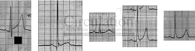
However, there has been evidence suggesting that the early repolarisation pattern may be associated with a risk for VF, depending on the location of early repolarisation, magnitude of the J wave and degree of ST elevation.[2,11,12] In a large study on a community-based general population of 10,864 middle-aged subjects, an early repolarisation pattern with J-point elevation of at least 0.1 mV in the inferior leads of a resting ECG was associated with an increased risk of death from cardiac causes.[13] In addition, among patients with a history of idiopathic ventricular fibrillation, an increased prevalence of early repolarisation, (up to 23 %), defined as an elevation of the QRS–ST junction of at least 0.1 mV from baseline in the inferior or lateral lead, manifested as QRS slurring or notching, has been detected.[11,14] A higher prevalence of J-wave and/or QRS slurring (but not of ST elevation) has been found among athletes with cardiac arrest/sudden death than in controls.[15] A horizontal/descending type (defined as ≤0.1 mV elevation of the ST segment within 100 ms after the J point) in the inferior leads, as opposed to a rapidly ascending ST segment type, may help to identify those individuals who are clearly at risk (see Figure 2).[16,17] Coexistence of an anterior early repolarisation pattern (e.g. in leads V1–V3),[18] and early repolarisation in the inferior leads, especially in cases without other QRS complex abnormalities, predict the occurrence of VT/VF.[19]
Figure 2: Rapidly Ascending (A) and Horizontal (B) ST Segment in the Leads Deploying J Waves.

J waves marked with arrowhead. ‘Concave/rapidly ascending’ – when there is 0.1 mV elevation of the ST segment within 100 ms after the J point and the ST segment merged gradually with the T wave. ‘Horizontal/descending’ – when the ST-segment elevation is 0.1 mV within 100 ms after the J point and continues as a flat ST segment until the onset of the T wave. With permission from Rosso, et al., 2012.
Still, several obscure points remain with this syndrome. An early repolarisation pattern in the inferolateral leads occurs in 5 % of apparently healthy individuals;[13,14] it may not be consistently seen, and even the horizontal/descending ST type was seen in 3 % of controls.[16,17] In the Atherosclerosis Risk in Communities (ARIC) study, J-point elevation was associated with an increased risk of sudden cardiac death (SCD) in whites and in women, but not in blacks or men.[20] A pattern of J-wave and/or QRS slurring (but not of ST elevation) has been associated with cardiac arrest/sudden death in athletes,[15] but many healthy athletes have early repolarisation with a rapidly ascending pattern. Inferolateral early repolarisation pattern is seen in 25–35 % of competitive athletes, and inferior only in 4 %, and is considered a dynamic phenomenon related to physical activity.[8,21,22] Finally, a large genome-wide association study has been unable to identify genetic variants associated with the pattern, possibly reflecting the phenotypic heterogeneity that exists among these individuals.[23] It seems, therefore, that the majority of individuals with early repolarisation are at no or minimal risk for arrhythmic events.[2]
Diagnosis
Specific diagnostic criteria for early repolarisation pattern and ERS were presented by the Heart Rhythm Society (HRS), European Heart Rhythm Association (EHRA) and Asia Pacific Heart Rhythm Society (APHRS) in 2013, as follows.[24]
ERS is diagnosed in:
The presence of J-point elevation ≥1 mm in ≥2 contiguous inferior and/or lateral leads of a standard 12-lead ECG in a patient resuscitated from otherwise unexplained VF/ polymorphic VT;
A SCD victim with a negative autopsy and medical chart review with a previous ECG demonstrating J-point elevation ≥1 mm in ≥2 contiguous inferior and/or lateral leads of a standard 12-lead ECG; and
The presence of J-point elevation ≥1 mm in ≥2 contiguous inferior and/or lateral leads of a standard 12-lead ECG.
In addition, specific repolarisation patterns that have been previously discussed should be also taken into account. The Brugada syndrome is characterised by J-point or ST-segment elevation in the right precordial leads, and approximately 12 % of patients display typical early repolarisation abnormalities. However, it is typical that the ST-segment elevation is augmented in the right precordial leads by sodium-channel blockers; whereas in ERS, the early repolarisation pattern is usually attenuated.[25]
Therapy
The risk stratification and optimum management of these patients are not well defined and the recognition of the truly malignant forms is difficult. Electrophysiology testing does not appear useful for risk stratification. VF is infrequently induced (22 %) and has no predictive value for ICD therapy.[26] Patients with aborted sudden death in the absence of identifiable cause (idiopathic VF) are treated with ICD. Ablation of idiopathic VF, targeted to short coupled ventricular premature beats that originate predominantly from the Purkinje system and the right ventricular outflow track and trigger VF, has also been reported.[27] According to the 2013 HRS/EHRA/APHRS statement, ICD is indicated only in patients who have survived a cardiac arrest (I) and it might be considered (IIb) in symptomatic family members of ER syndrome patients with a history of syncope in the presence of ST segment elevation >1 mm in 2 or more inferior or lateral leads. Quinidine may also be used in addition to ICD (IIa), as well as isoproterenol to suppress electrical storms (IIa).[24]
Acknowledgements
Andrew Grace, Section Editor–Arrhythmia Mechanisms/Basic Science acted as Editor for this article. This article is adapted from pp.530-533 Ch.61 Early repolarization syndromes: New Concepts, (updated) from ‘Clinical Cardiology: Current Practice Guidelines’ edited by Katritsis, Gersh, & Camm (2013): Oxford University Press, with kind permission. © Oxford University Press, 2013.
References
- 1.Macfarlane PW, Haissaguerre M, Huikuri HV et al. The early repolarization pattern: a consensus paper. J Am Coll Cardiol. 2015;66:470. doi: 10.1016/j.jacc.2015.05.033. [DOI] [PubMed] [Google Scholar]
- 2.Obeyesekere MN, Klein GJ, Nattel S et al. A clinical approach to early repolarization. Circulation. 2013;127:1620–9. doi: 10.1161/CIRCULATIONAHA.112.143149. [DOI] [PubMed] [Google Scholar]
- 3.Demidova MM, Martin-Yebra A, van der Pals J et al. Transient and rapid QRS-widening associated with a J-wave pattern predicts impending ventricular fibrillation in experimental myocardial infarction. Heart Rhythm. 2014;11:1195–201. doi: 10.1016/j.hrthm.2014.03.048. [DOI] [PubMed] [Google Scholar]
- 4.Antzelevitch C, Yan GX. J-wave syndromes: Brugada and early repolarization syndromes. Heart Rhythm. 2015;12:1852–66. doi: 10.1016/j.hrthm.2015.04.014. [DOI] [PMC free article] [PubMed] [Google Scholar]
- 5.Hu D, Barajas-Martinez H, Pfeiffer R et al. Mutations in SCN10A are responsible for a large fraction of cases of Brugada syndrome. J Am Coll Cardiol. 2014;64:66. doi: 10.1016/j.jacc.2014.04.032. [DOI] [PMC free article] [PubMed] [Google Scholar]
- 6.Watanabe H, Makiyama T, Koyama T et al. High prevalence of early repolarization in short QT syndrome. Heart Rhythm. 2010;7:647–52. doi: 10.1016/j.hrthm.2010.01.012. [DOI] [PubMed] [Google Scholar]
- 7.Surawicz B, Macfarlane PW. Inappropriate and confusing electrocardiographic terms: J-wave syndromes and early repolarization. J Am Coll Cardiol. 2011;57:1584–6.. doi: 10.1016/j.jacc.2010.11.040. [DOI] [PubMed] [Google Scholar]
- 8.Biasco L, Cristoforetti Y, Castagno D et al. Clinical, electrocardiographic, echocardiographic characteristics and long term follow up of elite soccer players with J-point elevation. Circ Arrhythm Electrophysiol. 2013;6:1178–84. doi: 10.1161/CIRCEP.113.000434. [DOI] [PubMed] [Google Scholar]
- 9.Ilkhanoff L, Soliman EZ, Prineas RJ et al. Clinical characteristics and outcomes associated with the natural history of early repolarization in a young, biracial cohort followed to middle age: the coronary artery risk development n young adults (CARDIA) study. Circ Arrhythm Electrophysiol. 2014;7:392, 9. doi: 10.1161/CIRCEP.113.000874. [DOI] [PMC free article] [PubMed] [Google Scholar]
- 10.Nakagawa M, Ezaki K, Miyazaki H et al. Electrocardiographic characteristics of patients with false tendon: possible association of false tendon with J waves. Heart Rhythm. 2012;9:782–8. doi: 10.1016/j.hrthm.2011.12.022. [DOI] [PubMed] [Google Scholar]
- 11.Derval N, Simpson CS, Birnie DH et al. Prevalence and characteristics of early repolarization in the CASPER registry: cardiac arrest survivors with preserved ejection fraction registry. J Am Coll Cardiol. 2011;58:722–8. doi: 10.1016/j.jacc.2011.04.022. [DOI] [PubMed] [Google Scholar]
- 12.Wu SH, Lin XX, Cheng YJ et al. Early repolarization pattern and risk for arrhythmia death: a meta-analysis. J Am Coll Cardiol. 2013;61:645–50. doi: 10.1016/j.jacc.2012.11.023. [DOI] [PubMed] [Google Scholar]
- 13.Tikkanen JT, Anttonen O, Junttila MJ et al. Long-term outcome associated with early repolarization on electrocardiography. N Engl J Med. 2009;361:2529–37. doi: 10.1056/NEJMoa0907589. [DOI] [PubMed] [Google Scholar]
- 14.Haissaguerre M, Derval N, Sacher F et al. Sudden cardiac arrest associated with early repolarization. N Engl J Med. 2008;358:2016–23. doi: 10.1056/NEJMoa071968. [DOI] [PubMed] [Google Scholar]
- 15.Cappato R, Furlanello F, Giovinazzo V et al. J wave, QRS slurring, and ST elevation in athletes with cardiac arrest in the absence of heart disease: marker of risk or innocent bystander? Circ Arrhythm Electrophysiol. 2010;3:305–11. doi: 10.1161/CIRCEP.110.945824. [DOI] [PubMed] [Google Scholar]
- 16.Rosso R, Glikson E, Belhassen B et al. Distinguishing ‘benign’ from ‘malignant early repolarization’: the value of the ST-segment morphology. Heart Rhythm. 2012;9:225–9. doi: 10.1016/j.hrthm.2011.09.012. [DOI] [PubMed] [Google Scholar]
- 17.Tikkanen JT, Junttila MJ, Anttonen O et al. Early repolarization electrocardiographic phenotypes associated with favorable long-term outcome. Circulation. 2011;123:2666–73. doi: 10.1161/CIRCULATIONAHA.110.014068. [DOI] [PubMed] [Google Scholar]
- 18.Kamakura T, Kawata H, Nakajima I et al. Significance of non-type 1 anterior early repolarization in patients with inferolateral early repolarization syndrome. J Am Coll Cardiol. 2013;62:1610–8. doi: 10.1016/j.jacc.2013.05.081. [DOI] [PubMed] [Google Scholar]
- 19.Junttila MJ, Tikkanen JT, Kenttä T et al. Early repolarization as a predictor of arrhythmic and nonarrhythmic cardiac events in middle-aged subjects. Heart Rhythm. 2014;11:1701–6. doi: 10.1016/j.hrthm.2014.05.024. [DOI] [PubMed] [Google Scholar]
- 20.Olson KA, Viera AJ, Soliman EZ et al. Long-term prognosis associated with J-point elevation in a large middle-aged biracial cohort: the ARIC study. Eur Heart J. 2011;32:3098–106. doi: 10.1093/eurheartj/ehr264. [DOI] [PMC free article] [PubMed] [Google Scholar]
- 21.Noseworthy PA, Weiner R, Kim J et al. Early repolarization pattern in competitive athletes: clinical correlates and the effects of exercise training. Circ Arrhthm Electrophysiol. 2011;4:432–40. doi: 10.1161/CIRCEP.111.962852. [DOI] [PMC free article] [PubMed] [Google Scholar]
- 22.Quattrini FM, Pelliccia A, Assorgi R et al. Benign clinical significance of J-wave pattern (early repolarization) in highly trained athletes. Heart Rhythm. 2014;11:1974–82. doi: 10.1016/j.hrthm.2014.07.042. [DOI] [PubMed] [Google Scholar]
- 23.Sinner MF, Porthan K, Noseworthy PA et al. A meta-analysis of genome-wide association studies of the electrocardiographic early repolarization pattern. Heart Rhythm. 2012;9:1627–34. doi: 10.1016/j.hrthm.2012.06.008. [DOI] [PMC free article] [PubMed] [Google Scholar]
- 24.Priori SG, Wilde AA, Horie M et al. HRS/EHRA/APHRS expert consensus statement on the diagnosis and management of patients with inherited primary arrhythmia syndromes: document endorsed by HRS, EHRA, and APHRS in May 2013 and by ACCF, AHA, PACES, and AEPC in June 2013. Heart Rhythm. 2013;10:1932–63. doi: 10.1016/j.hrthm.2013.05.014. [DOI] [PubMed] [Google Scholar]
- 25.Kawata H, Noda T, Yamada Y, et al. Effect of sodium-channel blockade on early repolarization in inferior/lateral leads in patients with idiopathic ventricular fibrillation and Brugada syndrome. Heart Rhythm. 2012;9:77–83. doi: 10.1016/j.hrthm.2011.08.017. [DOI] [PubMed] [Google Scholar]
- 26.Mahida S, Derval N, Sacher F et al. Role of electrophysiological studies in predicting risk of ventricular arrhythmia in early repolarization syndrome. J Am Coll Cardiol. 2015;65:151–9. doi: 10.1016/j.jacc.2014.10.043. [DOI] [PubMed] [Google Scholar]
- 27.Knecht S, Sacher F, Wright M et al. Long-term follow-up of idiopathic ventricular fibrillation ablation: a multicenter study. J Am Coll Cardiol. 2009;54:522–8. doi: 10.1016/j.jacc.2009.03.065. [DOI] [PubMed] [Google Scholar]