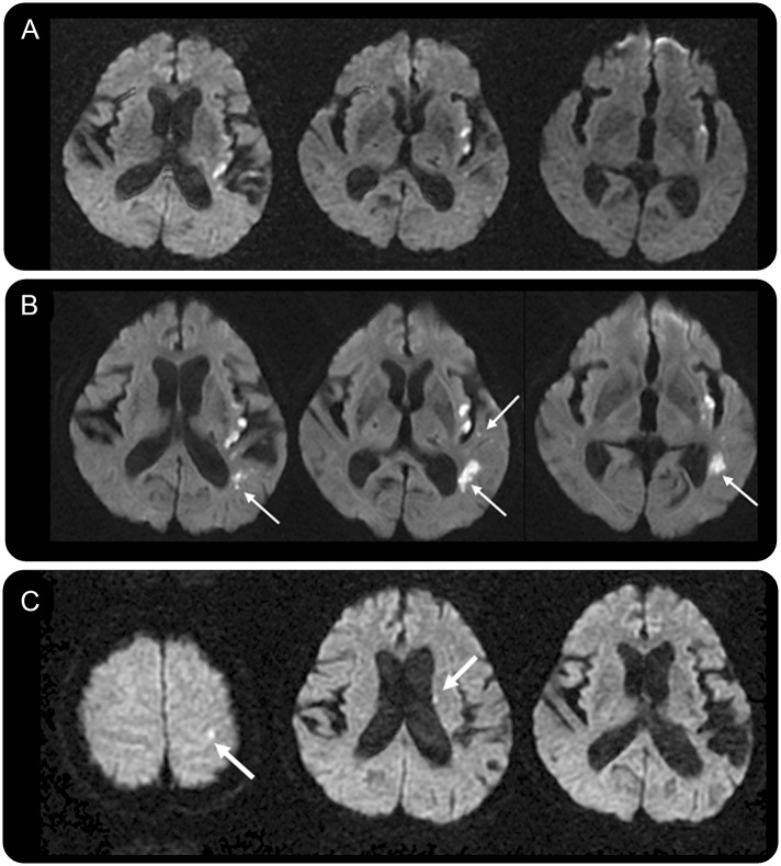
Figure 3. MRI recurrence and clinical recurrent ischemic stroke.
Images from a 69-year-old woman with sensory aphasia and dysarthria. (A) On the baseline diffusion-weighted imaging scan, there were multifocal infarcts in the left insular area. (B) Silent new ischemic lesions at 5 days (5D-SNIL) (thin arrows) were observed. (C) Twenty-three months later, the patient presented with right-sided weakness with new ischemic lesions in the left corona radiate and cortical area (thick arrows).

