Abstract
Lipomas are the most common benign mesenchymal tumors. They are located either subcutaneously or under the investing fascia in intramuscular or intermuscular regions. The reported frequency of intramuscular lipomas among all benign adipocytic tumors is 1.0%–5.0% and for intermuscular lipomas is 0.3%–1.9%. The frequency of these lesions is the same in all age groups, but in adults deep seated-lipomas are most commonly discovered between the ages of 30 and 60. The most common sites of involvement of intramuscular lipomas are the large muscles of the extremities, especially those of the thigh, shoulder, and upper arm. Intramuscular lipomas of the hand are extremely rare and only few cases have been reported in the literature. In cases with hand location, they may present with functional deficit or neurovascular compromise due to the effect of the mass. We report an unusual case of a large intramuscular lipoma of the thenar that was treated with surgical excision due to the impairment of hand function.
Keywords: Intramuscular, Lipoma, Thenar
Introduction
Lipomas are benign mesenchymal tumors of adipose tissue. Despite the fact that they are predominantly located in the subcutaneous tissue, intramuscular locations are relatively common. The most common sites of involvement of intramuscular lipomas are the large muscles of the extremities, especially those of the thigh, shoulder, and upper arm, while the hand location is extremely rare (1). In such cases they are presenting as slowly growing painless soft tissue masses and are usually associated with hand functional impairment. In the present report we describe an unusual case of thenar location. To our knowledge only a few cases have been reported in the English literature sharing the same location (2-4).
Case Report
A 59-year-old male patient was examined at our orthopedic office due to a long standing mass on his right hand. The patient reported a 10-year history of painless swelling on his hand that gradually enlarged over the last two years with consequent mild numbness on his right index finger. The clinical examination revealed a relatively large mass of approximately 5 cm in diameter in the first interdigital space of the right hand extending from the dorsal to the palmar aspect of the first metacarpal bone. On palpation the mass was painless, solid, soft, and mobile. The patient was unable to entirely flex his index finger and thumb and the thumb adduction was impaired. Also, grip strength of the right hand was reduced.
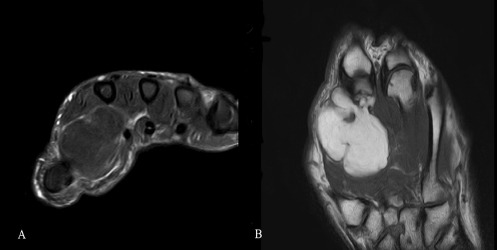
An MRI scan of the right hand was performed that showed a relatively large mass (dimensions: 4.9 * 3.7 * 2.9 cm) primary on the palmar aspect of the right hand with well-defined margins and low intensity signal at the T1 and fat suppression sequences [Figure 1]. No nerve and vessel entrapment was seen. MRI findings were consisted with the diagnosis of intramuscular lipoma of the right hand.
Figure 1.

T2 weighted images with fat suppression (A) and T1 (B) weighted images of the intramuscular lipoma.
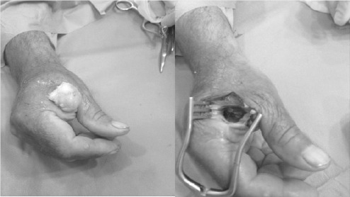
The patient was operated under local anesthesia (lidocaine 2%) after performing a median nerve block at the level of the carpal tunnel and a radial nerve block (cutaneous branch) at the level of the radial styloid process. An approximately 3 cm long longitudinal skin incision was made dorsally, exposing the mass that was carefully dissected and resected en-bloc from the surrounding tissues with care not to damage the digital arteries and nerves of the index finger and thumb [Figure 2]. The wound was closed primarily and the patient was dismissed with per-os antibiotics and painkillers.
Figure 2.

Surgical excision of the intramuscular lipoma.
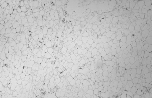
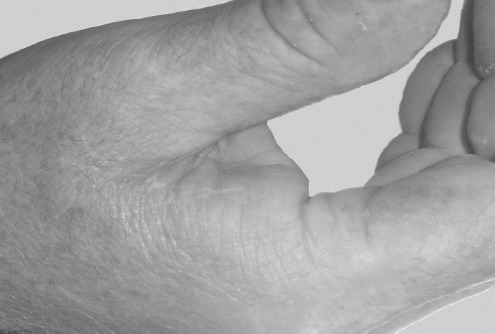
The histological examination of the specimen revealed a tumor consisting of well circumscribed mature adipocytes indicative of a lipoma [Figure 3]. One year postoperatively the patient had full range of motion and no numbness on the index finger and the thumb, with good grip power and no signs of recurrence [Figure 4].
Figure 3.

Histology showing a tumor consisted of mature adipocytes (haematoxylin and eosin stain, X200).
Figure 4.

One year postoperative image of the hand.
Discussion
The World Health Organization’s Committee for the Classification of Soft Tissue Tumors categorizes soft tissue lipomatous tumors into 9 entities: lipoma, lipomatosis, lipomatosis of nerve, lipoblastoma, angiolipoma, myolipoma of soft tissue, chondroid lipoma, spindle cell/pleomorphic lipoma, and hibernoma (5). Lipomas are common mesenchymal tumors that can develop in any fat-containing region. They account for approximately 16% of soft tissue mesenchymal tumors (6). Lipomas are either superficially located or can be deep-seated. The superficial location is the most common and involves the subcutaneous fat, whereas deep lesions present under the investing fascia. The deep lesions are mature fat lesions that may occur in intermuscular (affecting the intermuscular connective tissue) or intramuscular locations. Benign lipomatous lesions may also affect bones, joints and tendon sheaths. The reported frequency of intramuscular lipomas among all benign adipocytic tumors is 1.0%–5.0%, and that of intermuscular lipomas is 0.3%–1.9% (7). The frequency of these lesions is the same in all age groups, but in adults deep seated-lipomas are most commonly discovered between the ages of 30 and 60 (2). The thigh is the commonest location of intramuscular lipomas followed by the deltoid muscle. Intramuscular lipomas of the hand are very uncommon (1,4). There are two subgroups of intramuscular lipomas; infiltrative type and well-circumscribed type. The term infiltrating lipoma has been used due to the infiltrative nature of the tumor in histological examination and represents the commonest type (4).
In the vast majority of the cases intramuscular lipomas are asymptomatic. They generally present as slowly growing painless masses which may sometimes infiltrate deeply and wrap around nerves. They usually appear with large size at diagnosis. A lipoma of greater than 5 cm is classified as a “giant lipoma”. In cases with hand location, they may present with functional deficit or neurovascular compromise due to the mass effect (4). In particular cases of thenar location, they present as a painless mass over the thenar eminence. The overlying skin is normal without muscle adherence. They come to clinical attention due to the cosmetic concern and the impairment of hand functionality. Neurovascular compromises are rare. Some intramuscular thenar lipomas are small and thus difficult to palpate. In such cases imaging is necessary for diagnosis.
Ultrasound is initially used for the evaluation of these lesions. In cases of well circumscribed intramuscular lipomas, a well-defined hyperechoic ovoid mass is usually seen that is clearly delineated from the surrounding muscle. The appearance is similar to that of superficial lipomas. Some lesions appear isoechoic to the adjacent muscle tissue and this might lead to misdiagnosis or non-diagnosis. Areas of heterogeneity within the lesion can also be seen representing thin intrinsic septa. If encapsulated, the capsule is usually difficult to be identified on ultrasound. In infiltrative types of intramuscular lipomas, the muscle tissue is separated in a bland fashion by fat, producing a heterogeneous striated mass that might be interpreted as a non-lipoma lesion and mimic malignancy. The variety of these sonographic features, accounts for the limitation of sonography in accurately differentiating soft-tissue lipomas from other masses and necessitates the use of MRI imaging in order to establish a diagnosis (8).
An intramuscular lipoma predominantly appears in MRI as a fatty homogeneous mass with similar characteristics of typical lipoma, with high signal density on both T1 and T2 weighted images. However, some intramuscular lipomas present in MRI as inhomogeneous masses with regions of decreased signal that represent muscle or fibrous tissue within the lipoma (9). Uninodularity, presence of infiltrating margins and muscle fibers in intramuscular lipomas indicate a benign lesion rather than malignancy (9). Despite this, biopsies in the case of giant lipomas are deemed necessary to establish a definite diagnosis and to differentiate them from liposarcomas.
Despite the fact that intramuscular lipomas of thenar are entirely benign lesions with extremely low frequency of malignant transformation, the treatment of choice is surgical excision. The main reason for excision is the impairment of hand functionality and the cosmetic appearance. The lipoma is usually surrounded by a thin, fibrous capsule, which may allow the mass to be shelled-out. Careful dissection is necessary during the surgical procedure in order to avoid recurrence and damage of digital nerves and vessels. The reported recurrence after marginal excision is less than 5% and it occurs more frequently with deep and infiltrating lipomas, because of the modified surgical removal in order to avoid injuring adjacent neurovascular and muscular tissues (10).
References
- 1.Berlund P, Kalamaras M. A case report of trigger wrist associated with carpal tunnel syndrome caused by an intramuscular lipoma. Hand Surg. 2014;19(2):237–9. doi: 10.1142/S0218810414720174. [DOI] [PubMed] [Google Scholar]
- 2.Yadav SP, Jategaonkar PA, Haldar PJ. Giant hand lipoma revisited: report of a thenar lipoma & its literature review. J Hand Microsurg. 2013;5(2):84–5. doi: 10.1007/s12593-013-0093-3. [DOI] [PMC free article] [PubMed] [Google Scholar]
- 3.Kamra HT, Munde SL. Lipoma on palmar aspect of thumb: a rare case report. J Clin Diagn Res. 2013;7(8):1706–7. doi: 10.7860/JCDR/2013/5683.3261. [DOI] [PMC free article] [PubMed] [Google Scholar]
- 4.Lee YH, Jung JM, Baek GH, Chung MS. Intramuscular lipoma in thenar or hypothenar muscles. Hand Surg. 2004;9(1):49–54. doi: 10.1142/s0218810404002005. [DOI] [PubMed] [Google Scholar]
- 5.Fletcher CDM, Bridge JA, Hogendoorn PCW, Mertens F. WHO Classification of Tumours of Soft Tissue and Bone. Pathology and Genetics of Tumours of Soft Tissue and Bone. 4th ed. Lyon: IARC Press; 2013. [Google Scholar]
- 6.Rydholm A, Berg NO. Size, site and clinical incidence of lipoma: factors in the differential diagnosis of lipoma and sarcoma. Acta Orthop Scand. 1983;54(6):929–34. doi: 10.3109/17453678308992936. [DOI] [PubMed] [Google Scholar]
- 7.Fletcher CD, Martin-Bates E. Intramuscular and intermuscular lipoma: neglected diagnoses. Histopathology. 1988;12(3):275–87. doi: 10.1111/j.1365-2559.1988.tb01942.x. [DOI] [PubMed] [Google Scholar]
- 8.McFarland GB, Jr, Hoffer MM. Paralysis of the intrinsic muscles of the hand secondary to lipoma in Guyon’s tunnel. J Bone Joint Surg Am. 1971;53(2):375–6. [PubMed] [Google Scholar]
- 9.Matsumoto K, Hukuda S, Ishizawa M, Chano T, Okabe H. MRI findings in intramuscular lipomas. Skeletal Radiol. 1999;28(3):145–52. doi: 10.1007/s002560050491. [DOI] [PubMed] [Google Scholar]
- 10.Murphey MD, Carroll JF, Flemming DJ, Pope TL, Gannon FH, Kransdorf MJ. From the archives of the AFIP: benign musculoskeletal lipomatous lesions. Radiographics. 2004;24(5):1433–66. doi: 10.1148/rg.245045120. [DOI] [PubMed] [Google Scholar]


