
Fig. 3.
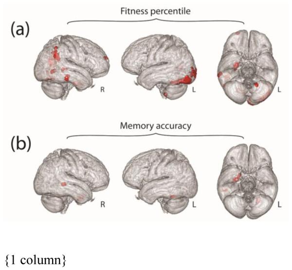
Results of exploratory whole-brain analysis. Parts (a) and (b) illustrate the results of an exploratory whole brain analysis, showing regions (red) where gray matter volume may be associated with fitness percentile or memory accuracy, respectively. Results are depicted within the group average brain.