Abstract
In drug discovery, it is very important to evaluate liver cells within an organism. Compared to 2D culture methods, the development of 3D culture techniques for liver cells has been successful in maintaining long-term liver functionality with the formation of a hepatic-specific structure. The key to performing drug testing is the establishment of a stable in vitro evaluation system. In this article, we report a Tapered Stencil for Cluster Culture (TASCL) device developed to create liver spheroids in vitro. The TASCL device will be applied as a toxicity evaluation system for drug discovery. The TASCL device was created with an overall size of 10 mm × 10 mm, containing 400 microwells with a top aperture (500 µm × 500 µm) and a bottom aperture (300 µm diameter circular) per microwell. We evaluated the formation, recovery, and size of HepG2 spheroids in the TASCL device. The formation and recovery were both nearly 100%, and the size of the HepG2 spheroids increased with an increase in the initial cell seeding density. There were no significant differences in the sizes of the spheroids among the microwells. In addition, the HepG2 spheroids obtained using the TASCL device were alive and produced albumin. The morphology of the HepG2 spheroids was investigated using FE-SEM. The spheroids in the microwells exhibited perfectly spherical aggregation. In this report, by adjusting the size of the microwells of the TASCL device, uniform HepG2 spheroids were created, and the device facilitated more precise measurements of the liver function per HepG2 spheroid. Our TASCL device will be useful for application as a toxicity evaluation system for drug testing.
Key words: Human hepatocellular carcinoma (HepG2) cells, Spheroid, Three-dimensional (3D) culture, Tapered stencil for cluster culture (TASCL), Medical evaluation
INTRODUCTION
From the last half of the 20th century, liver cells have been used in cell therapy (7,13) as well as drug discovery and development (3,14–19). When performing drug discovery, it is very important to evaluate liver cells within an organism (8,31). Although drug evaluation tests of liver cells based on the 2D monolayer culture method were a trend through the 1990s, tests using the 3D culture method were accelerated in the 2000s. Compared to the 2D culture method, the development of 3D culture techniques for assessing liver cells has been successful in maintaining the long-term liver function from the formation of a hepatic-specific structure (3,4,14,22,25).
In recent years, many research groups have made attempts to prepare human liver-like cells by inducing differentiation from stem cells (1,2,12) and using these cells for drug testing. However, compared with the liver functions of human primary hepatocytes, obtaining human liver-like cells of sufficient quality is challenging. Additionally, in advance, human induced pluripotent stem cell (iPSC)-derived constructs were transplanted into a cranial window of a nonobese diabetic/severe combined immunodeficient (NOD/SCID) mouse, the Takebe and Taniguchi groups (28,29) also initiated a new attempt to prepare to liver-like cells in mice. However, at present, it is not yet possible to fully reproduce the functions of human liver cells. Moreover, human primary hepatocytes isolated from livers and commercially available cryopreserved human hepatocytes tend to demonstrate large variations from lot to lot (20,21), and it is difficult to maintain reproducible results in liver cell evaluation tests. In recent years, drug interaction studies using human hepatocyte preparations from at least three donors have been recommended based on the U.S. Food and Drug Administration (FDA) guidance. In particular, the mRNA change by investigational drugs has been measured in cultured human hepatocytes from three donors (5). To solve this problem, toxicity screening tests using liver cell lines such as HepG2 (derived from human hepatocellular carcinoma of a 15-year-old male) and HepaRG™ (terminally differentiated hepatic cells derived from a hepatic progenitor cell line from female hepatocarcinoma; BioPredic International) cells are expected to maintain reproducible results in liver cell evaluation tests. Therefore, the key in performing drug testing is to establish a stable in vitro evaluation system. As a drug evaluation method using liver cells in the 2D culture method, it is relatively easier to obtain reproducible data between individual culture plates. On the other hand, the results obtained using 3D culture methods (spheroid culture) affect the reproducibility between individual culture plates. In particular, it should be mentioned as a cause that the size of spheroids formed with the 3D culture method is not uniform. Recently, several studies have reported the use of liver spheroids created according to the 3D culture method (3,4,6,14–19,31). Among them, a method was also reported to create a large number of cell clusters in 3D culture (9). We prepared uniform embryoid bodies (EBs) from iPSCs (26,27) at a large scale using a Tapered Stencil for Cluster Culture (TASCL) device and induced the cells to differentiate into liver cells from EBs (32). In addition, in a single seeding, we were successful in creating various sizes of 3D hepatocyte constructs using a combinatorial TASCL device (16).
In this article, we report the application of the TASCL device to prepare HepG2 spheroids in vitro. TASCL devices will be applied as toxicity evaluation systems for drug discovery. A TASCL device was created with an overall size of 10 mm × 10 mm, containing 400 microwells with a top aperture (500 µm × 500 µm) and bottom aperture (300 µm diameter circular) per a microwell. A six-well plate with an ultralow attachment surface (Corning Inc., Corning, NY, USA) under the TASCL device was used in the evaluation experiment. We assessed the formation, recovery, and size of the HepG2 spheroids in the TASCL device. In addition, we investigated the determination of live and dead cells and the albumin secretion activity of the HepG2 spheroids.
MATERIALS AND METHODS
Materials
Dulbecco’s modified Eagle’s medium (DMEM), Dulbecco’s phosphate-buffered saline without Ca2+ and Mg2+ supplementation (D-PBS-free), and antibiotics (penicillin and streptomycin) were purchased from Life Technologies, Inc. (Carlsbad, CA, USA). Fetal bovine serum (FBS) was obtained from Funakoshi Co, Ltd. (BIO-WEST, Nuaille, France). Paraformaldehyde (PFA) was purchased from Sigma-Aldrich (St. Louis, MO, USA). All other materials and chemicals not specified above were of the highest grade available.
Cells
HepG2 cells (male human hepatocellular carcinoma cells) were purchased from the American Type Culture Collection (ATCC, Manassas, VA, USA).
Fabrication of the TASCL Device
The TASCL device was created using polydimethylsiloxane (PDMS; SYLGARD® 184; Dow Corning, Midland, MI, USA) as previously described (10,11,16). The TASCL device was placed on a six-well cell culture plate (Falcon 353046; Becton Dickinson, Franklin Lakes, NJ, USA) and sterilized under ultraviolet (UV) for 12 h. The upper surface of the TASCL device was coated with an aqueous solution of poly(ethylene oxide)-poly(propylene oxide)-poly(ethylene oxide) triblock copolymer (1 wt% Anti-Link; Allvivo Vascular, Inc., Lake Forest, CA, USA) for 12 h at room temperature. The copolymer self-assembles on a hydrophobic surface and forms a hydrophilic layer that prevents cell adhesion. The bottom surface of the TASCL device is maintained intact due to the self-sealing ability of PDMS, whereas the top surface is hydrophilic. The array of 400 microwells was placed at the center of the TASCL device and surrounded by a trench to prevent the uncontrolled intrusion of cells from the boundary area.
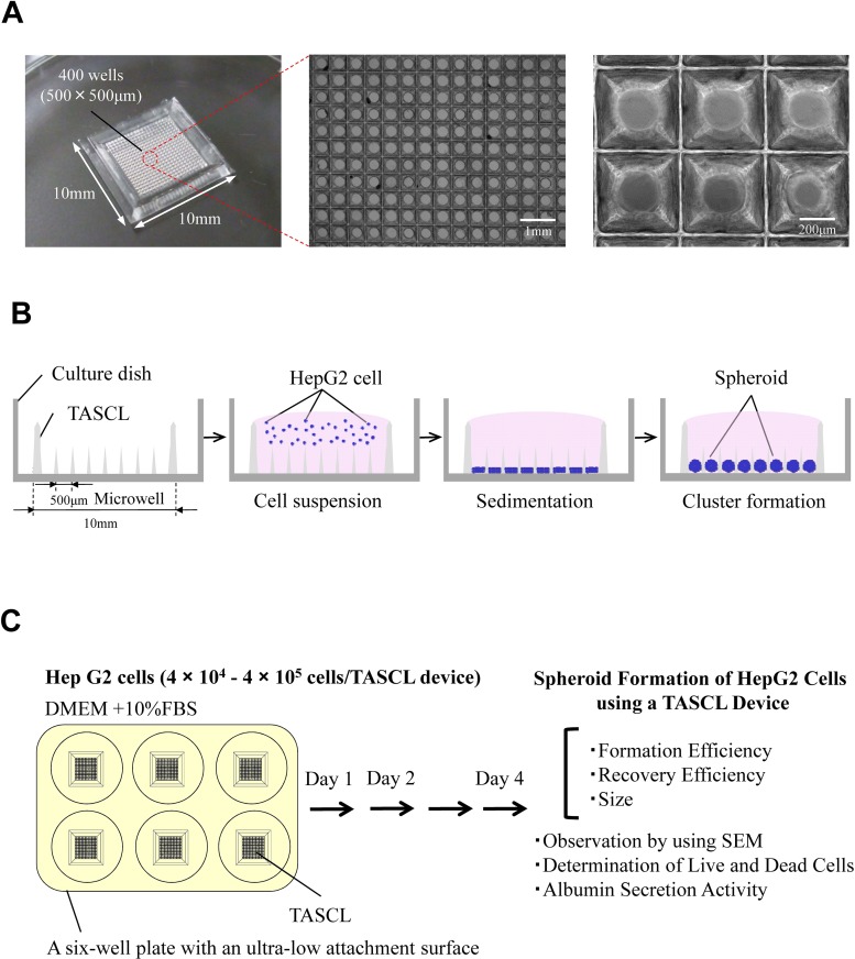
Figure 1A shows a schematic diagram of the TASCL device. A TASCL device was created with an overall size of 10 mm × 10 mm, containing microwells with a top aperture (500 µm × 500 µm) and bottom aperture (300 µm diameter circular) per microwell. The TASCL device can be easily installed on a commercially available culture dish and used to freely replace the cell adhesion surface matrix. In this study, we applied hydrophobic treatment of the surface inside the TASCL device and placed a six-well plate with an ultralow attachment surface (Corning Inc.) under the TASCL device.
Figure 1.
Fabrication of the Tapered Stencil for Cluster Culture (TASCL) device. (A) The TASCL device consisted of microwells measuring 10 mm × 10 mm with a thickness of 0.55 mm. Each microwell in the TASCL device had a top aperture (500 µm × 500 µm) and bottom aperture (300 µm diameter circular). The TASCL device was observed under phase-contrast microscopy. Scale bars: 1 mm and 200 µm. (B) A schematic diagram of cell seeding and the cluster formation process using the TASCL device. Human hepatocellular carcinoma cell (HepG2) spheroids can be created under multiple initial conditions simultaneously by injecting the cell suspension onto the TASCL device. (C) The study protocol for spheroid formation of HepG2 cells using the TASCL device. DMEM, Dulbecco’s modified Eagle’s medium; FBS, fetal bovine serum; SEM, scanning electron microscope.
Schematic Figure of Spheroid Formation in the TASCL Device
Figure 1B shows a schematic diagram of spheroid formation using the TASCL device. First, HepG2 cells are seeded at the same time by simply placing a droplet of cell suspension onto the TASCL device (cell suspension process). HepG2 cells are equally divided into the microwells of the TASCL device (sedimentation process), and HepG2 spheroids are formed after 1 day of culture (cluster formation process). HepG2 spheroids can be created under the single initial condition simultaneously by injecting the cell suspension onto the TASCL device.
Spheroid Formation of HepG2 Cells Using the TASCL Device
Figure 1C shows the study protocol for spheroid formation of HepG2 cells using the TASCL device. HepG2 cells at density of 4 × 10⁴, 2 × 10⁵, and 4 × 10⁵ cells/TASCL were inoculated on a TASCL device to form 3D spheroids. The viability of the HepG2 cells was greater than 95%, as determined by a trypan blue dye exclusion test. The final concentration of trypan blue (Gibco®, Life Technologies Inc.) was 0.2%. The cells were then cultured in 0.35 ml of DMEM supplemented with 10% FBS, 100 U/ml of penicillin, and 100 U/ml of streptomycin (culture medium) for 4 days. A six-well plate with an ultralow attachment surface was used as the bottom portion of the TASCL device. A total of 0.35 ml of HepG2 cells suspended in medium (4 × 10⁴, 2 × 10⁵, and 4 × 10⁵ cells/TASCL calculated to be 100, 500 and 1,000 cells/microwell each) was applied to each microwell. The culture medium on the TASCL was changed and recovered every 24 h. The cells were incubated at 37°C under a humidified 5% CO2 atmosphere and then cultured for 4 days in the TASCL, after which the albumin secretion activity was determined, as described below. The morphology of the HepG2 spheroids was observed under a phase-contrast microscope (Olympus, Tokyo, Japan).
Determination of Live and Dead Cells in the HepG2 Spheroids
HepG2 spheroids were stained with calcein acetoxymethyl ester [calcein AM, 4 mM in anhydrous dimethyl sulfoxide (DMSO)] and ethidium homodimer-1 [EthD-1, 2 mM in DMSO/H2O 1:4 (v/v)] (LIVE/DEAD® Viability/Cytotoxicity Kit, for mammalian cells, L3224; Molecular Probes®, Life Technologies Inc.) to detect live and dead cells. A total of 4 µl of the supplied 2 mM EthD-1 stock solution was transferred to 2 ml of D-PBS-free. Then, 1 µl of the supplied 4 mM calcein AM stock solution was added to 2 ml of the EthD-1 solution. The working solution containing 2 µM of calcein AM and 4 µM EthD-1 was then added directly to the HepG2 spheroids. After the cells were incubated for 20 min at 37°C, calcein AM-positive green fluorescence in the live cells and EthD-1-positive red fluorescence in the dead cells were judged to be present using a fluorescence microscope (Olympus, Tokyo, Japan). After trypsin (Gibco®, Life Technologies Inc.) treatment of the HepG2 spheroids, the cell viability was measured using a trypan blue exclusion assay under the conditions used for the experiments.
Quantitation of Albumin Secreted by the HepG2 Spheroids
HepG2 spheroids were cultured in the TASCL device, and the culture medium was changed every 24 h. The level of albumin production was assessed based on the accumulation of albumin in the culture medium after 24 h using a human albumin enzyme linked immunosorbent assay (ELISA) quantification kit, as instructed by the manufacturer (Bethyl Laboratories, Inc. Montgomery, TX, USA).
Scanning Electron Microscopic Observation of the HepG2 Spheroids
A scanning electron microscope (SEM) was used to visualize HepG2 spheroids in the TASCL device. HepG2 spheroids were fixed with 4% PFA and then cooled down to 4°C overnight. Afterward, the fixed samples were rinsed three times with D-PBS. A reagent for SEM sample preparation was prepared by transferring 20 µl of Hitachi’s Exclusive IL1000 Ionic Liquid for Electron Microscopy Applications (HILEM IL1000; Hitachi High-Technologies Corp., Tokyo, Japan) to 160 µl of Milli-Q water (Milli-Q Integral Water Purification System; Merck-Millipore, Darmstadt, Germany). The resulting approximately 10% IL1000 Ionic Liquid solution was then added directly to the samples at room temperature for 1 h. The samples were subsequently transferred to a filter paper and dehydrated at room temperature within 30 min. Carbon tape (Nissin Enamel Manufacturing Co. Ltd., Nagoya, Japan) was attached to a SEM stage (JEOL Ltd., Tokyo, Japan) surface, and the obtained samples were placed on the SEM stage. The morphology of the samples was investigated using a field emission scanning electron microscope (FE-SEM; JEOL JSM-7500F) at an acceleration voltage of 15 kV.
Statistical Analysis
Data are presented as the mean ± standard deviation (SD). Each experiment was repeated three times (n = 3). The statistical significance was determined using unpaired Student’s t-tests for comparisons between the 4 × 10⁴ and 4 × 10⁵ groups. A value of p < 0.05 was considered to be significant.
RESULTS
Formation of HepG2 Spheroids in the TASCL Device
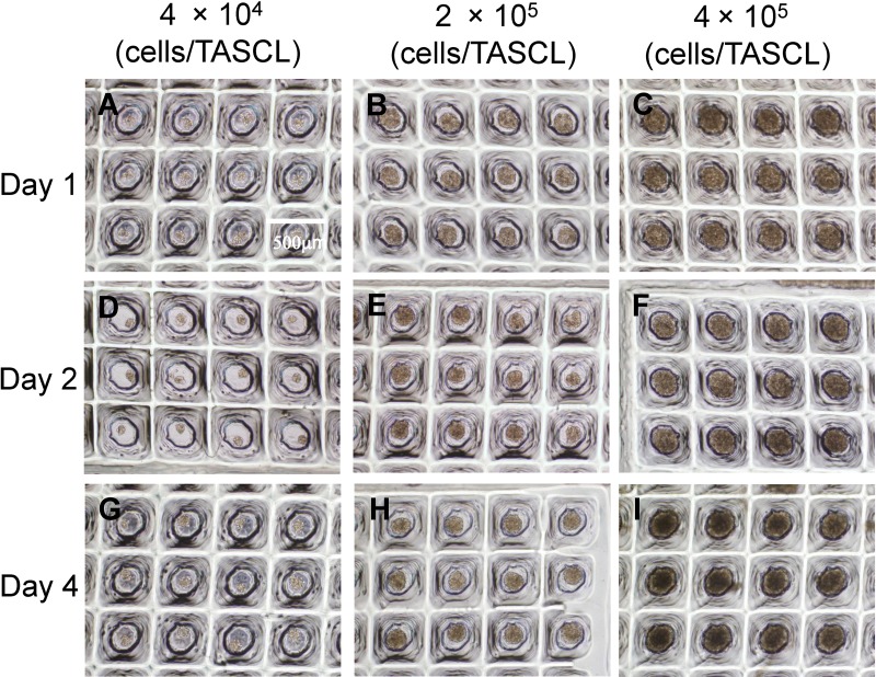
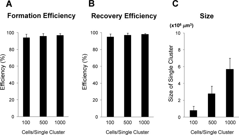
HepG2 cells at density of 4 × 10⁴, 2 × 10⁵, and 4 × 10⁵ cells/TASCL were inoculated on the TASCL device for 4 days starting from cell seeding. HepG2 cells at 4 × 10⁴, 2 × 10⁵, and 4 × 10⁵ cells/TASCL were counted to be 100, 500, and 1,000 cells/microwell each. After 1, 2, and 4 days of culture, HepG2 spheroids were observed in the TASCL device under a phase-contrast microscope (Fig. 2). The HepG2 spheroids formed in the TASCL device exhibited perfectly spherical aggregation on day 1 (Fig. 2A–C), and their spherical morphology was maintained after 2 and 4 days of culture (Fig. 2D–I). We also evaluated the formation efficiency, recovery, and size of the HepG2 spheroids in the TASCL device (Fig. 3). HepG2 spheroids forming a single cluster were counted as a successful spheroid on day 2. The formation and recovery were both nearly 100% (Fig. 3A and B). The size of the HepG2 spheroids formed in the TASCL device was determined based on the formula for the volume of a sphere. The size of the spheroids determined by their phase-contrast images in the HepG2 cells at density of 4 × 10⁴, 2 × 10⁵, and 4 × 10⁵ cells/TASCL was 0.8 × 106 µm3, 2.8 × 106 µm3, and 5.7 × 106 µm3, respectively (Fig. 3C) (The experiments were done three times, and data are shown as the mean ± SD.), indicating that the size of the HepG2 spheroids increased with an increase in the initial cell seeding density.
Figure 2.
Phase-contrast photomicrographs of the HepG2 spheroids created using the TASCL device. HepG2 cells at a density of 4 × 10⁴ (A, D, and G), 2 × 10⁵ (B, E, and H), and 4 × 10⁵ cells (C, F, and I) were seeded onto the TASCL device (calculated to be 100, 500 and 1,000 cells/microwell each) and then cultured for 4 days. The culture periods were as follows: day 1 (A, B, and C), day 2 (D, E, and F), and day 4 (G, H, and I). Scale bar: 500 µm.
Figure 3.
Efficiency of the formation, recovery, and size of HepG2 spheroids in the TASCL device. HepG2 cells at density of 4 × 10⁴, 2 × 10⁵, and 4 × 10⁵ cells/TASCL were inoculated on the TASCL device for 2 days. (A) The efficiency of spheroid formation was evaluated. (B) The efficiency of spheroid recovery was evaluated. (C) The size of the formed spheroids was measured in the case of 100, 500, and 1,000 cells/droplet. Data are shown as the mean ± standard deviation for triplicate experiments.
SEM Images of HepG2 Spheroids in the TASCL Device
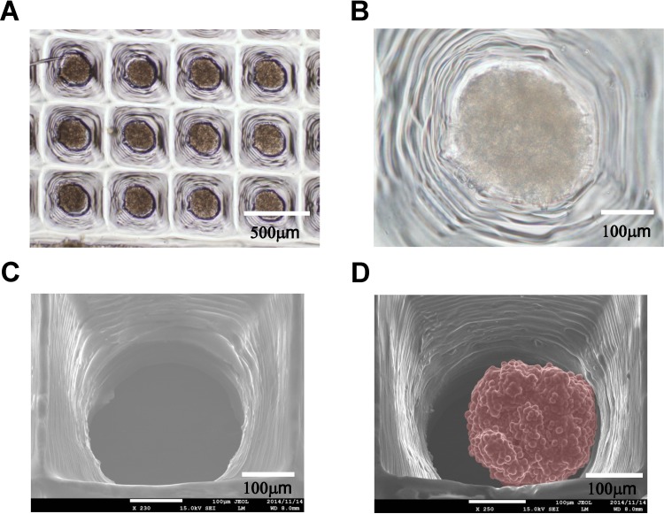
The morphology of the HepG2 spheroids was investigated using FE-SEM at an acceleration voltage of 15 kV. HepG2 cells at a density of 4 × 10⁵ cells/TASCL were seeded onto a TASCL device and cultured for 2 days (Fig. 4). Figures 4A and B show phase-contrast images of the resulting HepG2 spheroids. SEM images of the microwells in the TASCL device and HepG2 spheroids in the microwells were also observed (Figs. 4C and D). The spheroids in the microwells exhibited perfectly spherical aggregation (Fig. 4D) with a diameter and size of 218 ± 10.1 µm and 5.4 ± 0.69 × 106 µm3, respectively. The experiments were done three times, and data are shown as the mean ± SD.
Figure 4.
Scanning electron microscope (SEM) images of the HepG2 spheroids in the TASCL device. HepG2 cells at a density of 4 × 10⁵ were inoculated on the TASCL device for 2 days. (A, B) Phase-contrast photographs of the HepG2 spheroids observed from the microwell in a TASCL device. (C) SEM image observed of the microwell in a TASCL device. (D) SEM image of HepG2 spheroids (artificial red color) observed within the microwell of a TASCL device. The spheroids in the microwells exhibited perfectly spherical aggregation with a diameter and size of 218 ± 10.1 µm and 5.4 ± 0.69 × 106 µm3, respectively. Data are shown as the mean ± standard deviation for triplicate experiments.
Survival of HepG2 Spheroids in the TASCL Device
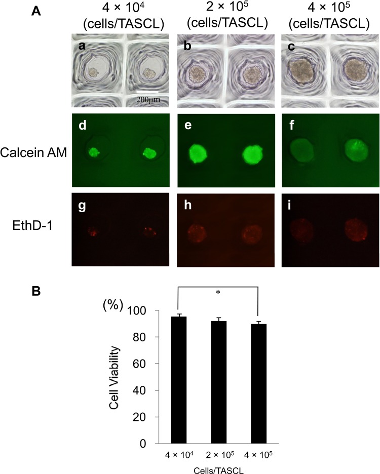
HepG2 spheroids were stained with calcein AM and EthD-1 to discriminate between live and dead cells. Calcein AM-positive green fluorescence in the live cells and EthD-1-positive red fluorescence in the dead cells were observed using a fluorescence microscope. HepG2 cells at density of 4 × 10⁴, 2 × 10⁵, and 4 × 10⁵ cells/TASCL were seeded onto the TASCL device and then cultured for 2 days (Fig. 5Aa, b, and c; phase contrast). Most cells observed in the HepG2 spheroids were viable (Fig. 5Ad, e, and f), although some dead cells were found (Fig. 5Ag, h, and i).
Figure 5.
The live/dead cells and single cell viability in the HepG2 spheroids created using the TASCL device. (A) HepG2 cells at a density of 4 × 10⁴, 2 × 10⁵, and 4 × 10⁵ cells were inoculated on the TASCL device for 2 days (phase contrast; a, b, and c). HepG2 spheroids were stained with calcein AM and EthD-1 to visualize live and dead cells. Calcein acetoxymethyl ester (calcein AM)-positive green fluorescence (d, e, and f) in live cells and ethidium homodimer-1 (EthD-1)-positive red fluorescence (g, h, and i) in dead cells were observed with a fluorescence microscope. (B) After trypsin treatment, single cell survival was measured using a trypan blue exclusion assay. Data are shown as the mean ± standard deviation of triplicate values. *p < 0.05.
We also measured the cell viability using a trypan blue exclusion assay under the conditions used for the experiments shown in Figure 5A. The cell viability of the HepG2 spheroids at density of 4 × 10⁴, 2 × 10⁵, and 4 × 10⁵ cells/TASCL was approximately 95%, 92%, and 89% (Fig. 5B) (p < 0.05 4 × 10⁴ compared with 4 × 10⁵ cells/TASCL), respectively, at 2 days after inoculation.
Liver Function of HepG2 Spheroids in the TASCL Device
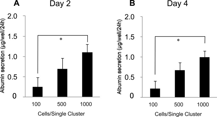
HepG2 cells at density of 4 × 10⁴, 2 × 10⁵, and 4 × 10⁵ cells/TASCL were seeded onto the TASCL device and then cultured for up to 4 days (Fig. 6). In order to examine the liver function of the HepG2 spheroids in the TASCL device, we also evaluated the amount of albumin secretion in the medium over 24 h on days 2 and 4 of culture. The albumin secretion activity of the HepG2 spheroids at density of 4 × 10⁴, 2 × 10⁵, and 4 × 10⁵ cells/TASCL on day 2 increased in a cell number-dependent manner (Fig. 6A) (p < 0.01 4 × 10⁴ compared with 4 × 10⁵ cells/TASCL). In the time-dependent change of culture, the activities on day 4 were almost the same (Fig. 6B).
Figure 6.
Albumin secretion in the HepG2 spheroids created using the TASCL device. HepG2 cells at density of 4 × 10⁴, 2 × 10⁵, and 4 × 10⁵ cells/TASCL were seeded onto the TASCL device and then cultured for 4 days. The amount of albumin secretion in the medium was measured by ELISA after 24 h of accumulation on day 2 (A) and day 4 (B) of culture. Data are shown as the mean ± standard deviation for triplicate experiments. *p < 0.01.
DISCUSSION
Liver cells are already being used successfully in cell transplantation with therapeutic approaches (7). In the past few decades, liver cells have been used for cell therapy in regenerative medicine (7,20,21) and drug toxicity screening tests for drug discovery (3,8). Cytotoxicity evaluation methods using drug discovery tests have changed from 2D culture methods to 3D culture methods. In comparison with 2D culture, 3D culture has an advantage that the cells can maintain their function for a long period (3,4,14,22). In the present study, we developed a toxicity evaluation system for assessing liver cells by utilizing the 3D culture method. Using HepG2 cells as a liver cell line, HepG2 spheroids partially formed on the TASCL device and mimicked the function and intrinsic morphology of liver tissue.
In our previous report (16), the creation of 3D hepatocyte constructs was performed with a combinatorial TASCL device [overall size of 10 mm × 10 mm, containing microwells with a top aperture (400 µm × 400 µm, 600 µm × 600 µm, 800 µm × 800 µm) and bottom aperture (40 µm × 40 µm, 80 µm × 80 µm, 160 µm × 160 µm) per microwell]. It was possible to create 3D hepatocyte constructs of various sizes in a single seeding by changing the upper and lower portions of the microwells of the TASCL device. On the other hand, in this report, we succeeded in constructing uniform-sized HepG2 spheroids by aligning the upper and lower portions of the microwells in a TASCL device [overall size of 10 mm × 10 mm, containing microwells with the top aperture (500 µm × 500 µm) and bottom aperture (300 µm diameter circular) per microwell] (Fig. 1). Additionally, in our previous report (16), the resulting liver constructs exhibited various cell morphologies when various culture substrates were used in the bottom portion of the TASCL device. In the present study, we achieved the establishment of a HepG2 cell suspension culture in each microwell of the TASCL device using a nonadhesive cell culture plate as the bottom portion.
Thus far, several studies have been reported on the formation of liver spheroids using 3D culture devices (3,4,6,11,16–19,22–24,30–32). In this report, it was initially confirmed that the cells can form HepG2 spheroids with different sizes using a TASCL device under various cell seeding density conditions (Figs. 2 and 3). As the cell seeding density increased, the size of the resulting HepG2 spheroids in each microwell increased. Compared with culture substrates used previously, the present TASCL device is a 3D culture device that can create a large number of HepG2 spheroids at the same time using a simple operation. However, there have so far been few reports of such 3D culture devices.
The SEM images of the resulting HepG2 spheroids were nearly spherical aggregates (Fig. 4D). In addition to the number of cells seeded in each microwell, it was possible to create HepG2 spheroids with a nearly spherical structure by changing the sizes of the upper and lower portions of the TASCL device. In this experiment, microwells with the same size were used to align the culture conditions in the TASCL device. It is essential to design the TASCL device in accordance with the cell number per microwell.
HepG2 spheroids in the TASCL device were cultured for 2 days and stained with calcein AM and EthD-1 to visualize live and dead cells under a fluorescence microscope (Fig. 5). Most cells of the HepG2 spheroids were viable; however, some dead cells were found. As the size of the HepG2 spheroids increased, the cell viability was found to be decreased (p < 0.05 vs. 4 × 10⁴ compared with 4 × 10⁵ cells/TASCL) (Fig. 5B). The level of albumin production of HepG2 spheroids was measured for 2 and 4 days of culture (Fig. 6). The albumin activity was found to increase with an increase in the size of the HepG2 spheroids (p < 0.01 4 × 10⁴ compared with 4 × 10⁵ cells/TASCL) (Fig. 6A). These results suggest that the liver function can be evaluated using the TASCL device.
In a previous report (16), by changing the size of the microwells in the TASCL device, the device was utilized as a screening tool for creating cell aggregates with different sizes. In the present report, by an adjusted, standardized size of microwells of the TASCL device, uniform HepG2 spheroids were created, and the device facilitated more precise measurements of the liver function per HepG2 spheroid. Our TASCL device will be useful as a toxicity evaluation system for drug testing.
ACKNOWLEDGMENTS
We thank Professor Koji Ikuta at The University of Tokyo for help with FE-SEM measurement. We thank Ms. Rina Yokota and Ms. Yumie Koshidaka at Nagoya University for their technical assistance. This work was supported in part by JSPS KAKENHI Grant Nos. 22650127, 25560248, 25600054 (Grant-in-Aid for Challenging Exploratory Research), and JST PRESTO program. The authors declare no conflicts of interest.
REFERENCES
- 1. Basma H.; Soto-Gutiérrez A.; Yannam G. R.; Liu L.; Ito R.; Yamamoto T.; Ellis E.; Carson S. D.; Sato S.; Chen Y.; Muirhead D.; Navarro-Álvarez N.; Wong R. J.; Roy-Chowdhury J.; Platt J. L.; Mercer D. F.; Miller J. D.; Strom S. C.; Kobayashi N.; Fox I. J. Differentiation and transplantation of human embryonic stem cell-derived hepatocytes. Gastroenterology 136:990–999; 2009. [DOI] [PMC free article] [PubMed] [Google Scholar]
- 2. Chen Y.; Soto-Gutiérrez A.; Navarro-Alvarez N.; Rivas-Carrillo J. D.; Yamatsuji T.; Shirakawa Y.; Tanaka N.; Basma H.; Fox I. J.; Kobayashi N. Instant hepatic differentiation of human embryonic stem cells using activin A and a deleted variant of HGF. Cell Transplant. 15:865–871; 2006. [DOI] [PubMed] [Google Scholar]
- 3. Enosawa S.; Miyamoto Y.; Hirano A.; Suzuki S.; Kato N.; Yamada Y. Application of cell array 3D-culture system for cryopreserved human hepatocytes with low-attaching capability. Drug Metab. Rev. 39(Suppl. 1):342; 2007. [Google Scholar]
- 4. Enosawa S.; Miyamoto Y.; Kubota H.; Jomura T.; Ikeya T. Construction of artificial hepatic lobule-like spheroids on a three-dimensional culture. Cell Med. 3(1-3):19–23; 2012. [DOI] [PMC free article] [PubMed] [Google Scholar]
- 5. Fahmi O. A.; Kish M.; Boldt S.; Obach R. S. Cytochrome P450 3A4 mRNA is a more reliable marker than CYP3A4 activity for detecting pregnane X receptor-activated induction of drug-metabolizing enzymes. Drug Metab. Dispos. 38(9):1605–1611; 2010. [DOI] [PubMed] [Google Scholar]
- 6. Feng Z. Q.; Chu X.; Huang N. P.; Wang T.; Wang Y.; Shi X.; Ding Y.; Gu Z. Z. The effect of nanofibrous galactosylated chitosan scaffolds on the formation of rat primary hepatocyte aggregates and the maintenance of liver function. Biomaterials 30(14):2753–2763; 2009. [DOI] [PubMed] [Google Scholar]
- 7. Fox I. J.; Chowdhury J. R.; Kaufman S. S.; Goertzen T. C.; Chowdhury N. R.; Warkentin P. I.; Dorko K.; Sauter B. V.; Strom S. C. Treatment of the Crigler-Najjar syndrome type I with hepatocyte transplantation. N. Engl. J. Med. 338(20):1422–1426; 1998. [DOI] [PubMed] [Google Scholar]
- 8. Hewitt N. J.; Lech M. J.; Houston J. B.; Hallifax D.; Brown H. S.; Maurel P.; Kenna J. G.; Gustavsson L.; Lohmann C.; Skonberg C.; Guillouzo A.; Tuschl G.; Li A. P.; LeCluyse E.; Groothuis G. M.; Hengstler J. G. Primary hepatocytes: Current understanding of the regulation of metabolic enzymes and transporter proteins, and pharmaceutical practice for the use of hepatocytes in metabolism, enzyme induction, transporter, clearance, and hepatotoxicity studies. Drug Metab. Rev. 39(1):159–234; 2007. [DOI] [PubMed] [Google Scholar]
- 9. Hori Y.; Rulifson I. C.; Tsai B. C.; Heit J. J.; Cahoy J. D.; Kim S. K. Growth inhibitors promote differentiation of insulin-producing tissue from embryonic stem cells. Proc. Natl. Acad. Sci. USA 99(25):16105–16110; 2002. [DOI] [PMC free article] [PubMed] [Google Scholar]
- 10. Ikeuchi M.; Ikuta K. Method for Producing Different Populations of Molecules or Fine Particles with Arbitrary Distribution Forms and Distribution Densities Simultaneously and in Quantity, and Masking Member Therefor. United States Patent Application, US 2011/0229953 A1.
- 11. Ikeuchi M.; Oishi K.; Noguchi H.; Shuji Hayashi.; Koji Ikuta. Soft tapered stencil mask for combinatorial 3D cluster formation of stem cells. Proc. µTAS 2010:641–643; 2010. [Google Scholar]
- 12. Ishikawa T, Banas A, Hagiwara K, Iwaguro H, Ochiya T. Stem cells for hepatic regeneration: the role of adipose tissue derived mesenchymal stem cells. Curr. Stem Cell Res. Ther. 5(2):182–189; 2010. [DOI] [PubMed] [Google Scholar]
- 13. Ishikawa T.; Banas A.; Teratani T.; Iwaguro H.; Ochiya T. Regenerative cells for transplantation in hepatic failure. Cell Transplant. 21(2–3):387–399; 2012. [DOI] [PubMed] [Google Scholar]
- 14. Janorkar A. V. Polymeric Scaffold Materials for Two-Dimensional and Three-Dimensional in Vitro Culture of Hepatocytes. In: Kulshrestha A. S.; Mahapatro A.; Henderson L. A., eds. Biomaterials. Washington, DC: ACS Publications; 2010:1–32. [Google Scholar]
- 15. Miyamoto Y.; Enosawa S.; Takeuchi T.; Takezawa T. Cryopreservation in situ of cell monolayers on collagen vitrigel membrane culture substrata: Ready-to-use preparation of primary hepatocytes and ES cells. Cell Transplant. 18:619–626; 2009. [DOI] [PubMed] [Google Scholar]
- 16. Miyamoto Y.; Ikeuchi M.; Noguchi H.; Yagi T.; Hayashi S. Three-Dimensional in vitro Hepatic Constructs Formed Using Combinatorial Tapered Stencil for Cluster Culture (TASCL) device. Cell Med. 7:67–74; 2015. [DOI] [PMC free article] [PubMed] [Google Scholar]
- 17. Miyamoto Y.; Ikeya T.; Enosawa S. Preconditioned cell array optimized for a three-dimensional culture of hepatocytes. Cell Transplant. 18:677–681; 2009. [DOI] [PubMed] [Google Scholar]
- 18. Miyamoto Y.; Koshidaka Y.; Noguchi H.; Oishi K.; Saito H.; Yukawa H.; Kaji N.; Ikeya T.; Iwata H.; Baba Y.; Murase K.; Hayashi S. Polysaccharide functionalized magnetic nanoparticles for cell labeling and tracking: A new three-dimensional cell-array system for toxicity testing. In: Nagarajan R., ed. Nanomaterials for biomedicine. Washington, DC: ACS Publications; 2012:191–208. [Google Scholar]
- 19. Miyamoto Y.; Koshidaka Y.; Noguchi H.; Oishi K.; Saito H.; Yukawa H.; Kaji N.; Ikeya T.; Suzuki S.; Iwata H.; Baba Y.; Murase K.; Hayashi S. Observation of positively-charged magnetic nanoparticles inside HepG2 spheroids using electron microscopy. Cell Med. 5(2–3):89–96; 2013. [DOI] [PMC free article] [PubMed] [Google Scholar]
- 20. Miyamoto Y.; Suzuki S.; Nomura K.; Enosawa S. Improvement of hepatocyte viability after cryopreservation by supplementation of long-chain oligosaccharide in the freezing medium in rats and humans. Cell Transplant. 15:911–919; 2006. [DOI] [PubMed] [Google Scholar]
- 21. Miyamoto Y.; Teramoto N.; Hayashi S.; Enosawa S. An improvement in the attaching capability of cryopreserved human hepatocytes by a proteinaceous high molecule, Sericin, in the serum-free solution. Cell Transplant. 19:701–706; 2010. [DOI] [PubMed] [Google Scholar]
- 22. Otsuka H.; Hirano A.; Nagasaki Y.; Okano T.; Horiike Y.; Kataoka K. Two-dimensional multiarray formation of hepatocytes spheroids on a microfabricated PEG-brush surface. Chembiochem 5:850–855; 2004. [DOI] [PubMed] [Google Scholar]
- 23. Peshwa M. V.; Wu F. J.; Sharp H. L.; Cerra F. B.; Hu W. S. Mechanistics of formation and ultrastructural evaluation of hepatocyte spheroids. In Vitro Cell Dev. Biol. Anim. 32(4):197–203; 1996. [DOI] [PubMed] [Google Scholar]
- 24. Sakai Y.; Nakazawa K. Technique for the control of spheroid diameter using microfabricated chips. Acta Biomater. 3(6):1033–1040; 2007. [DOI] [PubMed] [Google Scholar]
- 25. Sullivan J. P.; Gordon J. E.; Bou-Akl T.; Matthew H. W.; Palmer A. F. Enhanced oxygen delivery to primary hepatocytes within a hollow fiber bioreactor facilitated via hemoglobin-based oxygen carriers. Artif. Cells Blood Substit. Immobil. Biotechnol. 35(6):585–606; 2007. [DOI] [PubMed] [Google Scholar]
- 26. Takahashi K.; Tanabe K.; Ohnuki M.; Narita M.; Ichisaka T.; Tomoda K.; Yamanaka S. Induction of pluripotent stem cells from adult human fibroblasts by defined factors. Cell 131(5):861–872; 2007. [DOI] [PubMed] [Google Scholar]
- 27. Takahashi K.; Yamanaka S. Induction of pluripotent stem cells from mouse embryonic and adult fibroblast cultures by defined factors. Cell 126(4):663–676; 2006. [DOI] [PubMed] [Google Scholar]
- 28. Takebe T.; Sekine K.; Enomura M.; Koike H.; Kimura M.; Ogaeri T.; Zhang R. R.; Ueno Y.; Zheng Y. W.; Koike N.; Aoyama S.; Adachi Y.; Taniguchi H. Vascularized and functional human liver from an iPSC-derived organ bud transplant. Nature 499(7459):481–484; 2013. [DOI] [PubMed] [Google Scholar]
- 29. Takebe T.; Zhang R. R.; Koike H.; Kimura M.; Yoshizawa E.; Enomura M.; Koike N.; Sekine K.; Taniguchi H. Generation of a vascularized and functional human liver from an iPSC-derived organ bud transplant. Nat. Protoc. 9(2):396–409; 2014. [DOI] [PubMed] [Google Scholar]
- 30. Ungrin M. D.; Joshi C.; Nica A.; Bauwens C.; Zandstra P. W. Reproducible, ultra high-throughput formation of multicellular organization from single cell suspension-derived human embryonic stem cell aggregates. PLoS One 3(2):e1565; 2008. [DOI] [PMC free article] [PubMed] [Google Scholar]
- 31. Walker T. M.; Rhodes P. C.; Westmoreland C. The differential cytotoxicity of methotrexate in rat hepatocyte monolayer and spheroid cultures. Toxicol. In Vitro 14(5):475–485; 2000. [DOI] [PubMed] [Google Scholar]
- 32. Yukawa H.; Ikeuchi M.; Noguchi H.; Miyamoto Y.; Ikuta K.; Hayashi S. Embryonic body formation using the tapered soft stencil for cluster culture device. Biomaterials 32(15):3729–3738; 2011. [DOI] [PubMed] [Google Scholar]


