Abstract
AIM: To investigate the optimal magnetic pressure and provide a theoretical basis for choledochojejunostomy magnetic compressive anastomosis (magnamosis).
METHODS: Four groups of neodymium-iron-boron magnets with different magnetic pressures of 0.1, 0.2, 0.3 and 0.4 MPa were used to complete the choledochojejunostomy magnamosis. Twenty-six young mongrel dogs were randomly divided into five groups: four groups with different magnetic pressures and 1 group with a hand-suture anastomosis. Serum bilirubin levels were measured in all groups before and 1 wk, 2 wk, 3 wk, 1 mo and 3 mo after surgery. Daily abdominal X-ray fluoroscopy was carried out postoperatively to detect the path and the excretion of the magnet. The animals were euthanized at 1 or 3 mo after the operation, the burst pressure was detected in each anastomosis, and the gross appearance and histology were compared according to the observation.
RESULTS: The surgical procedures were all successfully performed in animals. However, animals of group D (magnetic pressure of 0.4 MPa) all experienced complications with bile leakage (4/4), whereas half of animals in group A (magnetic pressure of 0.1 MPa) experienced complications (3/6), 1 animal in the manual group E developed anastomotic stenosis, and animals in group B and group C (magnetic pressure of 0.2 MPa and 0.3 MPa, respectively) all healed well without complications. These results also suggested that the time required to form the stoma was inversely proportional to the magnetic pressure; however, the burst pressure of group A was smaller than those of the other groups at 1 mo (187.5 ± 17.7 vs 290 ± 10/296.7 ± 5.7/287.5 ± 3.5, P < 0.05); the remaining groups did not differ significantly. A histologic examination demonstrated obvious differences between the magnamosis groups and the hand-sewn group.
CONCLUSION: We proved that the optimal range for choledochojejunostomy magnamosis is 0.2 MPa to 0.3 MPa, which will help to improve the clinical application of this technique in the future.
Keywords: Choledochojejunostomy, Magnetic compressive anastomosis, Optimal range, Pressure intensity, Magnetic pressure
Core tip: This study introduced a magnetic anastomosis device and verified the feasibility of magnetic compression anastomosis (magnamosis) in choledochojejunostomy; moreover, 3D printing technology was used to design and produce magnetic shells of different sizes to explore the optimum magnetic pressure range in choledochojejunostomy. The result of this study provided a more efficient and accurate theoretical basis for clinical application of choledochojejunostomy magnamosis in the future.
INTRODUCTION
Roux-en-Y choledochojejunostomy is known as a standard operation for the treatment of benign biliary stricture or malignant biliary obstruction and a well-developed approach[1]. However, the currently used manual anastomosis technique is time-consuming and associated with a high risk of complications, such as stricture recurrence and anastomotic leak[2]. Staple anastomosis has been introduced to solve this problem, but many limitations remain, such as foreign residues, low histocompatibility, and anastomosis diameter mismatch[3].
The compressive anastomosis technique was invented two centuries ago, and Denans proposed the concept of compressive anastomosis as early as 1826[4]. A new device was invented by Murphy in 1892, which has been referred to as “Murphy’s button” and extensively used in intestinal anastomosis[5,6]. In 1978, Obora[7] first used magnets instead of mechanical devices; this technique cleverly avoids physical contact by using magnetic force, which is field-mediated. With advantages of simpleness, saving time, and low cost, the magnetic compressive anastomosis (magnamosis) has attracted many surgeons to solve a variety of surgical problems. Jansen et al[8] first successfully used magnetic rings for colorectal anastomosis in 1981. Mimuro et al[9] and Akita et al[10] reported many cases of the successful application of magnamosis for biliary strictures and biliary anastomoses in liver transplantation. In 2003, the Ventrica company launched magnetic devices used for vascular side-to-side anastomosis, and these devices were clinically successful[11,12]. Magnamosis has been proven to be a safe surgical technique that is equivalent or superior to anastomosis created by the hand-sewn or stapling technique[13].
Although magnetic approaches have shown promise in choledochojejunostomy, the compressive pressure and magnet specification are based on experience, and significant differences have been reported in these parameters. Furthermore, these parameters often lack systematic research, and a weaker magnetic force may create local ulceration or abscesses due to slow and contained perforation. Stronger attraction can cause severe ischemia and/or lacerating/shearing trauma, which leads to free perforation. We hypothesized that an appropriate range of magnetic pressure can create a viable and durable choledochojejunostomy, and we further proposed that the magnetic pressure affects the quality of anastomosis.
MATERIALS AND METHODS
Magnetic device preparation
The device used for the end-to-side choledochojejunostomy consisted of two parts: the biliary part and the enteric part. Both parts featured magnetic rings constructed of sintered-type neodymium-iron-boron (NdFeB, N45); the surface field was approximately 2500 GS. These magnetic materials were all plated with titanium film on the external surfaces to maintain material stability and biocompatibility. According to a previous design[14], two magnets for the biliary part were constructed with the following respective outer diameter, inner diameter and height: 6 mm × 2.5 mm × 6 mm and 7 mm × 3 mm × 6 mm. The two magnets for the enteric part were designed with the following respective outer diameter, inner diameter and height: 10 mm × 3 mm × 6 mm and 11 mm × 3 mm × 6 mm. The force-displacement curve of the biliary-enteric magnet pair with outside diameters of 6 mm-10 mm and 7 mm-11 mm were measured using a universal tensile testing machine (UTM6202, Suns Technology Stock Co., Ltd., Shenzhen, China) (Figure 1). The magnetic forces of different magnet pairs (6-10 mm, 6-11 mm, 7-10 mm, and 7-11 mm) at separation distances of 0 mm, 2 mm and 4 mm are given in Table 1.
Figure 1.
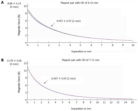
Magnetic force displacement curve. The magnetic force measured by a universal tensile testing machine is shown as a function of intermagnet separation (in mm) for 2 types of magnet pairs used for choledochojejunostomy magnamosis; 5 samples were tested for each magnet. A: Magnet pair with an outside diameter of 6 mm (biliary part) to 10 mm (enteric part); B: Magnet pair with an outside diameter of 7 mm (biliary part) to 11 mm (enteric part). OD: Outside diameter.
Table 1.
Magnetic force of magnet pairs at the distance of 0 mm, 2 mm or 4 mm
| Separation (mm) |
Outside diameter of biliary - enteric magnet pairs (mm) |
|||
| 6-10 | 6-11 | 7-10 | 6-11 | |
| 0 | 8.86 ± 0.14 | 8.24 ± 0.46 | 14.63 ± 0.24 | 13.79 ± 0.06 |
| 2 | 4.06 ± 0.07 | 4.04 ± 0.16 | 5.82 ± 0.09 | 6.01 ± 0.05 |
| 4 | 2.15 ± 0.03 | 2.30 ± 0.06 | 2.99 ± 0.05 | 3.27 ± 0.04 |
Three dimensional printing
To optimize the magnetic pressure for choledochojejunostomy, four different pressures were tested: 0.1 MPa, 0.2 MPa, 0.3 MPa, and 0.4 MPa (1 MPa = 1 N/mm2). According to P = F/S, the pressure can be changed by adjusting the crimping area at a constant force. Based on these calculations, we designed a series of magnetic outer shells with different crimping areas to meet the required predetermined pressure values. The shells were made of photosensitive resin and produced using a three dimensional (3-D) printer (Figure 2). The shells feature an internal drainage duct with an inside diameter of 1.5 mm at the middle, which allowed bile to flow through and the enteric part to precisely couple with the biliary part.
Figure 2.
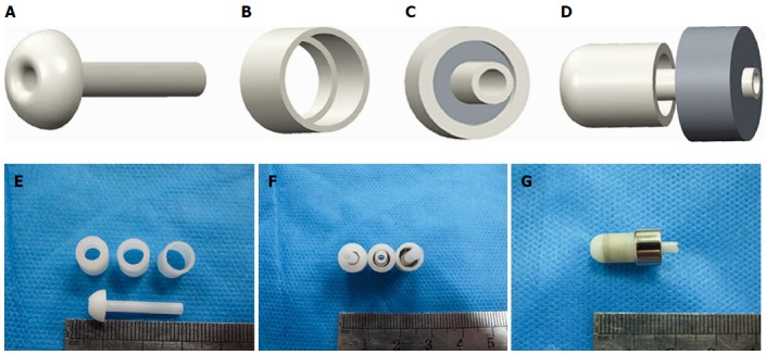
Mode pattern and real pictures of magnet devices. A: Lateral view of an internal drainage tube; B: View of the biliary part magnet shell; C: Antapical view of the combined biliary part; D: Lateral view of biliary part approach to enteric part; E: The internal drainage tube and shells of different crimping areas; F: Magnets with different pressures; G: Biliary part and enteric part coupled together.
Animals and grouping
Twenty-six mongrel male dogs older than 1 year and weighing more than 15 kg were provided by the Experimental Animal Center (SYXK-SHAN 2014-003) of the School of Medicine of Xi’an Jiaotong University. Male animals were selected because they cannot menstruate or become pregnant. The animals were randomly assigned to five groups: A (n = 6) - choledochojejunostomy with 0.1 MPa magnetic pressure, B (n = 6) - choledochojejunostomy with 0.2 MPa magnetic pressure, C (n = 6) - choledochojejunostomy with 0.3 MPa magnetic pressure, D (n = 4) - choledochojejunostomy with 0.4 MPa magnetic pressure, and E (n = 4) - choledochojejunostomy with traditional suture. The end-to-side enteroenteric anastomosis for the Roux-en-Y choledochojejunostomy of each group was completed using 3-0 non-absorbable sutures. One and 3 mo after surgery, postoperative complications, the bursting pressure of anastomoses, gross appearance, and pathology were evaluated.
Animal ethical approval
The experimental protocol was approved by the Animal Experimentation Committee of Xi’an Jiaotong University (No. XJTULAC201-398), and the animals were acclimatized to laboratory conditions (23 °C, 12 h/12 h light/dark, 50% humidity, ad libitum access to food and water) for 2 wk prior to experimentation. They were euthanized using a barbiturate overdose (intravenous injection, 150 mg/kg pentobarbital sodium) for tissue collection, and all dogs received humane care in compliance with the Guide for the Care and Use of Laboratory Animals published by the National Institutes of Health.
Surgical procedure
First, a common bile duct dilation animal model was established in all dogs. After being fasted for 12 h and water deprived for 4 h, the dogs were anesthetized by an intraperitoneal injection of 30 g/L pentobarbital (1 mL/kg). Penicillin (2400000 U) was injected intramuscularly 30 min before surgery to avoid postoperative infection. After disinfection with povidone iodine, sterile towels were placed and an abdominal midline incision was made. By fully exposing the hepatoduodenal ligament, the common bile duct could be easily identified. 3-0 Mersilk (Ethicon; Johnson & Johnson Medical Ltd., Shanghai, China) sutures were used to ligate the distal end of the common bile duct near the duodenum (Figure 3A). Water was freely available after recovery from anesthesia, but food was not provided until 2 d later. Penicillin (2400000 U) was injected twice daily after surgery for 3 consecutive days.
Figure 3.
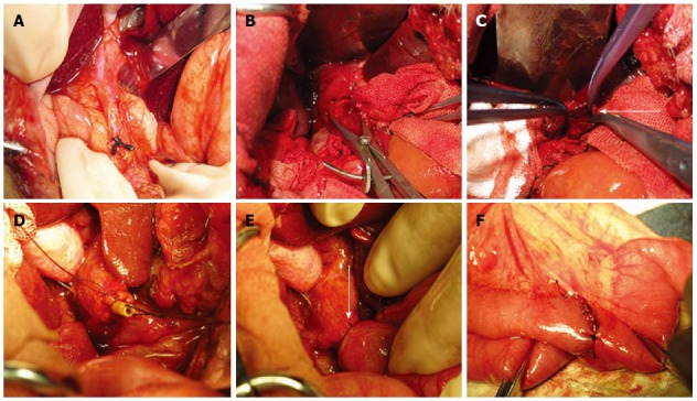
Surgical procedure. Images illustrating the surgical procedure with a magnet pair. A: The ligation of the distal end of common bile duct near the duodenum. B: Obvious dilatation of the common bile duct can be observed 10 d after ligation; C: Opened common bile duct before placing the biliary part magnet (arrow); D: The biliary part magnet was fixed to the stump of the common bile duct by a purse string; E: The choledochojejunostomy was constructed with the magnet pair (arrow); F: Suture enteroenteric anastomosis between the proximal end of the jejunum and the distal 50 cm of the Roux-en-Y limb.
Ten days after ligation, the dogs showed obvious symptoms of biliary obstruction such as dark-yellow urine and clay-colored stools. Serum bilirubin also significantly increased, confirming that the animal model was successfully established (Figure 3B).
The repair and reconstruction process was then performed. After the same presurgical procedures described in the common bile duct ligation operation, a second laparotomy was performed. The jejunum was dissociated and cut off approximately 15 cm away from the ligament of Treitz, and the distal end was closed with a suture. An end-to-side anastomosis was created between the proximal end and jejunum 50 cm away from the distal end with a double-layer suture of 3-0 Mersilk (Ethicon; Johnson & Johnson Medical Ltd., Shanghai, China).
In groups A, B, C and D, duct parts of the devices of different magnetic pressures were inserted into the proximal end of the common bile duct, and the stump of the bile duct was purse-string fixed with 5-0 Vicryl (Ethicon; Johnson & Johnson, Somerville, New Jersey, United States) and ligated onto the internal drainage tube. The jejunum was then punched 5 cm distal to the raised loop, and the enteric part magnet was inserted into the jejunum and coupled with the duct part magnet under the guidance of a drainage tube through the punch hole. After confirming that the common bile duct wall and the intestinal wall were clamped between the two magnets, the stump of the jejunum was then closed with 3-0 non-absorbable sutures.
Group E was subjected to a hand-sewn biliary-enteric bypass using 5-0 Vicryl, and full-thickness puncture and the mucosa-to-mucosa contact of the duct and jejunum were confirmed. The abdominal wall was then closed layer by layer (Figure 3C-E).
No food was allowed until the third day after the operation, and water was freely available. Penicillin sodium (2400000 U) was postoperatively injected intravenously twice daily for 3 d.
Blood test and follow-up
Blood samples for total bilirubin tests were collected at each time point, including before the ligation of the common bile duct and 10 d after ligation; 1, 2 and 3 wk after choledochojejunostomy and 1 and 3 mo after surgery.
Postoperative complications mainly included biliary stenosis and bile leakage. Biliary stenosis is reflected by recurrent jaundice and a rebound in the total bilirubin. Bile leakage can be judged by the animal state and postoperative cholangiography results. In case of death, dogs were carefully autopsied to determine the exact cause.
X-ray examination
After choledochojejunostomy, a plain abdominal X-ray was immediately taken to confirm the accurate coupling of the two magnets. The X-ray test and cholangiography were carried out every day after surgery to verify the passage of the magnets until they disappeared in the photograph. The time required to shed the magnets from the anastomosis was accurately recorded for different magnet pressures (Figure 4).
Figure 4.
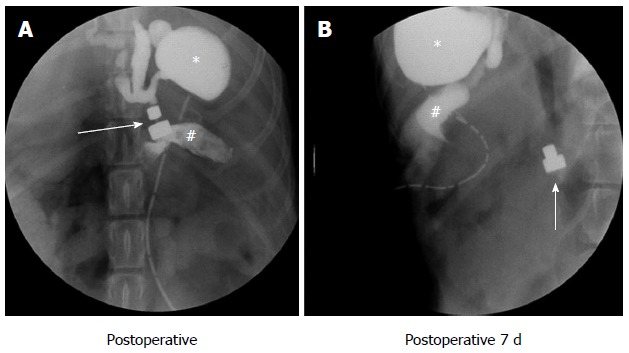
Postoperative abdominal X-ray. The path of the magnet pairs was monitored via an abdominal X-ray examination. The arrows indicates the magnetic pair, the asterisk indicates the gallbladder and the pound sign indicates the jejunum. A: Abdominal X-ray examination immediately after surgery; the two parts of the magnet pair coupled very well, exhibited good patency and were leak free; B: Daily abdominal radiography until the magnets were shed from the anastomosis.
Bursting strength test
The burst pressure was measured in each anastomosis. The two ends of the anastomotic specimen were ligated using hemostatic forceps or silk sutures, and the third end was attached to the sphygmomanometer and submerged in water. The intraluminal pressure was then gradually increased, and the readings were recorded as air first rose in bubbles to the surface of the water (Figure 5). The maximum hydraulic pressure when the specimen ruptured was recorded.
Figure 5.
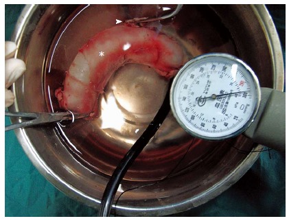
Bursting pressure test. Mechanical interrogation of anastomotic integrity. The arrowhead indicates the common bile duct, and the asterisk indicates the jejunum. The pointer on the sphygmomanometer is almost at the maximum, but bubbles were not observed.
Tissue harvest
Dogs from each group were sacrificed at 1 and 3 mo (half of animals were sacrificed at 1 mo, and the other half were sacrificed at 3 mo), and the anastomotic specimens were harvested. After a gross observation, the specimens were cut into sections, fixed in 10% neutral formalin for subsequent mounting and stained on slides.
Statistical analysis
The statistical methods of this study were reviewed by Qian Li from the First Affiliated Hospital at Xi’an Jiaotong University.
For descriptive statistics, the data were evaluated by analysis of variance (ANOVA) and student’s t-test. In all of the tests, the significant level was set at P < 0.05. The data were analyzed using the SPSS 17.0 software.
RESULTS
Total bilirubin
To accurately assess the patency of anastomosis, the initial bilirubin levels were ensured to be normal in each group. These levels significantly increased within 10 d after ligation, decreased during postoperative 1 wk and returned to normal within 1 mo. Bile leakage occurred in group A, which resulted in a faster bilirubin decrease than in the other groups (ANOVA, P < 0.05); in the manual group with stenosis formation, the bilirubin levels slightly increased 3 mo after a decline to normal levels (ANOVA, P < 0.01) (Figure 6).
Figure 6.
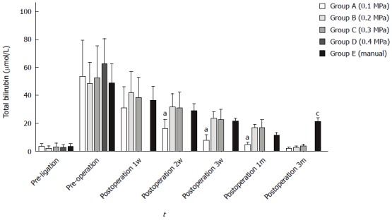
Serum total bilirubin between each group at different time points. Group A exhibited a faster decrease 2 wk, 3 wk and 1 mo after surgery (ANOVA, aP < 0.05); group E exhibited a significant increase within 3 mo (ANOVA, cP < 0.05); data were not obtained from group D due to a high mortality rate.
Postoperative complications
Four dogs in group A experienced postoperative bile leakage, and 3 animals died of anastomosis leakage because of a failure of coupling. In addition, all dogs in group D died within one week because of severe bile leakage. The total mortality rate of 26 dogs was 27% (7/26), and the incidences of postoperative complications was 34.6% (9/26) (Table 2).
Table 2.
Postoperative outcomes of dogs and stoma molding time
| Group | n | Complications, n | Death, n | Anastomotic molding time (d), mean ± SD |
| A | 6 | 4 | 3 | 9.3 ± 0.6 |
| B | 6 | 0 | 0 | 6.3 ± 0.82 |
| C | 6 | 0 | 0 | 4.5 ± 0.84 |
| D | 4 | 4 | 4 | / |
| E | 4 | 1 | 0 | / |
Stoma molding time
Daily abdominal radiography was performed strictly to monitor the exact time of anastomosis formation. We found that the magnet shedding time decreased as the pressure gradually increased; the mean times were 9.3 ± 0.6 d for group A, 6.3 ± 0.82 d for group B and 4.5 ± 0.8 d for group D, and these times significantly differed between groups (ANOVA P < 0.05) (Table 2).
Bursting strength
The burst pressures at 1 and 3 mo are displayed in Figure 7. At 1 mo, the burst pressure of group A was 187.5 ± 17.7 mmHg, and this pressure significantly differed from those of groups B (290 ± 10 mmHg), C (296.7 ± 5.7 mmHg) and E (287.5 ± 3.5 mmHg) (P < 0.05). All dogs in group D died of complications; therefore, the exact burst pressure could not be measured. Within 3 mo, significant differences could not be observed between groups A (275 mmHg), B (296.7 ± 5.7 mmHg), C (295 ± 5 mmHg) and E (297.5 ± 3.5 mmHg) (P = 0.052).
Figure 7.
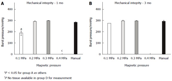
Anastomosis mechanical integrity at 1 (A) and 3 mo (B). The burst pressure at 1 mo was significantly lower in group A than in other groups (ANOVA, P < 0.05). The burst pressure did not differ between groups at 3 mo (ANOVA, P = 0.052).
Gross appearance of anastomosis
One month after the anastomosis, significant differences were observed between the traditional hand-sewn group and groups with magnamosis (Figure 8A1-E1). The suture knots remained evident in the manual group, and the mucosal surfaces appeared uneven and rough due to the interference of suture (Figure 8E1). By contrast, the mucosa of the magnamosis groups was smoother and flatter, but differences were evident between groups: an obvious anastomotic line in the mucosa can be observed in group A (Figure 8A1), whereas the mucosa was smoother in groups B and C, and the anastomosis line was not easily identified (Figure 8B1-C1). Animals in group D all experienced perforation (Figure 8D1, D2). Within 3 mo, all animals exhibited improved gross appearances, and the inner surfaces of anastomoses healed very well (Figure 8A2-C2). Even in the manual group, almost all the suture knots disappeared from the mucosa (Figure 8E2).
Histological studies
Histological observations revealed that all groups healed very well: the submucosal layer, muscular layer and collagen fibers were appropriately organized, and a continuous epithelium migrating from the bile duct wall to the jejunum wall could be observed. Within one month, the mucous of group E sloughed and exhibited higher levels of lymphocyte infiltration compared with other groups due to foreign body stimulation (Figure 9E1). Little difference could be observed between groups A-C, and all showed mild inflammation in the submucosal region (Figure 9A1-C1). Within 3 mo, healing was completed at the anastomosis site in all groups, and the serosal, submucosal, and mucosal layers were continuous without ischemia or necrosis (Figure 9A2-E2). However, the level of lymphocytes in group E remained higher than those in the magnamosis groups (Figure 9E2).
DISCUSSION
Since the introduction of the concept of magnetic compressive anastomosis (magnamosis), it has been used to resolve certain clinical dilemmas in some case reports[15-17], including a wide range of applications in diseases of the biliary system, mainly in the treatment of biliary strictures and obstructions. Although success is not uncommon, these approaches have lacked a theoretical basis to regulate their use. Because magnamosis relies on transluminal compression, the magnetic pressure must be sufficient to effect ischemia with central necrosis such that a new channel is formed rather than an ulcer or fistula. However, the pressure must allow the surrounding non-compressed tissue to have sufficient time to remodel.
Existing research has reported that an optimized range for the bilioenteric compression force is 0.18 to 0.3 N (18-31 g) and the associated pressure can vary between 1 and 3.5 N/mm2 (1 MPa = 1 N/mm2)[18]. However, these data are based on the author’s retrospective study of previously published studies with the help of the MAGDA online tool (http://magda.ucc.ie). Because the MAGDA tool is too idealistic to simulate the actual magnetic force and the pressure, the accuracy of this conclusion remains to be demonstrated. Thus, we adjusted the magnitude of compression and topology of the mated surfaces in this study to explore the relationship between ischemic processes and pressure and identified the most appropriate range of pressure.
Our research demonstrated the feasibility of magnamosis in performing choledochojejunostomies. The most prominent advantages of this technique are a low incidence of stenosis and the absence of postoperative residual foreign bodies. However, due to the low pressure intensity in group A, segments of tissue sandwiched between two magnets appeared viable, and the remaining ischemic and necrotic tissue eventually formed partially free perforations to cause leakage and death. Furthermore, a very long molding time was required in only three cases. In group D, excessive pressure led to larger cutting forces, which exerted the greatest effect toward the center but did not allow sufficient time for the surrounding tissue to remodel; therefore, all dogs died. In groups B and C, the moderate pressure optimized the effect of anastomosis without any postoperative complications.
To accurately monitor the anastomotic molding time under different pressures, abdominal X-ray examinations should routinely be conducted after surgery. The results showed that the formation time was negatively correlated with the pressure - group C showed the fastest molding time; in one case, the magnets pressed on ischemic necrotic tissue and fell off on the third day. Fortunately, bile leakage was not observed under careful monitoring, the device was found in the feces two days later, and the progression of the dog was uneventful in the following days.
Although the burst pressures at 1 mo and 3 mo after surgery were similar in each group, the suture produced significant inflammation in the hand-sewn group. This inflammation easily led to fibrin deposition around the stoma and severely limited the expansion of anastomosis due to fluctuations in pressure, which increases the risk of stenosis. The burst pressure of group A was slightly lower than those of the other groups 1 mo after surgery; therefore, a lower pressure is the key factor to influence tissue anastomosis. Histopathology showed that layers of tissue did not fully heal, and obvious breakage was observed, which led to the formation of leakage. The anastomoses healed well in the other two groups with sufficient burst pressure, indicating that the magnetic pressures in these groups were ideal to complete the choledochojejunostomy magnamosis.
In conclusion, the inappropriate selection of compression characteristics may incur difficulties. This study proved that the magnetic pressure for choledochojejunostomy anastomosis can vary between 0.1 MPa to 0.3 MPa, and the optimized pressure range is 0.2 MPa to 0.3 MPa. Designing and selecting the appropriate magnet specifications will help both physicians and engineers. Further investigation remains necessary, including finite-element modeling and an analysis of optimized pressure ranges in additional, different tissues, such as gastro-enteric, entero-enteric and vascular tissues. Only such studies can provide a more rational theoretical basis for magnamosis and improve and accelerate its clinical application.
COMMENTS
Background
The magnetic compression technique (MCT), which is a simple and effective way of anastomosis, has been applied in gastroenterostomy and bilioenterostomy since it was first proposed in 1978. The authors have designed and successfully applied magnetic devices for choledochojejunostomy anastomosis. However, the blind use of these devices will result in complications. Therefore, the authors examined the effect of magnetic pressure on the effectiveness of anastomosis devices in this study. These devices were tested in animal models to determine the optimal pressure intensity in choledochojejunostomy magnamosis. The result provided a more reliable theoretical basis for the scientific and clinical application of these devices in the future.
Research frontiers
MCT utilizes a magnetic field force to achieve organ compression anastomosis, which has been widely researched and applied in bilioenterostomy and hollow organ anastomosis. However, few studies have examined the optimal magnetic pressure, and a theoretical basis is consequently lacking for these devices.
Innovations and breakthroughs
3D printing technology features advantage of high precision and easy operation. Thus, the authors combined MCT with 3D printing technology to design and produce devices used for choledochojejunostomy magnamosis with different magnetic pressures and verified the relationship between pressure gradient and tissue healing to lay a foundation for the further clinical application of these devices.
Applications
The scientific and theoretical basis provided in this study has greatly improved the safety and reliability of choledochojejunostomy magnamosis, which can be used for the treatment of obstructive jaundice as a minimally invasive approach that provides stable stomas and allows the patients to be implant free in the long term.
Terminology
MCT is a novel procedure utilizing magnetic forces for suture-less anastomoses in hollow organs. Combined with endoscopic or interventional techniques, some conventional laparotomies may turn to be solved in a simplified minimal invasive procedure.
Peer-review
The research group performed animal experiments to examine choledochojejunostomy using MCT. Combined with 3D printing technology, the magnetic devices were cleverly designed. The animal study was well designed and executed with great innovativeness.
Footnotes
Supported by the National Natural Science Foundation of China, No. 51275387; the Project of Development and Innovation Team of Ministry of Education, No. IRT1279; and the Science and Technology Co-ordination and Innovation Project, Shaanxi Province of China, No. 2011KTCQ03-12.
Institutional review board statement: The entire study was carried out in strict accordance with protocols approved by the Xi’an Jiaotong University Biomedical Ethics Committee (Ethics Permit No. XJTULAC201-398).
Institutional animal care and use committee statement: All procedures involving animals were reviewed and approved by the Xi’an Jiaotong University Institutional Care and Use Committee, and performed according to the Guidelines for Animal Experimentation of Xi’an Jiaotong University (SYXK-SHAN 2014-003) and the Guide for the Care and Use of Laboratory Animals Published by the US National Institutes of Health.
Conflict-of-interest statement: The authors report no declarations of interest. Yi Lv has received research funding from the Ministry of Education of China and the Science and Technology Co-ordination and Innovation Project of Shaanxi province of China. Ya-Xiong Liu has received research funding from the National Natural Science Foundation of China. Hao-Hua Wang and Feng Ma are the employees in professor Lv’s research group. Fei Xue, Jian-Peng Li and Jian-Wen Lu are the graduate students professor Lv’s research group in Xi’an Jiaotong University. Hong-Chang Guo is the graduate student in Xi’an Polytechnic University and a member in professor Liu’s research group.
Data sharing statement: Technical appendix, statistical code, and dataset available from the corresponding author at luyi169@126.com. Participants gave informed consent for data sharing.
Open-Access: This article is an open-access article which was selected by an in-house editor and fully peer-reviewed by external reviewers. It is distributed in accordance with the Creative Commons Attribution Non Commercial (CC BY-NC 4.0) license, which permits others to distribute, remix, adapt, build upon this work non-commercially, and license their derivative works on different terms, provided the original work is properly cited and the use is non-commercial. See: http://creativecommons.org/licenses/by-nc/4.0/
Peer-review started: June 6, 2015
First decision: September 29, 2015
Article in press: December 12, 2015
P- Reviewer: Harrison MR S- Editor: Yu J L- Editor: Wang TQ E- Editor: Liu XM
References
- 1.Ahrendt SA, Pitt HA. A history of the bilioenteric anastomosis. Arch Surg. 1990;125:1493–1500. doi: 10.1001/archsurg.1990.01410230087016. [DOI] [PubMed] [Google Scholar]
- 2.Nealon WH, Urrutia F. Long-term follow-up after bilioenteric anastomosis for benign bile duct stricture. Ann Surg. 1996;223:639–645; discussion 645-648. doi: 10.1097/00000658-199606000-00002. [DOI] [PMC free article] [PubMed] [Google Scholar]
- 3.Cirocchi R, Covarelli P, Mazieri M, Fabbri B, Bisacci C, Fabbri C, Bisacci R. [Choledochojejunostomy using a mechanical stapler] Chir Ital. 1999;51:177–179. [PubMed] [Google Scholar]
- 4.Hardy KJ. Non-suture anastomosis: the historical development. Aust N Z J Surg. 1990;60:625–633. doi: 10.1111/j.1445-2197.1990.tb07444.x. [DOI] [PubMed] [Google Scholar]
- 5.Classical articles in colonic and rectal surgery. Cholecysto-intestinal, entero-intestinal anastomosis, and approximation without sutures. Dis Colon Rectum. 1981;24:51–59. [PubMed] [Google Scholar]
- 6.Booth CC. What has technology done to gastroenterology? Gut. 1985;26:1088–1094. doi: 10.1136/gut.26.10.1088. [DOI] [PMC free article] [PubMed] [Google Scholar]
- 7.Obora Y, Tamaki N, Matsumoto S. Nonsuture microvascular anastomosis using magnet rings: preliminary report. Surg Neurol. 1978;9:117–120. [PubMed] [Google Scholar]
- 8.Jansen A, Brummelkamp WH, Davies GA, Klopper PJ, Keeman JN. Clinical applications of magnetic rings in colorectal anastomosis. Surg Gynecol Obstet. 1981;153:537–545. [PubMed] [Google Scholar]
- 9.Mimuro A, Tsuchida A, Yamanouchi E, Itoi T, Ozawa T, Ikeda T, Nakamura R, Koyanagi Y, Nakamura K. A novel technique of magnetic compression anastomosis for severe biliary stenosis. Gastrointest Endosc. 2003;58:283–287. doi: 10.1067/mge.2003.354. [DOI] [PubMed] [Google Scholar]
- 10.Akita H, Hikita H, Yamanouchi E, Marubashi S, Nagano H, Umeshita K, Dono K, Tsutsui S, Hayashi N, Monden M. Use of a metallic-wall stent in the magnet compression anastomosis technique for bile duct obstruction after liver transplantation. Liver Transpl. 2008;14:118–120. doi: 10.1002/lt.21273. [DOI] [PubMed] [Google Scholar]
- 11.Klima U, Falk V, Maringka M, Bargenda S, Badack S, Moritz A, Mohr F, Haverich A, Wimmer-Greinecker G. Magnetic vascular coupling for distal anastomosis in coronary artery bypass grafting: a multicenter trial. J Thorac Cardiovasc Surg. 2003;126:1568–1574. doi: 10.1016/s0022-5223(03)01314-x. [DOI] [PubMed] [Google Scholar]
- 12.Klima , Macvaugh Iii, Maringka , Kirschner , Haverich Clinical experience with the Ventrica distal anastomotic device in coronary surgery. Minim Invasive Ther Allied Technol. 2004;13:26–31. doi: 10.1080/13645700410024805. [DOI] [PubMed] [Google Scholar]
- 13.Jamshidi R, Stephenson JT, Clay JG, Pichakron KO, Harrison MR. Magnamosis: magnetic compression anastomosis with comparison to suture and staple techniques. J Pediatr Surg. 2009;44:222–228. doi: 10.1016/j.jpedsurg.2008.10.044. [DOI] [PubMed] [Google Scholar]
- 14.Li J, Lü Y, Qu B, Zhang Z, Liu C, Shi Y, Wang B. Application of a new type of sutureless magnetic biliary-enteric anastomosis stent for one-stage reconstruction of the biliary-enteric continuity after acute bile duct injury: an experimental study. J Surg Res. 2008;148:136–142. doi: 10.1016/j.jss.2007.09.014. [DOI] [PubMed] [Google Scholar]
- 15.Zaritzky M, Ben R, Zylberg GI, Yampolsky B. Magnetic compression anastomosis as a nonsurgical treatment for esophageal atresia. Pediatr Radiol. 2009;39:945–949. doi: 10.1007/s00247-009-1305-7. [DOI] [PubMed] [Google Scholar]
- 16.Marubashi S, Nagano H, Yamanouchi E, Kobayashi S, Eguchi H, Takeda Y, Tanemura M, Maeda N, Tomoda K, Hikita H, et al. Salvage cystic duct anastomosis using a magnetic compression technique for incomplete bile duct reconstruction in living donor liver transplantation. Liver Transpl. 2010;16:33–37. doi: 10.1002/lt.21934. [DOI] [PubMed] [Google Scholar]
- 17.Ashikawa K, Komoriyama H, Kumano R, Yamanouchi E. Magnetic compression anastomosis for rectal anastomotic stenosis: report of a case. Brit J Surg. 2010;97:S79–S80. [Google Scholar]
- 18.Lambe T, Ríordáin MG, Cahill RA, Cantillon-Murphy P. Magnetic compression in gastrointestinal and bilioenteric anastomosis: how much force? Surg Innov. 2014;21:65–73. doi: 10.1177/1553350613484824. [DOI] [PubMed] [Google Scholar]


