Abstract
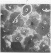
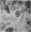
The phenomenen of autofluorescence of bone is due primarily to the collagen itself rather than to incidental substances adsorbed on to it. Fluorescent microscopy is a convenient method of dating the different microanatomical components of bone, and in this respect the same conclusions can be reached from the study of microradiographs or autofluorescent photographs, which is proof of the intimate relation between the development of matrix and the progression of mineralization.
Full text
PDF


Images in this article
Selected References
These references are in PubMed. This may not be the complete list of references from this article.
- AMPRINO R. Rapporti fra processi di ricostruzione e distribuzione dei minerali nelle ossa. I. Ricerche eseguite col metodo di studio dell'assorbimento dei raggi roentgen. Z Zellforsch Mikrosk Anat. 1952;37(2):144–183. [PubMed] [Google Scholar]
- Armstrong W. G., Horsley H. J. Isolation of fluorescent components from ox-bone human dentine and gelatin. Nature. 1966 Aug 27;211(5052):981–981. doi: 10.1038/211981a0. [DOI] [PubMed] [Google Scholar]
- Prentice A. I. Bone autofluorescence and mineral content. Nature. 1965 Jun 12;206(989):1167–1167. doi: 10.1038/2061167a0. [DOI] [PubMed] [Google Scholar]


