Figure 3. Comparison of the crystal structure of the ferredoxin-ferredoxin fusion to the design model.
The crystal structure (5CW9) aligns well with the design model over both the helices (A) and the fused beta sheet (B).
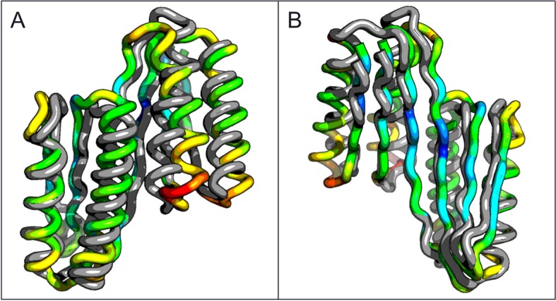
Figure 3—figure supplement 1. Circular dichroism spectra of ferrrodoxin-ferrodoxin at 25°C.
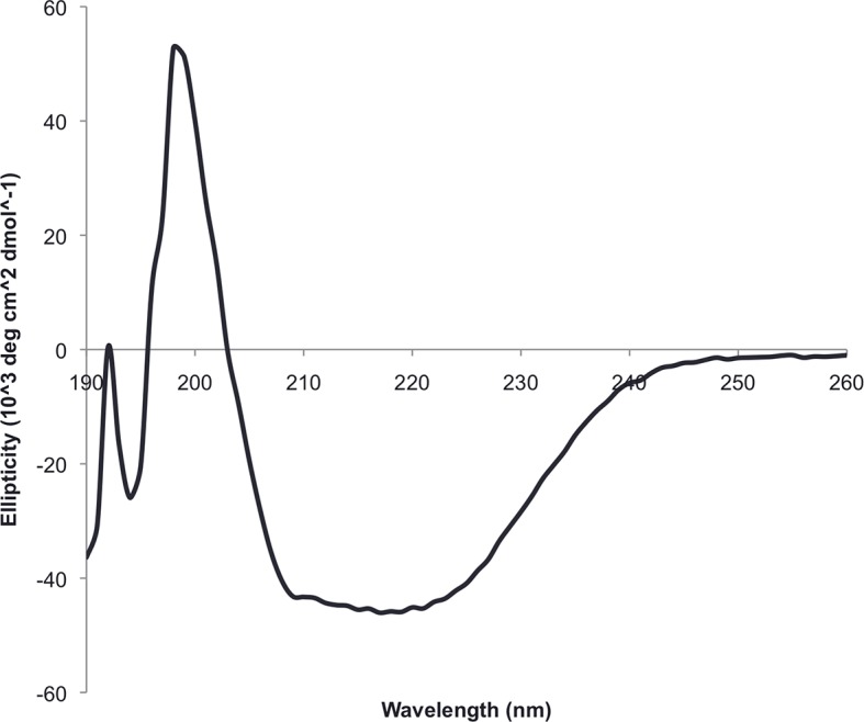
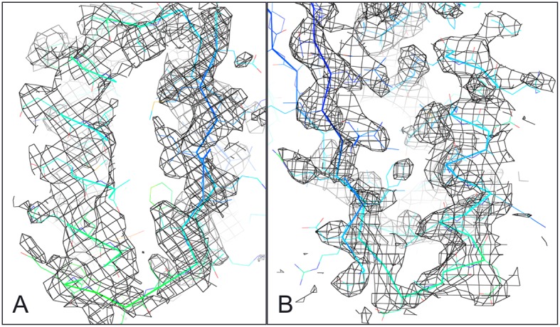
Figure 3—figure supplement 2. Ferredoxin-Ferredoxin 2Fo-Fc omit map superimposed with crystal structure shows core packing of host (A) and insert (B) domains.