



Abstract
Background
Data from prior studies have yielded inconsistent results regarding the association of serum testosterone levels with the risk of cardiovascular disease (CVD). There are no clinical trial data on the effects of testosterone replacement therapy on plaque progression.
Objective
We designed a study to investigate the effect of testosterone therapy on coronary artery plaque progression using serial coronary computed tomographic angiography (CCTA). In this paper we describe the study design, methods, and characteristics of the study population.
Methods
The Cardiovascular Trial of The Testosterone Trials (TTrials) (NCT00799617), a double-blind, placebo-controlled trial of one year of testosterone therapy in men ≥65 years with clinical manifestations of androgen deficiency and unequivocally low serum testosterone concentrations (<275 ng/dL). CCTA performed at baseline and after 12 months of therapy will determine testosterone effects on the progression of total volume of non-calcified plaque. All scans are evaluated at a central reading center by an investigator blinded to treatment assignment.
Results
One hundred sixty-five men were enrolled. The average age is 71.1 years and average BMI 30.7. About 9% of men had a history of myocardial infarction, 6% angina, and 10% coronary artery revascularization. A majority reported hypertension and/or high cholesterol; 31.8% reported diabetes. Total non-calcified plaque at baseline showed a slight but non-significant trend of lower plaque volume with higher serum testosterone concentrations (p=0.12).
Conclusions
The Cardiovascular Trial will test the hypothesis that testosterone therapy inhibits coronary plaque progression as assessed by serial CCTA.
Keywords: cardiovascular disease, coronary CTA, testosterone, coronary artery plaque progression, randomized controlled trial
Introduction
As demonstrated by both cross-sectional [1] and longitudinal studies [2,3], men’s serum testosterone concentration falls gradually, beginning at age 20. While the reported prevalence rates vary in different studies, one widely quoted sentinel study reported that by the eighth decade, approximately 30% of U.S. men have concentrations of total testosterone lower than normal for younger men and 70% have free testosterone concentration lower than normal for younger men [3].
Whether the age-related decline in testosterone concentrations contributes to, or is independent of, atherosclerosis progression with age is currently unknown. Although testosterone was once considered to be a risk factor for cardiovascular disease, the data are mixed. Several recent studies in men show an inverse association between serum testosterone concentration and cardiovascular disease, the metabolic syndrome or diabetes [4–6], an association independent of traditional CVD risk factors. A recent randomized trial, however, demonstrated increased CV risk in participants receiving testosterone replacement [7], and recent meta-analyses have shown conflicting results.
The Cardiovascular Trial, a component of the The Testosterone Trials (TTrials), was designed to assess the effects of testosterone treatment on several markers of cardiovascular disease in men ≥65 years old with symptoms and signs consistent with androgen deficiency and low serum testosterone concentrations [8]. These include progression of coronary atherosclerosis as assessed by serial coronary computed tomographic angiography (CCTA); cardiovascular risk factors such as blood pressure, lipids and lipoproteins; and markers of glucose metabolism, inflammation, coagulation and platelet function, endothelial function, and myocardial damage as assessed by high-sensitivity troponin.
MATERIALS AND METHODS
Design
The TTrials are a coordinated group of multi-center, double-blind placebo-controlled trials [8] to evaluate the effects of testosterone treatment in older men with low testosterone levels for no apparent reason other than age, on clinical endpoints that have been shown to improve with testosterone therapy in men with disease-induced hypogonadism. The TTrials are registered at http://clinicaltrials.gov/ (NCT00799617).
The TTrials include 12 clinical sites geographically distributed across the United States and is sponsored by the National Institute on Aging (NIA), National Institute of Neurological Disorders and Stroke (NINDS), Eunice Kennedy Shriver National Institute of Child Health and Human Development (NICHD), National Heart, Lung, and Blood Institute (NHLBI), and AbbVie, Inc. Men ≥65 years who had a mean serum testosterone concentration <275 ng/dL on two morning measures, who had subjective complaints and objective evidence of sexual dysfunction, physical dysfunction and/or reduced vitality (clinical conditions thought to be potentially improved by testosterone supplementation), and who were not at elevated risk for prostate, cardiovascular or hematologic problems associated with high levels of testosterone, were recruited; those eligible and consenting were assigned to one year of treatment with testosterone therapy or placebo [8,9]. The Cardiovascular Trial, designed to determine the effect of testosterone on the progression of coronary atherosclerosis, was conducted at 9 of the 12 TTrials sites. These sites all have ≥64-slice CCTA scanners and sufficient experience using them, as determined by a questionnaire completed at each site.
The TTrials and the Cardiovascular Trial protocols were approved by institutional review boards of the participating sites. All participants provided written informed consent before trial-related procedures were conducted. Participant safety and trial conduct were overseen by an Independent Data and Safety Monitoring Board appointed by the National Institute on Aging.
Treatment
Participants received AndroGel®, testosterone in an alcohol-water gel, or matched placebo gel, administered transdermally daily, for one year. Treatment allocation used a minimization algorithm with a random component [8,10]. The testosterone dose was adjusted by the (unblinded) data coordinating center as needed to maintain testosterone concentration levels between 500 and 800 ng/ml; similar adjustments were performed on matched placebo participants to maintain blinding. Regular monitoring of participants for potential adverse effects of testosterone was performed.
Eligibility criteria
Inclusion and exclusion criteria for the TTrials have been published [8]. Age, serum testosterone concentration, biochemical and clinical conditions that could affect the interpretation of the results, and conditions that could be worsened or exacerbated by testosterone therapy were the major factors used in assessing eligibility. In addition to the testosterone concentration and symptom requirements mentioned earlier, TTrials participants had to have a normal baseline renal function (estimated glomerular filtration rate (eGFR) >60 ml/min/1.73m2) to be eligible for the Cardiovascular Trial (Table 1). Men were excluded if their weight was >300 pounds, they had known allergy to iodinated contrast medium, were unable to breath-hold for 10 seconds, or had a prior diagnosis of tachycardia or irregular heart rhythm (eg, atrial fibrillation), or coronary artery bypass graft surgery by patient report or medical records.
Table 1.
Inclusion and Exclusion Criteria for The Cardiovascular Trial of The Testosterone Trials.
| Inclusion Criterion |
|
| Exclusion Criteria |
|
Follow-up
Men entered into The Cardiovascular Trial underwent a coronary CT angiography (CCTA) scan without and with contrast to allow calculation of non-calcified plaque volume, total plaque volume, subcutaneous fat and coronary artery calcium (CAC) score. The Month 12 Visit included re-evaluation of medical history for development of allergy to iodinated contrast medium and review of renal function based on month 12 e-GFR calculation (> 60 ml/min/1.73m2) . All participants still enrolled at 12 months with an eGFR > 60 and no new contraindication had a second CCTA scan to repeat the same measures obtained at the baseline visit.
Primary endpoint
The primary trial endpoint is percent change in non-calcified coronary plaque volume over the 12 month treatment period. The rationale for using noncalcified plaque volume is that prior trials have shown significant reduction in the amount of noncalcified plaque volume among statin users; calcified plaque is generally not considered amenable to reversal, and may even be increased by statins [11]. Experienced readers in the CCTA core lab blinded to the assigned treatment visually evaluated the CCTA scans for the presence, extent, and severity of coronary artery plaque. CCTA imaging and measurement of coronary plaque were conducted using standardized procedures and technology for longitudinal measurements of atherosclerosis change. The baseline and follow up scans were compared side by side (with blinding to both treatment arm and date of study [baseline vs. 12 month] to ensure like segments were compared and measured. The area of each coronary plaque visualized in at least 2 adjacent slices (reconstructed slice thickness 0.6 mm) was determined on all affected slices [12]. The total plaque per segment was summed over all segments with plaque. The degree of coronary artery stenosis was assessed by using axial images, multi-planar reconstructions and curved multiplanar images to assess the degree of luminal narrowing in all assessable coronary segments. The coronary artery stenosis severity was classiffied into 4 groups (0%, 1–29%, 30–69%, and ≥70% stenosis). The most narrowed diameter in each segment was determined even if the plaque was eccentric. A segment stenosis score was generated based on the degree of underlying stenotic disease in each segment (0: no plaque, 1: 1–29% stenosis, 2: 30–49% stenosis, 3: 50–69% stenosis, 4: ≥70% stenosis). The extent scores of all 17 individual segments were summed to yield a Total Stenosis Score (TSS) ranging from 0 to 68 [12]. In addition, each coronary territory (right coronary artery, left main, left anterior descending and left circumflex artery) was also scored according to presence of the most significant lesion.
Composition of coronary atherosclerotic plaque was evaluated from both axial source images and multi-planar reconstruction images of the long axis at each site of the coronary arteries. Each coronary segment was classified as either normal (no plaque), containing non-calcified plaque, containing partially calcified plaque with either predominantly non-calcified plaque (<50% of plaque area occupied by calcium) or containing calcified plaque (>50% of plaque area occupied by calcium) [13]. Calcified atherosclerotic plaque was defined as any discernible structure that: 1) had a CT density greater than the contrast-enhanced lumen; 2) was clearly assignable to the coronary artery wall; and, 3) was identified in at least 2 independent planes. Non-calcified atherosclerotic plaque was defined as any discernible structure that: 1) had a CT density less than the contrast-enhanced coronary lumen but greater than the surrounding connective tissue; 2) was clearly assignable to the coronary artery wall; and, 3) was identified in at least 2 independent planes [13]. Details of the CCTA methods are described elsewhere [11–16].
Secondary Outcomes
Secondary CCTA measures include changes in total plaque volume, coronary artery calcium (CAC) score and subcutaneous fat. HgA1C and HOMA-IR will also be assessed as secondary endpoints in participants without diabetes at baseline. CAC measures will include presence of any CAC (dichotomous variable) and CAC score, as well as changes in LV mass between testosterone and placebo.
Scanning Methodology
Non-Calcified plaque volume
Plaque volume was assessed by experienced readers at the CCTA core lab in all affected coronary segments on CCTA using semi-automated software (QAngio CT Research Edition 2.1.2, Medis medical imaging systems b.v., the Netherlands) (Figure 1) [17]; at baseline and follow up visits. First, an automatic tree extraction algorithm was used to obtain all the 3-dimensional centerlines of the coronary tree [18]. Based on these centerlines, straightened multi-planar reformatted (MPR) volumes are created of all vessels. Next, the lumen border contours and vessel wall borders are assessed using spatial first- and second-derivative gradient filters in longitudinal cross sections in combination with knowledge of the expected CCTA intensity values in the arteries [17–19].
Figure 1.
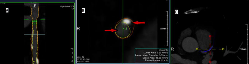
Cardiac computed tomography angiography image using QAngio CT Research Edition 2.1.2 software.
A – Straightened multi-planar reformatted volume of the vessel with lumen border contours and vessel wall borders of the of the left main and proximal left anterior descending coronary artery segment.
B – Transversal cross-section perpendicular to the centerlines of figure 1A. One arrow (→) points to the contrast at the lumen of the vessel and the other arrow (←) points to a mixed plaque with calcified and no calcified composition.
C – Axial cross-section demonstrating the left main and the proximal left anterior descending coronary artery segment.
Thereafter lumen and vessel contour are detected in the individual transversal cross-sections perpendicular to the centerlines, whereby the locations from the longitudinal analyses are taken into account. This method is insensitive to differences in attenuation values between data sets and independent of window and level settings. As previously described [19], 740HU and 220 HU were used as window and width levels to identify luminal and vessel outlines. Once automated software completed the vessel trace, expert readers manually corrected areas if additionally needed. The plaque area of each coronary plaque visualized in at least 2 adjacent slices (slice thickness 0.6 mm) was determined on all slices and plaque volume assessed by multiplying the area and slice thickness. The plaque volume was measured in any coronary artery segment ≥1.5 mm diameter. For each lesion, minimal lumen diameter was summed and reported as non-calcified plaque including fibrous plaque, fibrous fatty plaque, low attenuation plaque, and dense calcified plaque, which all variables have been widely used for quantitative plaque assessment in numerous previous studies [14,20–22]. Plaque Composition is based upon predefined fixed intensity cutoff values on the Hounsfield units (HU). These thresholds are based upon studies by comparing CCTA with virtual histology by intravascular ultrasound or histological examination in our lab and others [23–27]. The fixed HU cut-off values for classifying are: −30 to 30 HU for low attenuation plaque, 31–130 HU for fibrous fatty plaque, 131–350 HU for fibrous plaque, and >350 HU for dense calcified plaque. These values were initially based on a previous report by Brodoefel et al. [28] and empirically optimized using three representative training sets [17,29,30]. The excellent inter- and intra-observer variability for the lumen and vessel segmentation has been described previously [27,29]. On a per-patient basis, the total plaque volume is calculated by the sum of all plaques measured in available segments at baseline and follow-up. As previously described [19], the total plaque volume, total plaque burden [(total plaque volume (TPV)/total vessel volume) × 100] and normalized total plaque volume (TPVNorm) [total plaque volume/total segment length) × (the mean total segment length in the whole population)] were assessed at baseline and follow-up. The change between serial CCTAs in these variables was also assessed (Figure 2).
Figure 2.

Association of serum testosterone with noncalcified plaque volume in men participating in the Cardiovascular Trial of the Testosterone Trials.
CAC Scanning
For CAC scanning, each scan extended from 1 cm below the carina to the bottom of the heart to include the entire coronary tree. Scan parameters were obtained as follows: prospective electrocardiogram-triggering (typically 65–80%), 35 cm field-of-view, 512×512 matrix size, and peak tube voltage of 120 kVp. Slice thickness was 3 mm. Acquired images were then transferred to the CCTA core lab for analysis. All coronary calcium score measurements were performed on a GE AW system (AW Volume Share™, GE Medical Systems, Milwaukee, WI) to quantify coronary artery calcification using the Agatston method [31]. The coronary calcium was identified in 3 contiguous voxels by using a cutoff point of 130 Hounsfield Units (HU) resulting in a minimum calcified lesion area of 1.02 mm2. The area density method was used to compute the lesion score, in which the lesion area was multiplied by an attenuation factor. This attenuation factor is derived from the maximal HU within the area as described by Agatston, ranging from 1 to 4. A total coronary artery calcium (CAC) score was obtained by aggregating individual lesion scores from each of 4 anatomic sites (left main, left anterior descending, circumflex, and right coronary arteries).
CCTA quality assurance/control
The CCTA-core lab read all CTA scans, blinded to all demographics, testosterone randomization and risk factors, in near real time (within one week of acquisition) during enrollment and scanning period for stenosis evaluation and assessability. Three level 3 experienced cardiac CT readers evaluated all studies in the trial. The CCTA-core lab also evaluated the prevalence of slab, blurring, and partial volume averaging artifacts on all scans. The contrast-to-noise ratio of the distal left anterior descending artery is calculated for each scan as a quantitative metric of image quality.
Sample size
We reviewed data on 63 older men from an observational study using CT angiography techniques similar to those employed in this trial. Of these men, 35 were taking statins and 28 were not. Scans were performed at two time points approximately one year apart. In this cohort, the standard deviations of the changes from the first to the second scan were similar in the 2 groups; we used the larger of the 2 values (a SD of 26) for our calculations, which indicated that 140 men, 70/arm, would provide 80% power to detect a difference of 12 mm3 in non-calcified plaque volume, somewhat less than the difference of 14 mm3 between statin users and nonusers seen in the prior study. Since it was difficult to know the number of men who would enroll, as this was dependent on the proportion of men to be enrolled in the TTrials who would be eligible for and consent to participate in this trial, we also calculated power for a total sample size of 120, 60/arm; that number would provide 80% power to detect a 13 mm3 difference between treatment arms after one year of treatment.
Statistical analysis
The primary hypothesis was that testosterone treatment would decrease progression of non-calcified coronary artery plaque volume, as assessed by computed tomography angiography. Efficacy will be measured through a multivariate linear model that adjusts for baseline non-calcified plaque volume and all balancing variables used in the minimization procedure: study site, indicator variables of participation in each primary efficacy trial, baseline testosterone concentration (≤ or >200 ng/dL), age (≤ or > 75), use of anti-depressants, and use of PDE inhibitors. All subjects with a baseline and 12-month plaque volume measurement will be included in the analysis. Significance will be assessed through the two-sided Wald test and confidence interval for the treatment effect.
Safety analysis and evaluation of adverse events
Safety Measures Related to CT Angiography include the amount of radiation absorbed by the body tissues and the exposure to iodinated contrast agents. Beta-blockers were used to control the heart rate and thus maintain the radiation dose as low as reasonably achievable. Other radiation reduction methods included prospective triggering, limited field of view, limited scan length, iterative reconstruction and reduced tube voltage whenever possible based on body habitus [32–35].
Results and Discussion
Recruitment
One hundred ninety-nine men consented to be screened for the trial. Of these, 170 were eligible, and 165 had a baseline scan. Five men could not be scanned due to high heart rates or lack of venous access.
Demographics
One hundred sixty-five men were entered into the Cardiovascular Trial and had a baseline CCTA. Baseline information about these men is displayed in Table 2. The average age of participants was 71. Most men were overweight or obese; 61% had a body mass index ≥ 30. Hypertension and/or hyperlipidemia were present in approximately 2/3 of participants, and approximately one-third were diabetic. A prior MI was reported for 9%; 10% had had a prior revascularization; and 6% had a history of angina. The mean volume of non-calcified plaque was 91.0 mm3, with a SD of 95.9 mm3.
Table 2.
Baseline Characteristics of Men in The Cardiovascular Trial
| Characteristic* | Mean ± SD or N(%) |
|---|---|
| Age (Years) | 71.1±5.7 |
| 65–74 | 135 (79/4%) |
| 75–84 | 25 (14.7%) |
| ≥85 | 10 (5.9%) |
| BMI (kg/m2) | 30.7±3.6 |
| ≥30 | 104 (61.2%) |
| <30 | 66 (38.8%) |
| Prior MI | 15 (8.9%) |
| Angina | 10 (5.9%) |
| Heart failure | 1 (0.6%) |
| Atrial fibrillation | 2 (1.2%) |
| Revascularization | 17 (10.0%) |
| Stroke | 3 (1.8%) |
| TIA | 3 (1.8%) |
| Peripheral Arterial Disease | 2 (1.2%) |
| Leg pain/cramping | 13 (7.7%) |
| Angioplasty/stent | 2 (1.2%) |
| Hypertension | 112 (65.9%) |
| High cholesterol | 111 (65.3%) |
| Diabetes | 54 (31.8%) |
| Taking antidiabetics | 47 (27.7%) |
| Ever smoker: cigarettes | 108 (63.5%) |
| Current smoker: cigarettes | 11 (6.5%) |
| Current smoker: cigars | 6 (3.5%) |
| COPD | 12 (7.1%) |
| Serum Total Testosterone (ng/dL) | 237.3±59.5 |
| Non-calcified plaque volume (mm3) | 91.0±95.9 |
by self-report
Association of coronary plaque volume with serum testosterone concentration
We assessed whether there was any notable association between serum testosterone values and non-calcified coronary plaque volume at baseline. These data are shown in Figure 2. There was a trend towards higher values of testosterone associated with lower plaque volume, but this association was not statistically significant using a linear regression model (p=0.12).
Discussion
Observational studies have found associations of low testosterone concentrations in older men with adverse cardiovascular outcomes [4–6]. Further, in a study of 40–79 year-old community-dwelling men in Rancho Bernardo, baseline serum testosterone concentrations were inversely correlated with blood pressure [4], prevalence of diabetes and the risk of future diabetes [5]. In the same cohort, men with a testosterone level less than 250 ng/dL had a significantly increased risk of mortality compared to men with higher levels, independent of other covariates. In a larger cohort study from England, testosterone levels in men were inversely associated with cardiovascular risk with a graded stepwise association throughout the entire range of testosterone [6]. This association was independent of traditional risk factors. Some [35,36], but not other [37,38] retrospective studies of testosterone treatment have raised concern that exogenous testosterone might increase risk of CVD events, and results of meta-analysis have been mixed [39–46]. The conclusions of a critical review of 31 placebo-controlled trials of testosterone therapy in older men published in 2004 by the Institute of Medicine (IOM) were that the effects of testosterone therapy, including effects on coronary atherosclerosis and cardiovascular risk in this population, were uncertain [39]. However, a randomized trial of testosterone in older men with limited mobility [7] was stopped early due to the increased frequency of cardiovascular adverse events in the testosterone arm;
Strengths of the Study
The Cardiovascular Trial has many strengths. We selected CCTA as the imaging modality to assess coronary atherosclerosis in this trial. Until recently, estimating the plaque progression was only possible using intravascular ultrasound (IVUS), an invasive approach that shows only proximal coronary artery plaque volumes and correlates poorly with the individual total plaque burden [9,40]. It is also invasive and expensive, with well-defined associated morbidity. Therefore IVUS studies generally recruit patients with acute coronary syndromes, to justify the first invasive angiogram and IVUS. CCTA has emerged as a promising non-invasive tool to examine directly the coronary artery wall, determine the degree of plaque burden and assess the degree of coronary artery stenosis [41]. In addition, based on the tissue-specific x-ray attenuation characteristics, CCTA also provides additional information about atherosclerotic plaque composition. It is able to differentiate calcified from predominantly non-calcified plaques and ones that contain a large lipid pool [11,42] and allows volumetric analysis of plaque volumes. Plaque characterization (i.e. determining the vulnerability of plaque rupture by examining its tissue components) is now possible using CCTA. New ≥64 slice CCTA technology is extremely accurate in detecting lesions obstructing more than 50% of the lumen, with sensitivity, specificity, and positive and negative predictive values all better than 90% in patients without known CAD; it also has an important role in characterizing the vulnerable non-obstructive plaque [43]. Tissue density measured by CCTA can be used to characterize atherosclerotic plaque composition.
The Cardiovascular Trial involve strict standardization and quality control in assessing the imaging data, with all readings done by the same readers, who were blind to treatment assignment. The equivocal results of prior testosterone trials was once thought to be due to selection of men who did not have sufficiently low testosterone concentrations. This pitfall was avoided in the TTrials since the eligibility criterion for serum testosterone was set at a level low enough to ensure that men were unequivocally testosterone deficient. In addition, the serum testosterone concentration is measured at months 1, 2, 3, 6 and 9 to allow the assessment of medication compliance as well as dosage adjustments to maintain the serum testosterone concentration of the men assigned to the testosterone arm within the normal range for young men.
Limitations of the study
The TTrials had a long list of inclusion and exclusion factors [8], so this study will not be representative of all men over the age of 65. In particular, men with a recent MI, serum testosterone within the normal range or < 100 ng/dL, and men without functional concerns were excluded. In addition, the size of the study and the one-year duration are insufficient to assess the effect of testosterone on clinical cardiovascular outcomes.
The continuing uncertainty over the benefits of testosterone therapy in elderly men emphasizes the need for initiation of randomized trials such as the TTrials and the associated Cardiovascular Trial to test this treatment rigorously. Larger trials focused on clinical outcomes will be needed to fully assess the impact of testosterone therapy on cardiovascular health.
Acknowledgments
Supported by the National Institutes of Health grant U01 AG030644 and AbbVie; R01 AG037679 (Bone Trial); T32-DK007571; 5-U01-AG030644, Subaward No. 558836; UAB Diabetes Research and Training Center (DRCT), Grant Number DK-079626 from the National Institute for Diabetes, Digestive and Kidney Diseases, National Institutes of Health; Funding for Rancho Bernardo Study has been supported by National Institutes of Health/National Institute on Aging grants AG07181 and AG028507 and the National Institute of Diabetes and Digestive and Kidney Diseases, grant DK31801
TTrials Research Group Members
Peter J Snyder MD1, Susan S Ellenberg, PhD2, Glenn R Cunningham MD3, Alvin M Matsumoto MD4, Shalender Bhasin MD5, Elizabeth Barrett-Connor MD6, Thomas M Gill MD7, John T Farrar MD PhD8, David Cella PhD9, Raymond C Rosen PhD10, Susan M Resnick PhD11, Ronald S Swerdloff MD12, Jane A Cauley DrPH13, Denise Cifelli MS8, Laura Fluharty MPH8, Marco Pahor MD14, Kristine E Ensrud MD MPH15, Cora E Lewis MD MPSH16, Mark E Molitch MD17, Jill P Crandall MD18, Christina Wang MD12, Matthew J Budoff MD19, Nanette K Wenger MD20, Emile R Mohler 3rd MD21, Diane E Bild, MD MPH22, Nakela L Cook, MD MPH23, Tony M Keaveny PhD24, David L Kopperdahl PhD25, David Lee PhD25, Ann V Schwartz, PhD MPH26, Thomas W Storer PhD27, William B Ershler MD28, Cindy N Roy PhD29, Leslie J Raffel MD30, Sergei Romashkan MD PhD31, Evan Hadley MD31
1Division of Endocrinology, Diabetes, and Metabolism, Perelman School of Medicine, University of Pennsylvania, Philadelphia, PA, USA
2Department of Biostatistics and Epidemiology, Perelman School of Medicine, University of Pennsylvania, Philadelphia, PA, USA
3Division of Diabetes, Endocrinology and Metabolism, Baylor College of Medicine and St. Luke’s Episcopal Hospital, Houston, TX, USA
4Geriatric Research, Education, and Clinical Center, Division of Gerontology & Geriatric Medicine, Department of Veterans Affairs Puget Sound Health Care System, Department of Internal Medicine, University of Washington School of Medicine, Seattle, WA, USA
5Research Program in Men’s Health: Aging and Metabolism, Brigham and Women’s Hospital, Harvard Medical School, Boston, MA, USA
6Division of Epidemiology, Department of Family and Preventive Medicine, University of California San Diego, La Jolla, CA, USA
7Division of Geriatric Medicine, Yale School of Medicine, New Haven, CT, USA
8Center for Clinical Epidemiology & Biostatistics, Perelman School of Medicine, University of Pennsylvania, Philadelphia, PA, USA
9Department of Medical Social Sciences, Northwestern University Feinberg School of Medicine, Chicago, IL, USA
10New England Research Institutes, Watertown, MA, USA
11Laboratory of Behavioral Neuroscience, National Institute on Aging, National Institutes of Health, Baltimore, MD, USA
12Division of Endocrinology, Harbor-University of California at Los Angeles Medical Center; Los Angeles Biomedical Research Institute ;Torrance, CA; David Geffen School of Medicine at University of California, Los Angeles, Los Angeles, CA, USA
13Department of Epidemiology, University of Pittsburgh, Graduate School of Public Health, Pittsburgh, PA, USA
14Department of Aging & Geriatric Research, University of Florida, Gainesville, FL, USA
15Department of Medicine, Division of Epidemiology & Community Health, University of Minnesota, Minneapolis VA Health Care System, Minneapolis, MN, USA
16Division of Preventive Medicine, University of Alabama at Birmingham, Birmingham, AL, USA
17Division of Endocrinology, Metabolism and Molecular Medicine, Northwestern University, Feinberg School of Medicine, Chicago, IL, USA
18Divisions of Endocrinology and Geriatrics, Albert Einstein College of Medicine, Bronx, NY, USA
19Division of Cardiology, Los Angeles Biomedical Research Institute, Harbor- University of California at Los Angeles Medical Center, Torrance, CA, USA
20Division of Cardiology, Department of Medicine, Emory University School of Medicine, Emory Heart and Vascular Center, Atlanta, GA, USA
21Division of Cardiovascular Disease and Section of Vascular Medicine, Perelman School of Medicine, University of Pennsylvania, Philadelphia, PA, USA
22Division of Cardiovascular Sciences, National Heart, Lung, and Blood Institute, Bethesda, MD, USA
23Clinical Applications and Prevention Branch, Division of Cardiovascular Sciences, Centers for Cardiovascular Outcomes Research, National Heart, Lung, and Blood Institute, Bethesda, MD, USA
24Departments of Mechanical Engineering and Bioengineering, University of California and ON Diagnostics, Berkeley, CA, USA
25O.N. Diagnostics, Berkeley, CA, USA
26Department of Epidemiology and Biostatistics, University of California, San Francisco, CA, USA
27Section of Endocrinology, Diabetes, and Nutrition, Boston Medical Center, Boston, MA, USA
28Division of Hematology/Oncology, Institute for Advanced Studies in Aging, Falls Church, VA, USA
29Division of Geriatric Medicine and Gerontology, Johns Hopkins University School of Medicine, Baltimore, MD, USA
30Medical Genetics Institute, Cedars-Sinai Medical Center, Los Angeles, CA, USA
31Division of Geriatrics and Clinical Gerontology, National Institute on Aging, Bethesda, MD, USA
Footnotes
Conflict of Interest Reporting
SSE reports a conference grant from AbbVie during the conduct of the study; RSS reports grants and consulting from AbbVie, Clarus, Ardana, Besins Health, and Endo Pharma; CEL was supported by the National Institute for Diabetes, Digestive and Kidney Diseases, National Institutes of Health (DK079626) to the UAB Diabetes Research and Training Center; PJS reports grants from NIH and AbbVie for the conduct of this study and has consulted for Watson Laboratories; MJB reports grants from NIH and grant support from General Electric
References
- 1.Vermeulen A, Rubens R, Verdonck L. Testosterone secretion and metabolism in male senescene. J Clin Endocrinol Metab. 1972;34:730–735. doi: 10.1210/jcem-34-4-730. [DOI] [PubMed] [Google Scholar]
- 2.Feldman HA, Longcope C, Derby CA, Johannes CB, Araujo AB, Coviello AD, et al. Age trends in the level of serum testosterone and other hormones in middle-aged men: longitudinal results from the Massachusetts male aging study. J Clin Endocrinol Metab. 2002;87(2):589–598. doi: 10.1210/jcem.87.2.8201. [DOI] [PubMed] [Google Scholar]
- 3.Harman S, Metter E, Tobin J, Pearson J, Blackman M. Longitudinal effects of aging on serum total and free testosterone levels in healthy men. Baltimore Longitudinal Study of Aging. J Clin Endocrinol Metab. 2001;86(2):724–731. doi: 10.1210/jcem.86.2.7219. [DOI] [PubMed] [Google Scholar]
- 4.Barrett-Connor E, Khaw KT. Endogenous sex hormones and cardiovascular disease in men. A prospective population-based study. Circulation. 1988;78(3):539–545. doi: 10.1161/01.cir.78.3.539. [DOI] [PubMed] [Google Scholar]
- 5.Oh JY, Barrett-Connor E, Wedick NM, Wingard DL. Endogenous sex hormones and the development of type 2 diabetes in older men and women: the Rancho Bernardo study. Diabetes Care. 2002;25(1):55–60. doi: 10.2337/diacare.25.1.55. [DOI] [PubMed] [Google Scholar]
- 6.Smith GD, Ben-Shlomo Y, Beswick A, Yarnell J, Lightman S, Elwood P. Cortisol, testosterone, and coronary heart disease: prospective evidence from the Caerphilly study. Circulation. 2005;112(3):332–340. doi: 10.1161/CIRCULATIONAHA.104.489088. [DOI] [PubMed] [Google Scholar]
- 7.Basaria S, Coviello AD, Travison TG, Storer TW, Farwell WR, Jette AM, et al. Adverse events associated with testosterone administration. [Multicenter Study Randomized Controlled Trial Research Support, N.I.H., Extramural Research Support, U.S. Gov’t, Non-P.H.S.] N Engl J Med. 2010;363(2):109–122. doi: 10.1056/NEJMoa1000485. [DOI] [PMC free article] [PubMed] [Google Scholar]
- 8.Snyder PJ, Ellenberg SS, Cunningham GR, Matsumoto AM, Bhasin S, Barrett-Connor E, et al. The Testosterone Trials: Seven coordinated trials of testosterone treatment in elderly men. Clin Trials. 2014;11(3):362–375. doi: 10.1177/1740774514524032. [DOI] [PMC free article] [PubMed] [Google Scholar]
- 9.Cauley JA, Fluharty L, Ellenberg SS, Gill TM, Ensrud KE, Barrett-Connor E, et al. Recruitment and Screening for the Testosterone Trials. J Gerontol A Biol Sci Med Sci. 2015 doi: 10.1093/gerona/glv031. [DOI] [PMC free article] [PubMed] [Google Scholar]
- 10.Taves DR. Minimization: a new method of assigning patients to treatment and control groups. Clin Pharmacol Ther. 1974;15(5):443–453. doi: 10.1002/cpt1974155443. [DOI] [PubMed] [Google Scholar]
- 11.Zeb I, Li D, Nasir K, Malpeso J, Batool A, Flores F, et al. Effect of statin treatment on coronary plaque progression – a serial coronary CT angiography study. Atherosclerosis. 2013;231(2):198–204. doi: 10.1016/j.atherosclerosis.2013.08.019. [DOI] [PubMed] [Google Scholar]
- 12.Pagali SR, Madaj P, Gupta M, Nair S, Hamirani YS, Min JK, et al. Interobserver variations of plaque severity score and segment stenosis score in coronary arteries using 64 slice multidetector computed tomography: a substudy of the ACCURACY trial. J Cardiovasc Comput Tomogr. 2010;4(5):312–318. doi: 10.1016/j.jcct.2010.05.018. [DOI] [PubMed] [Google Scholar]
- 13.Leber AW, Knez A, Becker A, Becker C, von Ziegler F, Nikolaou K, et al. Accuracy of multidetector spiral computed tomography in identifying and differentiating the composition of coronary atherosclerotic plaques: a comparative study with intracoronary ultrasound. J Am Coll Cardiol. 2004;43(7):1241–1247. doi: 10.1016/j.jacc.2003.10.059. [DOI] [PubMed] [Google Scholar]
- 14.Iwasaki K, Matsumoto T, Aono H, Furukawa H, Samukawa M. Prevalence of subclinical atherosclerosis in asymptomatic diabetic patients by 64-slice computed tomography. Coron Artery Dis. 2008;19(3):195–201. doi: 10.1097/MCA.0b013e3282f3fbef. [DOI] [PubMed] [Google Scholar]
- 15.Carr JJ, Nelson JC, Wong ND, McNitt-Gray M, Arad Y, Jacobs DR, Jr, et al. Calcified coronary artery plaque measurement with cardiac CT in population-based studies: standardized protocol of Multi-Ethnic Study of Atherosclerosis (MESA) and Coronary Artery Risk Development in Young Adults (CARDIA) study. Radiology. 2005;234(1):35–43. doi: 10.1148/radiol.2341040439. [DOI] [PubMed] [Google Scholar]
- 16.Budoff MJ, Dowe D, Jollis JG, Gitter M, Sutherland J, Halamert E, et al. Diagnostic performance of 64-multidetector row coronary computed tomographic angiography for evaluation of coronary artery stenosis in individuals without known coronary artery disease: results from the prospective multicenter ACCURACY (Assessment by Coronary Computed Tomographic Angiography of Individuals Undergoing Invasive Coronary Angiography) trial. J Am Coll Cardiol. 2008;52(21):1724–1732. doi: 10.1016/j.jacc.2008.07.031. [DOI] [PubMed] [Google Scholar]
- 17.de Graaf MA, Broersen A, Kitslaar PH, Roos CJ, Dijkstra J, Lelieveldt BP, et al. Automatic quantification and characterization of coronary atherosclerosis with computed tomography coronary angiography: cross-correlation with intravascular ultrasound virtual histology. Int J Cardiovasc Imaging. 2013;29(5):1177–1190. doi: 10.1007/s10554-013-0194-x. [DOI] [PubMed] [Google Scholar]
- 18.Yang G, Kitslaar P, Frenay M, Broersen A, Boogers MJ, Bax JJ, et al. Automatic centerline extraction of coronary arteries in coronary computed tomographic angiography. Int J Cardiovasc Imaging. 2012;28(4):921–933. doi: 10.1007/s10554-011-9894-2. [DOI] [PMC free article] [PubMed] [Google Scholar]
- 19.Boogers MJ, Broersen A, van Velzen JE, de Graaf FR, El-Naggar HM, Kitslaar PH, et al. Automated quantification of coronary plaque with computed tomography: comparison with intravascular ultrasound using a dedicated registration algorithm for fusion-based quantification. Eur Heart J. 2012;33(8):1007–1016. doi: 10.1093/eurheartj/ehr465. [DOI] [PubMed] [Google Scholar]
- 20.Budoff MJ, Gopal A, Gopalakrishnan D. Cardiac computed tomography: diagnostic utility and integration in clinical practice. Clin Cardiol. 2006;29(9 Suppl 1):I4–14. doi: 10.1002/clc.4960291303. [DOI] [PubMed] [Google Scholar]
- 21.Ahmadi N, Nabavi V, Hajsadeghi F, Flores F, French WJ, Mao SS, et al. Mortality incidence of patients with non-obstructive coronary artery disease diagnosed by computed tomography angiography. Am J Cardiol. 2011;107(1):10–16. doi: 10.1016/j.amjcard.2010.08.034. [DOI] [PubMed] [Google Scholar]
- 22.Choi TY, Malpeso J, Li D, Sourayanezhad S, Budoff MJ. Radiation dose reduction with increasing utilization of prospective gating in 64-multidetector cardiac computed tomography angiography. J Cardiovasc Comput Tomogr. 2011;5(4):264–270. doi: 10.1016/j.jcct.2011.06.005. [DOI] [PubMed] [Google Scholar]
- 23.Madaj PM, Budoff MJ, Li D, Tayek JA, Karlsberg RP, Karpman HL. Identification of noncalcified plaque in young persons with diabetes: an opportunity for early primary prevention of coronary artery disease identified with low-dose coronary computed tomographic angiography. Acad Radiol. 2012;19(7):889–893. doi: 10.1016/j.acra.2012.03.013. [DOI] [PMC free article] [PubMed] [Google Scholar]
- 24.Cheng VY, Nakazato R, Dey D, Gurudevan S, Tabak J, Budoff MJ, et al. Reproducibility of coronary artery plaque volume and composition quantification by 64-detector row coronary computed tomographic angiography: an intraobserver, interobserver, and interscan variability study. J Cardiovasc Comput Tomogr. 2009;3(5):312–320. doi: 10.1016/j.jcct.2009.07.001. [DOI] [PubMed] [Google Scholar]
- 25.Papadopoulou SL, Garcia-Garcia HM, Rossi A, Girasis C, Dharampal AS, Kitslaar PH, et al. Reproducibility of computed tomography angiography data analysis using semiautomated plaque quantification software: implications for the design of longitudinal studies. Int J Cardiovasc Imaging. 2013;29(5):1095–1104. doi: 10.1007/s10554-012-0167-5. [DOI] [PubMed] [Google Scholar]
- 26.Motoyama S, Kondo T, Anno H, Sugiura A, Ito Y, Mori K, et al. Atherosclerotic plaque characterization by 0.5-mm-slice multislice computed tomographic imaging. Circ J. 2007;71(3):363–366. doi: 10.1253/circj.71.363. [DOI] [PubMed] [Google Scholar]
- 27.Motoyama S, Kondo T, Sarai M, Sugiura A, Harigaya H, Sato T, et al. Multislice computed tomographic characteristics of coronary lesions in acute coronary syndromes. J Am Coll Cardiol. 2007;50(4):319–326. doi: 10.1016/j.jacc.2007.03.044. [DOI] [PubMed] [Google Scholar]
- 28.Brodoefel H, Reimann A, Burgstahler C, Schumacher F, Herberts T, Tsiflikas I, et al. Noninvasive coronary angiography using 64-slice spiral computed tomography in an unselected patient collective: effect of heart rate, heart rate variability and coronary calcifications on image quality and diagnostic accuracy. Eur J Radiol. 2008;66(1):134–141. doi: 10.1016/j.ejrad.2007.05.013. [DOI] [PubMed] [Google Scholar]
- 29.Brodoefel H, Reimann A, Heuschmid M, Tsiflikas I, Kopp AF, Schroeder S, et al. Characterization of coronary atherosclerosis by dual-source computed tomography and HU-based color mapping: a pilot study. Eur Radiol. 2008;18(11):2466–2474. doi: 10.1007/s00330-008-1019-5. [DOI] [PMC free article] [PubMed] [Google Scholar]
- 30.Brodoefel H, Burgstahler C, Tsiflikas I, Reimann A, Schroeder S, Claussen CD, et al. Dual-source CT: effect of heart rate, heart rate variability, and calcification on image quality and diagnostic accuracy. Radiology. 2008;247(2):346–355. doi: 10.1148/radiol.2472070906. [DOI] [PubMed] [Google Scholar]
- 31.Agatston AS, Janowitz WR, Hildner FJ, Zusmer NR, Viamonte M, Jr, Detrano R. Quantification of coronary artery calcium using ultrafast computed tomography. J Am Coll Cardiol. 1990;15(4):827–832. doi: 10.1016/0735-1097(90)90282-t. [DOI] [PubMed] [Google Scholar]
- 32.Burgstahler C, Reimann A, Beck T, Kuettner A, Baumann D, Heuschmid M, et al. Influence of a lipid-lowering therapy on calcified and noncalcified coronary plaques monitored by multislice detector computed tomography: results of the New Age II Pilot Study. Invest Radiol. 2007;42(3):189–195. doi: 10.1097/01.rli.0000254408.96355.85. [DOI] [PubMed] [Google Scholar]
- 33.Gopal A, Budoff MJ. A new method to reduce radiation exposure during multi-row detector cardiac computed tomographic angiography. Int J Cardiol. 2009;132(3):435–436. doi: 10.1016/j.ijcard.2007.08.072. [DOI] [PubMed] [Google Scholar]
- 34.Hausleiter J, Meyer T, Hadamitzky M, Huber E, Zankl M, Martinoff S, et al. Radiation dose estimates from cardiac multislice computed tomography in daily practice: impact of different scanning protocols on effective dose estimates. Circulation. 2006;113(10):1305–1310. doi: 10.1161/circulationaha.105.602490. [DOI] [PubMed] [Google Scholar]
- 35.Vigen R, O’Donnell CI, Baron AE, Grunwald GK, Maddox TM, Bradley SM, et al. Association of testosterone therapy with mortality, myocardial infarction, and stroke in men with low testosterone levels. JAMA. 2013;310(17):1829–1836. doi: 10.1001/jama.2013.280386. [DOI] [PubMed] [Google Scholar]
- 36.Finkle WD, Greenland S, Ridgeway GK, Adams JL, Frasco MA, Cook MB, et al. Increased risk of non-fatal myocardial infarction following testosterone therapy prescription in men. PLoS One. 2014;9(1):e85805. doi: 10.1371/journal.pone.0085805. [DOI] [PMC free article] [PubMed] [Google Scholar]
- 37.Shores MM, Smith NL, Forsberg CW, Anawalt BD, Matsumoto AM. Testosterone treatment and mortality in men with low testosterone levels. J Clin Endocrinol Metab. 2012;97(6):2050–2058. doi: 10.1210/jc.2011-2591. [DOI] [PubMed] [Google Scholar]
- 38.Baillargeon J, Urban RJ, Kuo YF, Ottenbacher KJ, Raji MA, Du F, et al. Risk of Myocardial Infarction in Older Men Receiving Testosterone Therapy. Ann Pharmacother. 2014;48(9):1138–1144. doi: 10.1177/1060028014539918. [DOI] [PMC free article] [PubMed] [Google Scholar]
- 39.Liverman C, Blazer D, editors. Testosterone and aging Clinical research directions. Washington, DC: The National Academies Press; 2004. [PubMed] [Google Scholar]
- 40.Newby DE, Fox KA. Invasive assessment of the coronary circulation: intravascular ultrasound and Doppler. Br J Clin Pharmacol. 2002;53(6):561–575. doi: 10.1046/j.1365-2125.2002.01582.x. [DOI] [PMC free article] [PubMed] [Google Scholar]
- 41.Cury RC, Pomerantsev EV, Ferencik M, Hoffmann U, Nieman K, Moselewski F, et al. Comparison of the degree of coronary stenoses by multidetector computed tomography versus by quantitative coronary angiography. Am J Cardiol. 2005;96(6):784–787. doi: 10.1016/j.amjcard.2005.05.020. [DOI] [PubMed] [Google Scholar]
- 42.Achenbach S, Moselewski F, Ropers D, Ferencik M, Hoffmann U, MacNeill B, et al. Detection of calcified and noncalcified coronary atherosclerotic plaque by contrast-enhanced, submillimeter multidetector spiral computed tomography: a segment-based comparison with intravascular ultrasound. Circulation. 2004;109(1):14–17. doi: 10.1161/01.cir.0000111517.69230.0f. [DOI] [PubMed] [Google Scholar]
- 43.Hirai N, Horiguchi J, Fujioka C, Kiguchi M, Yamamoto H, Matsuura N, et al. Prospective versus retrospective ECG-gated 64-detector coronary CT angiography: assessment of image quality, stenosis, and radiation dose. Radiology. 2008;248(2):424–430. doi: 10.1148/radiol.2482071804. [DOI] [PubMed] [Google Scholar]
- 44.Corona G, Maseroli E, Rastrelli G, Isidori AM, Sforza A, Mannucci E, et al. Cardiovascular risk associated with testosterone-boosting medications: a systematic review and meta-analysis. Expert Opin Drug Saf. 2014;13(10):1327–1351. doi: 10.1517/14740338.2014.950653. [DOI] [PubMed] [Google Scholar]
- 45.Xu L, Freeman G, Cowling BJ, Schooling CM. Testosterone therapy and cardiovascular events among men: a systematic review and meta-analysis of placebo-controlled randomized trials. BMC Med. 2013;11:108. doi: 10.1186/1741-7015-11-108. [DOI] [PMC free article] [PubMed] [Google Scholar]
- 46.Fernandez-Balsells MM, Murad MH, Lane M, Lampropulos JF, Albuquerque F, Mullan RJ, et al. Clinical review 1: Adverse effects of testosterone therapy in adult men: a systematic review and meta-analysis. J Clin Endocrinol Metab. 2010;95(6):2560–2575. doi: 10.1210/jc.2009-2575. [DOI] [PubMed] [Google Scholar]