Abstract
Adherents claim many benefits from the practice of yoga, including promotion of bone health and prevention of osteoporosis. However, no known studies have investigated whether yoga enhances bone mineral density. Furthermore, none have estimated reaction forces applied by yoga practitioners. The purpose of this study was to collect ground reaction force (GRF) data on a variety of hatha yoga postures that would commonly be practiced in fitness centers or private studios. Twelve female and eight male volunteers performed a sequence of 28 hatha yoga postures while GRF data were collected with an AMTI strain-gauge force platform. The sequence was repeated six times by each study subject. Four dependent variables were studied: peak vertical GRF, mean vertical GRF, peak resultant GRF, and mean resultant GRF. Univariate analysis was used to identify mean values and standard deviations for the dependent variables. Peak vertical and resultant values of each posture were similar for all subjects, and standard deviations were small. Similarly, mean vertical and resultant values were similar for all subjects. This 28 posture yoga sequence produced low impact GRF applied to upper and lower extremities. Further research is warranted to determine whether these forces are sufficient to promote osteogenesis or maintain current bone health in yoga practitioners.
Keywords: Mind-body, weight-bearing, strength, flexibility
INTRODUCTION
Yoga is a popular form of exercise practiced in private studios, fitness clubs, and recreation centers across the nation. Hatha yoga is one of several yoga practices that aims to link the body (through postures or asanas), the mind (through concentration), and the breath (15). Thus, a common yoga session in one of the above settings might focus on breathing and meditation, stretching and flexibility exercises, weight-bearing balance and strength postures, or a combination of all three.
There is a limited amount of research that highlights yoga’s potential physical benefits, some of which include increasing strength and flexibility (3); improving grip strength and reducing pain in patients with carpal tunnel syndrome (6); and reduction in pain and disability of patients with osteoarthritis of the knee (12). Despite a lack of controlled trials to support their position, some yoga adherents publish the claim in books and magazines that practicing yoga promotes healthy bones and may also prevent osteoporosis (20). This assertion is intuitive due to the fact that many of the postures (asanas) practiced in yoga are weight-bearing, wherein the body mass is supported by large muscle contractions from any combination of the limbs against the force of gravity.
Researchers believe that exercise exerts its beneficial effects on the bones by both the ground reaction force (GRF), related to the gravitational force, and the force of muscle contractions (13). A wide variety of exercise interventions appear to corroborate this opinion wherein researchers use bone mineral density (BMD) as measured by dual energy x-ray absorptiometry (DEXA) as a clinical measurement of bone health. For example, a weight training regimen for post-menopausal women using variable resistance machines increased BMD at the femoral neck and lumbar spine while BMD at the same measured sites decreased in controls (14). A significant increase in BMD measured at the femoral neck, Ward’s triangle, and greater trochanter compared to baseline BMD occurred in both young and elderly men and women following a 6-month resistance training program (18). In contrast to this type of exercise, high-impact aerobics classes consisting of stepping and jumping exercises resulted in significantly increased BMD of the greater trochanter in men and women 50 to 73 years of age compared to the non-exercising control group (23). Similarly, an 18-month high-impact aerobic exercise program involving pre-menopausal women produced significantly greater BMD at numerous weight-bearing sites compared to controls (8). An exercise trial that compared low- and moderate-impact exercises such as walking, stair climbing, and light jogging with weight lifting and rowing produced significant increases in BMD of the whole body, proximal femur, and lumbar spine which were similar for both groups of postmenopausal women (11). A year-long randomized controlled trial with adult male and female Crohn’s patients evaluated a low-impact exercise program carried out at home and found that increases in BMD were significantly associated with exercise session compliance. Moreover, BMD increases at the greater trochanter in the exercise group were statistically significant (12). Finally, a year-long intervention comparing low-impact and high-impact exercise programs showed that BMD of the lumbar spine was maintained in both groups of early postmenopausal women (7). In summary, the preceding examples are representative of the many forms of both high- and low-impact exercise that appear to benefit bone health. Yoga’s weight bearing postures most closely resemble weight or strength training exercises but none of these study designs included yoga exercises, nor can inferences be made about yoga’s effect on bone health based on this similarity alone. Nevertheless, it raises the question of whether the yoga postures would yield similar benefits to BMD as those reported in the low-impact exercise trials.
Availability of controlled exercise trials that quantify low- and high-impact exercises using GRF data are extremely limited. A sample of such trials is outlined in table 1. The GRF in these studies was normalized to body weight and thus identified possible intensities of exercise that may benefit bone health (1, 2, 7, 8). Only Grove and Londeree (7) attempted to further specify an exercise dose for improving bone health by defining low- and high-impact with a GRF value normalized to body weight (BW). They used exercises that produced GRF greater than or equal to two times BW for their high-impact group of subjects and exercises that generated a GRF less than or equal to 1.5 times BW for the low-impact group.
Table 1.
Summary of studies by exercise type and influence on bone mineral density (BMD). Intensity expressed as a ratio to body weight (BW).
| Author & Year | Population | Study duration | Exercise type | Ground reaction force | Results |
|---|---|---|---|---|---|
| Grove & Londeree, 1992 | early post menopausal women | 1yr | High impact vs low impact | ≥2xBW ≤1.5×BW |
Both groups maintained BMD lumbar spine L2–L4 |
| Bassey & Ramsdale, 1995 | postmenopausal women | 1yr | Heel drops vs low impact/flexibility | 2.5–3.0×BW | No change in BMD for either group at femur or spine |
| Heinonen et al., 1996 | pre-menopausal women | 18mo | High impact step & aerobics vs control | 2.1–5.6×BW | Significant increase in BMD at femoral neck for impact group |
| Bassey et al., 1998 | pre- and postmenopausal women | 18mo | Vertical jump from 8.5cm 6days/week | 3.0–4.0×BW | Only premenopausal women had significant increase in BMD of proximal femur |
A typical yoga session involves little or no jumping; thus, yoga would most likely be defined as low-impact. The asanas, with the exception of the seated poses, employ one to four limbs to support body mass against gravity. Force is generated as practitioners perform and hold these positions, accelerate into and out of the postures, and transfer weight between extremities. These forces may be similar to those measured in low-impact exercises and walking regimens, examples of which are listed in Table 2 (7, 9, 10, 17). With the exception of the research conducted by Grove and Londeree, these studies were not designed to measure the effect of exercise on BMD but to identify the GRF of given exercises. Thus, the data may be useful in categorizing different types of exercise as high- or low-impact and in providing a reference for comparison to forces generated by the yoga asanas.
Table 2.
Mean ground reaction force (GRF) by exercise type expressed in relation to body weight (BW).
| Author and year | Exercise | GRF × BW |
|---|---|---|
| Grove & Londeree 1992 | Jumping jacks | 3.29 |
| Running in place | 2.47 | |
| Knee to elbow with jump | 2.79 | |
| Slow walk | 1.19 | |
| Fast walk | 1.49 | |
| Heel jack without jump | 1.34 | |
| Charleston | 1.32 | |
| Johnson et al., 1992 | Walking | 1.13 |
| Slow jog | 2.26 | |
| Low impact marching | 1.74 | |
| High impact double-hop knee lift | 3.14 | |
| Step push-off | 1.27 | |
| Step down | 1.51 | |
| Kato & Bassey, 2002 | Two-footed jump, intermittent | 4.22 ± 0.24 |
| Two-footed jump, continuous | 4.08 ± 0.17 | |
| Heel drops | 3.38 ± 0.17 | |
| Rousanaglou & Boudolos, 2005 | Step touch | 2.0 |
| Leap with triple step | 2.75 |
Despite the tremendous body of research available on many forms of exercise and bone health, there are neither randomized, controlled trials of a yoga regimen and its effect on BMD, nor studies that identify yoga as high- or low-impact or quantify its force generating qualities. Therefore, the primary objective of this study was to collect GRF data on a variety of hatha yoga asanas as a starting point to address the claim that yoga is beneficial to the bones and to answer the following research question: Are GRF measurements from yoga comparable to GRF measurements from other types of low-impact exercise? The researcher hypothesized that GRF results would be similar to low-impact forces of less than two times (< 2 × BW) and that the data would produce a similar range of GRF for all subjects.
METHODS
Participants
Male and female yoga instructors and intermediate-level yoga practitioners were recruited from local private studios and fitness centers where they either taught or attended hatha yoga classes. These individuals possessed the expertise and stamina required to execute the sequence of 28 hatha yoga asanas six times during the single data-collection period. Twelve women ages 22 to 49 (28.3 ± 7.4 years) and eight men 24 to 55 (34.4 ± 11.5 years) volunteered to participate. Weight and height ranges for women were 48.4 to 88.1 kg and 152.4 to 177.2 cm (61.2 ± 20 kg; 167.3 ± 7.7 cm). Ranges for men were 68.9 to 86.5 kg and 170.2 to 186 cm (77.1 ± 9.5 kg; 178.2 ± 5.7 cm). One male and eight female subjects were yoga instructors.
Equipment
A single researcher used a 40×60 cm AMTI (Watertown, MA, USA) force platform to measure GRF (1000 Hz). After recording the GRF in Newtons, the unfiltered data were exported into Matlab (MathWorks, Natick, MA, USA), where peak and means were determined for vertical and resultant GRF traces that corresponded to each trial. The Matlab algorithm also normalized all GRF values to the body weight of each study subject.
Procedures
The study was approved by the local Institutional Review Board. Potential participants were educated by phone or in person about the study prior to data collection. Subjects then reported to the Biomechanics Lab at BYU and signed a consent form. Body mass and height were measured using an electronic scale and stadiometer. Subjects then participated in a practice session and warm up of the entire yoga sequence as they would be executing it for the study, with instructions and demonstration provided by the researcher to assure uniform execution by study subjects of each posture and the order in which they should be performed.
The perimeter of the force platform was marked on a sticky yoga mat, which was positioned over the force platform, to keep participants’ hands and feet oriented to the platform area. Pilot tests demonstrated that placement of the mat over the force platform did not alter the characteristics of the recorded GRF. The force platform was zeroed prior to each data collection session. The yoga sequence consisted of 28 hatha yoga postures typically incorporated into beginning or intermediate-level yoga classes (table 3). Weight bearing hatha yoga postures were selected for the sequence rather than any of the seated or flexibility poses, in order to measure GRF as subjects moved from one asana to the other.
Table 3.
Sequence of 28 hatha yoga postures.
| Name in Sanskrit (English) |
|---|
|
postures repeated in the sequence for smooth transitions
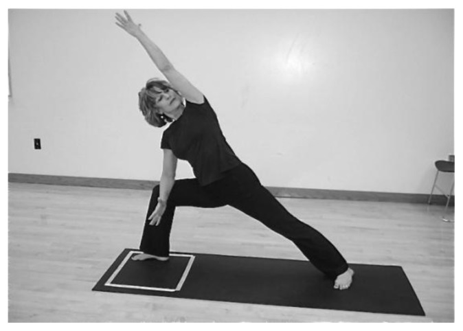
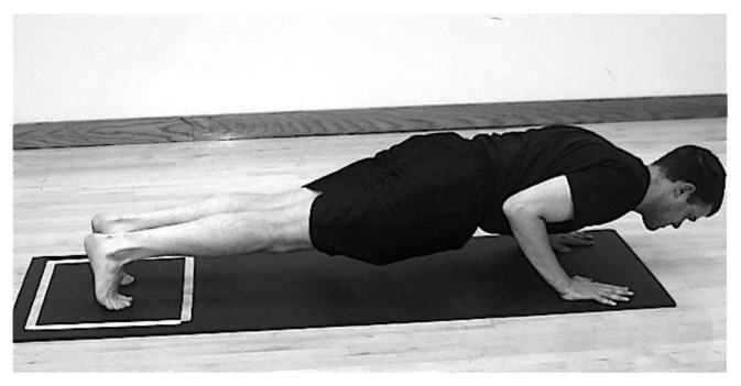
This 28-posture sequence was performed in its entirety six times (six rounds) during the single data collection period, three times under condition one, and three times under condition two. Because one stationary force platform was available for data collection, these two conditions were used in order to obtain GRF measurements from each extremity employed in a given posture. In condition one, the subject began the sequence by standing on the force platform. Depending upon which posture was being performed, variables were measured from the lower or upper extremities (i.e., the subject began in standing position so the force in both legs was measured; upon stepping back to plank pose, the force in the arms was measured; upon stepping forward with one leg into any of the warrior poses, the force was measured in the leg that was over the platform). In condition two, the subject began the sequence by standing in front of the platform, so that, depending upon which asana was being executed, forces could be measured in one or both lower extremities as the subject stepped back onto the force platform (figures 1 and 2). Collecting data using these two conditions yielded measurements from all of the extremities used to perform a given posture using one, stationary force platform. The researcher opted to measure the sequence three times for each condition in order to detect any inconsistencies in a given subjects’ performance and to obtain an average value for each asana. Each posture was performed within a five-second interval. Preliminary testing of timed intervals ranging from three to seven seconds showed that the five-second interval was appropriate for complete execution of each asana. The researcher verbally cued the subject both to begin the sequence and when to begin each successive posture. Subjects were allowed to rest as needed between rounds.
Figure 1.
Condition 1. Force plate perimeter is outlined on yoga mat and subject begins sequence by standing over the force platform. Forces are measured in either one or both lower and upper extremities.
Figure 2.
Condition 2. Subject begins sequence standing in front of force platform and steps back with one or both lower extremities.
Variables
The independent variable, the yoga sequence, was the same for all subjects and was executed under two different conditions, as described previously. Four dependent variables were measured for each condition: peak vertical GRF, mean vertical GRF, peak resultant GRF, and mean resultant GRF. All GRF values were normalized to body weight.
Statistical analysis
Data were analyzed using Microsoft Office Excel 2007 and SPSS Statistics 17.0 software (SPSS Inc. Chicago, IL). Mean values and standard deviations for all four variables on each posture were calculated across all subjects.
RESULTS
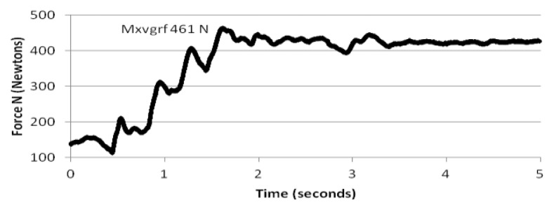
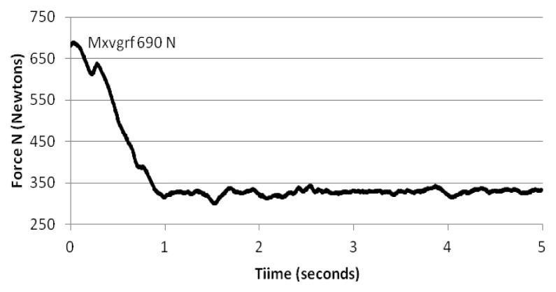
Figures 3a through 8c provide a representative sample of the yoga sequence and include explanations of the movement into the postures and graphs depicting results in Newtons of the maximum vertical GRF exerted by one of the subjects. Force measurements for all of the dependent variables were obtained in Newtons and normalized to BW on each individual subject.
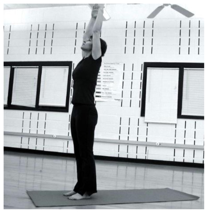
Figure 3a.
Tadasana (mountain). Subject begins with arms at sides.
Figure 8c.
Upward curves reflect weight shifting as hips lift upward and body mass is shared by lower extremities. Condition two.
Descriptive statistics (table 4) show that this yoga sequence produced a range of reaction forces less than 2 × BW for all subjects. All of the force measurements were normalized to BW by the custom algorithm. Univariate analysis identified mean values and standard deviations of the dependent variables (peak vertical GRF, mean vertical GRF, peak resultant GRF, and mean resultant GRF) for each individual posture and subject. All standard deviations were less than 0.5. Likewise, standard deviations across all subjects and postures were less than 0.5. As can be observed in table 4, peak vertical GRF and peak resultant GRF are identical or almost identical for all postures. Likewise, mean vertical GRF and mean resultant GRF are almost identical. Thus, results were consistent regardless of age, gender or body composition of the subjects.
Table 4.
Mean vertical and resultant ground reaction force (GRF) ranges by posture type, expressed as a ratio to body weight (BW).
| Posture type/supporting limb(s) | Limb(s) measured | Peak vertical GRF | Mean vertical GRF | Peak resultant GRF | Mean resultant GRF |
|---|---|---|---|---|---|
| Upright1 | Bilateral lower extremities | 0.98–1.47 × BW | 0.18–0.99 × BW | 1.00–1.49 × BW | 0.20–0.99 × BW |
| Upright2 | Forward leg | 0.66–1.07 | 0.52–0.68 | 0.68–1.07 | 0.54–0.70 |
| Rear leg | 0.40–0.75 × BW | 0.32–0.47 × BW | 0.45–0.86 × BW | 0.34–0.51 × BW | |
| Upright balancing3 | Balancing leg | 1.04–1.10 × BW | 0.84–0.99 × BW | 1.05–1.10 × BW | 0.84–0.99 × BW |
| Four limb support4 | Arms | 0.71–1.08 | 0.50–0.76 | 0.72–1.10 | 0.54–0.77 |
| Legs | 0.27–0.64 × BW | 0.21–0.48 × BW | 0.29–0.71 × BW | 0.22–0.53 × BW | |
| Three limbs5 | Arm | 0.67 × BW | 0.62 × BW | 0.67 × BW | 0.63 × BW |
| Legs | 0.45 × BW | 0.36 × BW | 0.49 × BW | 0.37 × BW | |
| Two limbs6 | Arm | 0.70 × BW | 0.66 × BW | 0.70 × BW | 0.64 × BW |
| Leg | 0.40 × BW | 0.36 × BW | 0.42 × BW | 0.38 × BW | |
| Two limbs7 | Arms | 1.05 × BW | 0.73 × BW | 1.06 × BW | 0.74 × BW |
Mountain, monkey, forward fold, chair, twisting chair, airplane
All warrior (except 3) and side angle asanas, triangle, pyramid
Half moon, tree, standing big toe, eagle, warrior 3
Crocodile, upward and downward facing dog, plank, dolphin
Side plank
Side plank on one leg
Crow
DISCUSSION
The main purpose of this study was to obtain GRF data from typical hatha yoga postures and determine if they are comparable to other forms of low-impact exercise. The mean values clearly demonstrate that the asanas in this particular study design produced low-impact forces which were less than those reported in table 2, with one exception. The highest peak vertical and resultant values in the present study resulted from the two-footed landing as participants leaped from plank to forward fold. The generated force is similar to that measured for low-impact exercises like walking and stepping down or pushing off of an 8-inch bench (9). It is important to note though, that the frequency of this two-footed landing in a typical yoga session is very small compared to the number of impacts sustained by walking or stepping. The lowest peak vertical and resultant values were measured from the feet in condition two for upward facing dog (table 4) where most of the weight is supported by the upper extremities.
Little is written about forces generated through the upper extremities and their influence on the skeleton. Indeed, neither a definition of high- or low-impact that applies to the upper extremities could be found by the researcher. All of the studies thus far cited measured only peak vertical GRF generated through the lower extremities. In contrast, execution of the yoga postures yielded vertical and resultant values for both upper and lower extremities.
It is important to consider that the upper extremities are not designed to absorb weight as are the lower extremities. Thus, what is considered low-impact for the legs, might with further study be considered high-impact for the upper extremities. In one study, a reaction force range (≥1.6 ≤3 × BW) was quantified for gymnasts performing the floor exercise round-off and Yurchenko vault round-off (19). Mean reaction force for the pommel horse measured 1.5 × BW, with forward and back handsprings averaging 2.9 and 3.6 × BW, respectively, in a study of male gymnasts (4). These researchers suggest that 1.5 to 3.6 × BW represents high-impact in the upper extremities. Another study examined the effect on BMD of lower impacts to the upper extremities. Researchers randomly assigned healthy adult females to impact either a firm or padded surface with their outstretched hand (22). A force platform, situated beneath the surface, measured the peak perpendicular reaction force during training sessions. Pre- and post-intervention BMD was obtained of the distal, ultradistal, and total radius. There were no statistically significant differences in BMD for either group from baseline to post-intervention. The mean reaction force ranged from 46.9% ± 33.6% of BW (minimum: 13.3%, maximum: 80.5%) in subjects who exercised against the firm wall. Compared to these three studies, the maximum GRF measured at the upper extremities in the present study were similar to the study by Wang, et al, and ranged from 0.63 to 0.70 × BW for one arm supported poses and 0.50 to 1.10 × BW for poses supported with both arms (Table IV). Still, a definition of low- or high-impact for the upper extremities is lacking.
Although most discussions about bone health center around the measurement of bone density using dual energy x-ray absorptiometry (DEXA), researchers recognize that some changes in the shape and strength of bones may be undetected by DEXA (5) and that a low or high BMD does not necessarily mean that bones are weak and prone to fracture or strong and healthy. Turner and Robling examined cross-sections of rat ulnas subjected to mechanical strain and noted that the very small change in BMD measured by DEXA did not accurately predict bone strength observed by a large increase in the amount of force the ulnas were able to sustain before failing (21). Other diagnostic means beside DEXA might eventually clarify how various types of exercises exert their beneficial effects on the skeleton.
At present, this study provides previously untested, unreported GRF data on hatha yoga asanas for both upper and lower extremities that are applicable to the real world since the individual postures in the design are common to beginner and intermediate-level hatha yoga classes and represent a variety of impacts.
Some of the study’s limitations warrant mentioning. Not all yoga sequences are performed at the tempo used in this design. Further accelerating the speed of execution could result in higher GRF since force equals mass times acceleration. Additionally, the results cannot be generalized to more vigorous styles of yoga which require a higher level of strength and expertise than was required with this design.
In conclusion, it is evident from a plethora of research on exercise and bone density that many kinds of weight-bearing activities, both high- and low-impact, are beneficial to individuals of all ages. Yoga’s low-impact, weight-bearing characteristics may positively influence bone health as many of its adherents believe, but controlled trials are needed to further understand the effect on the bones of vertical and resultant forces sustained by the extremities through yoga postures. Further research could also include cross sectional comparisons of BMD between yoga practitioners and athletes of other disciplines. Controlled trials might compare the effect of weight bearing yoga postures and weight lifting regimens on BMD and other parameters of bone health, including measurements at the wrist, forearm, shoulder, and spine in addition to the lower extremities. Researchers may want to explore the intensity of impact that is both safe and beneficial to the upper extremities, shoulder girdle and spine. Low-impact exercises and yoga practice could also be compared by measuring indicators of bone health and muscle strength. Motion analysis or electromyography, coupled with GRF measurements might explain how resultant forces, such as those measured in the present study, influence the skeleton. Research in any of the above areas would promote clearer understanding of yoga’s potential influence on bone health.
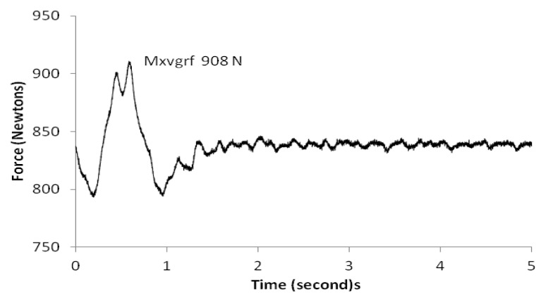
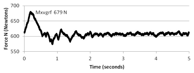
Figure 3b.
Maximum vertical ground reaction force (Mxvgrf) attained as arms extend overhead. Dual peaks reflect a force from each foot followed by stabilization. Condition one.
Figure 4a.
Uttanasana (forward fold). Subject flexes at the hip and brings arms to floor.
Figure 4b.
Maximum force achieved as subject shifts weight into heels of feet and brings upper body toward floor. Condition one.
Figure 5a.
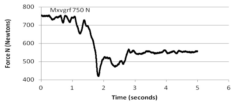
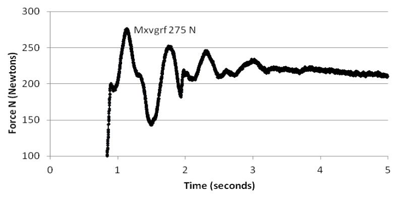
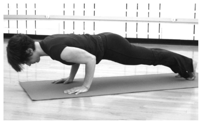
Dandasana (plank). Subject places hands on floor and steps from uttanasana into position.
Figure 5b.
Maximum force in upper extremities decreases once feet are in place. Condition one.
Figure 5c.
Peaks in force reflect movement of feet into position and stabilization of weight in lower extremities. Condition two.
Figure 6a.
Chaturanga dandasana (crocodile). Subject lowers body toward the floor.
Figure 6b.
Force diminishes as body lowers. Maximum force is generated through arms at cessation of descent. Condition one.
Figure 6c.
Maximum force in the feet as body begins to lower toward floor, followed by stable force curve measured at the feet. Condition two.
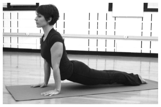
Figure 7a.
Urdva mukha svanasana (upward facing dog). Subject pushes upward from chaturanga. Hips and thighs contract and lift away from the floor so mass is supported in hands and feet.
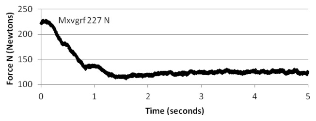
Figure 7b.
Maximum force is generated with upper extremity contraction which lifts torso upward. Condition one.
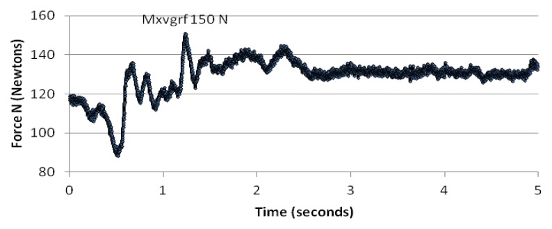
Figure 7c.
Multiple peaks reflect changing foot position from balls to tops of feet, followed by stabilization in lower extremities. Condition two.
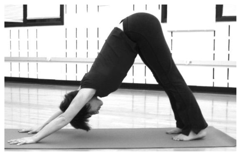
Figure 8a.
Adho mukha svanasana (downward facing dog). Subject lifts hips.
Figure 8b.
Maximum force generated in upper extremities as subject lifts hips upward and pushes against floor. Condition one.
ACKNOWLEDGEMENTS
The author thanks the Exercise Sciences faculty at Brigham Young University for its guidance and the Mary Lou Fulton Chair for funding the study.
REFERENCES
- 1.Bassey EJ, Ramsdale SJ. Weight bearing exercise and ground reaction forces: a 12-month randomized controlled trial of effects on bone mineral density in healthy postmenopausal women. Bone. 1995;16(4):469–476. doi: 10.1016/8756-3282(95)90193-0. [DOI] [PubMed] [Google Scholar]
- 2.Bassey EJ, Rothwell MC, Littlewood JJ, Pye DW. Pre and post- menopausal women have different bone mineral density responses to the same high-impact exercise. J Bone Miner Res. 1998;13(12):1805–1813. doi: 10.1359/jbmr.1998.13.12.1805. [DOI] [PubMed] [Google Scholar]
- 3.Cowen VS, Adams TB. Physical and perceptual benefits of yoga asana practice: results of a pilot study. Journal of Bodywork and Movement Therapies. 2005;9:211–219. [Google Scholar]
- 4.Daly RM, Rich PA, Klein R, Bass S. Effects of high-impact exercise on ultrasonic and biochemical indices of skeletal status: a prospective study in young male gymnasts. J Bone Miner Res. 1999;14(7):1222–1230. doi: 10.1359/jbmr.1999.14.7.1222. [DOI] [PubMed] [Google Scholar]
- 5.Forwood M. What does the animal model teach us about the effects of physical activity on growing bone? Pediatric Exercise Science. 2006;18:282–289. [Google Scholar]
- 6.Garfinkel MS, Singhal A, Katz WA, Allan DA, Reshetar R, Schumacher HR. Yoga-based intervention for carpal tunnel syndrome: a randomized trial. JAMA. 1998;280(18):1601–1603. doi: 10.1001/jama.280.18.1601. [DOI] [PubMed] [Google Scholar]
- 7.Grove KA, Londeree BR. Bone density in postmenopausal women: high impact vs low impact exercise. Med Sci Sports Exerc. 1992;24:1190–1194. [PubMed] [Google Scholar]
- 8.Heinonen A, Kannus P, Sievanen H. Randomised controlled trial of effect of high-impact exercise on selected risk factors for osteoporotic fractures. Lancet. 1996;348:1343–1347. doi: 10.1016/S0140-6736(96)04214-6. [DOI] [PubMed] [Google Scholar]
- 9.Johnson BF, Rupp JC, Berry SA, Rupp DA. Peak vertical ground reaction forces and time-to-peak force in bench-step aerobics and other activities. Med Sci Sports Exerc. 1992;24:S131. [Google Scholar]
- 10.Kato T, Bassey EJ. Ground reaction force in different types of high-impact exercise. Research Reports of Suzuka University of Medical Science. 2002;9:128–135. [Google Scholar]
- 11.Kohrt WM, Ehsani A, Birge S. Effects of exercise involving predominantly either joint-reaction or ground-reaction forces on bone mineral density in older women. J Bone Miner Res. 1997;12(8):1253–1261. doi: 10.1359/jbmr.1997.12.8.1253. [DOI] [PubMed] [Google Scholar]
- 12.Kolasinski S, Garfinkel M, Tsai AG, Matz W, Van Dyke A, Schumacher HR. Iyengar yoga for treating symptoms of osteoarthritis of the knees: a pilot study. Journal of Alternative and Complementary Medicine. 2005;11(4):689–693. doi: 10.1089/acm.2005.11.689. [DOI] [PubMed] [Google Scholar]
- 13.Mayoux-Benhamou MA, Leyge JF, Roux C, Revel M. Cross-sectional study of weight-bearing activity on proximal femur bone mineral density. Calcif Tissue Int. 1999;64:179–183. doi: 10.1007/s002239900599. [DOI] [PubMed] [Google Scholar]
- 14.Nelson ME, Fiataron MA, Morganti CM. Effects of high-intensity strength training on multiple risk factors for osteoporotic fractures. JAMA. 1999;272(24):1909–1914. doi: 10.1001/jama.1994.03520240037038. [DOI] [PubMed] [Google Scholar]
- 15.Raub JA. Psychophysiologic effects of hatha yoga on musculoskeletal and cardiopulmonary function: a literature review. Journal of Alternative and Complementary Medicine. 2002;8(6):797–812. doi: 10.1089/10755530260511810. [DOI] [PubMed] [Google Scholar]
- 16.Robinson RJ, Krzywicki T, Almond L, Al-Azzawi F, Abrams K, Iqbal SJ, Mayberry JF. Effect of a low-impact exercise program on bone mineral density in Crohn’s disease: A randomized controlled trial. Gastroenterology. 1998;115:36–41. doi: 10.1016/s0016-5085(98)70362-2. [DOI] [PubMed] [Google Scholar]
- 17.Rousanoglou EN, Boudolos KD. Ground reaction forces and heart rate profile of aerobic dance instructors during a low and high impact exercise programme. J Sports Med Phys Fitness. 2005;45:162–170. [PubMed] [Google Scholar]
- 18.Ryan AS, Ivey FM, Hurlbut DE, Martel GF, Lemmer JT, Sorkin JD, et al. Regional bone mineral density after resistive training in young and older men and women. Scand J Med Sci Sports. 2004;14:16–23. doi: 10.1111/j.1600-0838.2003.00328.x. [DOI] [PubMed] [Google Scholar]
- 19.Seeley MK, Bressel E. A comparison of upper-extremity reaction forces between the Yurchenko vault and floor exercise. Journal of Sports Science and Medicine. 2005;4:85–94. [PMC free article] [PubMed] [Google Scholar]
- 20.Sparrow L. Yoga for healthy bones A woman’s guide. Boston: Shambhala; 2004. [Google Scholar]
- 21.Turner CH, Robling AG. Designing exercise regimens to increase bone strength. Exerc Sport Sci Rev. 2003;31(1):45–50. doi: 10.1097/00003677-200301000-00009. [DOI] [PubMed] [Google Scholar]
- 22.Wang MY, Salem GJ. The relations among upper-extremity loading characteristics and bone mineral density changes in young women. Bone. 2004;34:1053–1063. doi: 10.1016/j.bone.2004.01.025. [DOI] [PubMed] [Google Scholar]
- 23.Welsh L, Rutherford OM. Hip bone mineral density is improved by high-impact aerobic exercise in postmenopausal women and men over 50 years. Eur J Appl Physiol. 1996;74:511–517. doi: 10.1007/BF02376766. [DOI] [PubMed] [Google Scholar]


