Abstract
Aim
To analyze the changes of different Lactobacillus species in ulcerative colitis patients and to further assess the therapeutic effects of selected Lactobacillus strains on dextran sulfate sodium (DSS)-induced experimental colitis in BALB/c mice.
Methods
Forty-five active ulcerative colitis (UC) patients and 45 population-based healthy controls were enrolled. Polymerase chain reaction (PCR) amplification and real-time PCR were performed for qualitative and quantitative analyses, respectively, of the Lactobacillus species in UC patients. Three Lactobacillus strains from three species were selected to assess the therapeutic effects on experimental colitis. Sixty 8-week-old BALB/c mice were divided into six groups. The five groups that had received DSS were administered normal saline, mesalazine, L. fermentum CCTCC M206110 strain, L. crispatus CCTCC M206119 strain, or L. plantarum NCIMB8826 strain. We assessed the severity of colitis based on disease activity index (DAI), body weight loss, colon length, and histologic damage.
Results
The detection rate of four of the 11 Lactobacillus species decreased significantly (P < 0.05), and the detection rate of two of the 11 Lactobacillus species increased significantly (P < 0.05) in UC patients. Relative quantitative analysis revealed that eight Lactobacillus species declined significantly in UC patients (P < 0.05), while three Lactobacillus species increased significantly (P < 0.05). The CCTCC M206110 treatment group had less weight loss and colon length shortening, lower DAI scores, and lower histologic scores (P < 0.05), while the CCTCC M206119 treatment group had greater weight loss and colon length shortening, higher histologic scores, and more severe inflammatory infiltration (P < 0.05). NCIMB8826 improved weight loss and colon length shortening (P < 0.05) with no significant influence on DAI and histologic damage in the colitis model.
Conclusions
Administration of an L. crispatus CCTCC M206119 supplement aggravated DSS-induced colitis. L. fermentum CCTCC M206110 proved to be effective at attenuating DSS-induced colitis. The potential probiotic effect of L. plantarum NCIMB8826 on UC has yet to be assessed.
Introduction
Ulcerative colitis (UC), a primary type of inflammatory bowel disease (IBD), is a chronic bowel disorder characterized by recurrent uncontrolled inflammation of the intestinal mucosa [1]. UC was perceived as a Western disease in the past, based on the high incidence and prevalence reported in industrialized countries (especially northern Europe and North America). However, the incidence and prevalence of UC has continued to increase in various regions around the world in recent decades; numerous developing countries traditionally considered as areas of low incidence are undergoing a dramatic rise in disease incidence [1,2]. Not surprisingly, UC is currently emerging as a global disease.
Increasing prevalence may partly account for the heavy health burden UC brings about; another crucial contributor could be patient requirements for lifetime care due to a lack of a guaranteed curative therapeutic regimen [3]. Conventional medical treatment of UC involves the administration of high-dose steroids and immunomodulators. Recently, significant advances have provided substantial insight into the etiology and pathogenesis of UC. The role of intestinal microbes in particular was shown to be of greater significance than previously considered [4]. Thus, novel therapies geared towards modulating the intestinal flora has become a research focus, a representative of which was the extensive use of probiotics in IBD, including UC [5].
The beneficial effects of probiotics in the induction of remission and the maintenance of UC have been examined in numerous animal experiments as well as in clinical trials [6–9]. As one of the most extensively commercially exploited probiotic bacteria, the effectiveness and safety of several Lactobacillus strains for supplementation have been evaluated in several studies [10–12]. However, to date, the understanding of these strains is still far from being sufficient for appropriate application in UC patients. The possible therapeutic efficacy of certain Lactobacillus strains in UC has yet to be assessed or identified further.
Thus, to identify more putative Lactobacillus species that may be of therapeutic value for UC, we undertook a study to investigate the changes of different Lactobacillus species in UC patients. Because species-specific primers can only be developed if there are known gene sequences containing highly variable regions of the universal Lactobacillus genes, we merely analyzed the changes in 11 common Lactobacillus species in UC patients compared with healthy controls using published Lactobacillus species-specific gene sequences. Based on this analysis, three Lactobacillus strains from three species were selected to assess the therapeutic effects on DSS-induced experimental colitis in BALB/c mice.
Materials and Methods
Participants
Eligible patients were recruited from the Gastroenterology Department of The Second Xiangya Hospital between April 2008 and May 2012. Forty-five active UC patients (26 males and 19 females; average age, 34 years) based on widely accepted diagnostic criteria, and 45 population-based healthy controls (30 males and 15 females; average age, 28 years) were enrolled in the study.
Participants were excluded if they had concurrent bacillary dysentery, ischemic enterocolitis, enterophthisis, or the use of antibiotics within 4 weeks prior to the sampling.
All human fecal samples were collected with written informed consent from patients prior to participation in the study. The protocols for the collection and analysis of the samples were approved by the Ethical Committee of the Second Xiangya Hospital of Central South University, in accordance with the current revision of the Helsinki Declaration.
Collection and storage of fecal samples
Fresh fecal samples were collected from 45 patients and 45 healthy controls. All samples were placed in sterile, anaerobic ice boxes and immediately stored at –80°C until processing within 2 h.
DNA extraction from fecal samples
DNA extraction was performed following the manufacturer’s instructions (QIAamp DNA Stool Mini Kit; Qiagen, Hilden, Germany). Ninety samples were processed by a single individual. The concentration of the DNA extracts was assessed by measuring the optical density at wavelengths of 230 nm, 260 nm, and 280 nm after dilution with deionized water. The purity of total DNA extracted was checked by electrophoresis on a 1% agarose gel.
PCR screening of Lactobacillus species in which the detection rate significantly differed between UC patients and healthy controls
The primers used in the investigation are listed in Table 1. BLAST searches were performed to determine the specificity of the primers. For DNA extracts from each fecal sample, PCR amplification reactions (in a total volume of 20 μL) were performed on a PCR thermal cycler (PC-808; Astec, Fukuoka, Japan). Each reaction was performed in a 20 μL reaction volume containing 2 μL of template DNA, 2 μL of 25 mM MgCl2, 0.5 μL of 10 mM dNTPs, 5 μL of 10×PCR buffer, 10 pmol of each primer, and 1.0 U of Taq DNA polymerase (Promega, Madison, WI, USA). PCR was performed using the following parameters: an initial denaturation at 95°C for 3 min, followed by 36 cycles of 94°C for 30 s; 56°C for 40 s; 72°C for 40 s; and a final elongation period at 72°C for 5 min. The amplifications were confirmed by standard electrophoresis of the PCR products (5 μL) using 2% agarose gels (Sigma–Aldrich, St. Louis, MO, USA) prepared in 0.5× TBE buffer (45 mM Tris base, 45 mM boric acid, and 1 mM EDTA [pH 8.0]) and visualized by silver staining. A Lactobacillus species was considered to be detectable in the fecal samples of participants when primed PCR products were obtained. Thus, we screened out the Lactobacillus species that had significantly different detection rates between the patients and controls.
Table 1. Sequencing primers for the Lactobacillus species used in this study.
| Species | Primer | Sequence (5ʹ-3ʹ) | Annealing temperature (°C) | Amplicon size (bp) |
|---|---|---|---|---|
| L. crispatus | LcrisF | AGCGAGCGGAACTAACAGATTTAC | 58 | 154 |
| LcrisR | AGCTGATCATGCGATCTGCTT | |||
| L. gallinarum | LgallF | CGGTAATGACGCTGGGGAC | 56 | 128 |
| LcrisR | AGCTGATCATGCGATCTGCTT | |||
| L. fermentum | LfermF | GCACCTGATTGATTTTGGTCG | 58 | 317 |
| LactoR | GTCCATTGTGGAAGATTCCC | |||
| L. johnsonii | LactoF | TGGAAACAGRTGCTAATACCG | 56 | 322 |
| LjohnR | CAGTTACTACCTCTATCTTTCTTCACTAC | |||
| L. salivarius | LsaliF | CGAAACTTTCTTACACCGAATGC | 58 | 332 |
| LactoR | GTCCATTGTGGAAGATTCCC | |||
| L. acidophilus | F_acid | GAAAGAGCCCAAACCAAGTGATT | 56 | 85 |
| R_acid | CTTCCCAGATAATTCAACTATCGCTTA | |||
| L. casei | F_case | CTATAAGTAAGCTTTGATCCGGAGATTT | 56 | 132 |
| R_case | CTTCCTGCGGGTACTGAGATGT | |||
| L. delbrueckii | F_delb | CACTTGTACGTTGAAAACTGAATATCTTAA | 56 | 94 |
| R_delb | CGAACTCTCTCGGTCGCTTT | |||
| L. paracasei | F_paca | ACATCAGTGTATTGCTTGTCAGTGAATAC | 56 | 88 |
| R_paca | CCTGCGGGTACTGAGATGTTTC | |||
| L. plantarum | F_plan | TGGATCACCTCCTTTCTAAGGAAT | 56 | 80 |
| R_plan | TGTTCTCGGTTTCATTATGAAAAAATA | |||
| L. reuteri | F_reut | ACCGAGAACACCGCGTTATTT | 56 | 93 |
| R_reut | CATAACTTAACCTAAACAATCAAAGATTGTCT | |||
| E. faecalis | Efs130F | AACCTACCCATCAGAGGG | 56 | 360 |
| Efs490R | GACGTTCAGTTACTAACG | |||
| Lactobacillus spp | F_alllact | TGGATGCCTTGGCACTAGGA | 58 | 92 |
| R_alllact | AAATCTCCGGATCAAAGCTTACTTAT | |||
| All bacteria | F_eub | TCCTACGGGAGGCAGCAGT | 58 | 466 |
| R_eub | GGACTACCAGGGTATCTAATCCTGTT |
Quantitative real-time PCR (qPCR) analysis of the Lactobacillus species in UC patients and healthy controls
The qPCR reaction was performed using an Applied Biosystems 7300 Real-Time system (Applied Biosystems, Foster City, CA, USA). Quantification assays and data analyses were performed using SDS version 1.2.3 software (Applied Biosystems). A 50 μL PCR reaction contained 5.0 μL of template DNA (50 ng), a 2.5 μL mixture of screened primers (10 pmol/L for each), 25.0 μL of 2× GoTaq qPCR Master Mix, 0.5 μL of 100× CXR reference dye, and sterile ddH2O. The qPCR reaction conditions were as follows: DNA polymerase activation at 95°C for 5 min, followed by 40 cycles of DNA melting at 95°C for 15 s and annealing at 56°C for 60 s. The specificity of the qPCR reaction was confirmed by melting-curve analysis.
The data were analyzed using the 2-ΔΔCt method. As reported in several studies [13,14], we used total bacteria as the endogenous control to normalize the data. The threshold cycle (Ct) indicates the fractional number at which the amount of amplified target reaches a fixed threshold. ΔCt was calculated as the difference between the Ct value of the primers specific for Lactobacillus species and the Ct value of the primers for total bacteria. ΔΔCt is defined as the difference between the ΔCt value of UC patients and the ΔCt value of the control group. The fold change of expression of each Lactobacillus species in UC patients relative to that of the healthy controls was calculated using log10 RQ where RQ is 2-ΔΔCt. Log10RQ correlates directly to up-regulation (positive value) and down-regulation (negative value). A value of 1 in log10RQ (relative quantification) represents a 10-fold increase in expression. Similarly, a value of -1 represents 10-fold decrease in expression compared to control.
Animal trial protocol
Sixty female (8-week-old) BALB/c mice weighing 20.0 ± 2.0 g were purchased from Hunan SJA Laboratory Animal Co. Ltd., Changsha, China. Each group consisted of ten mice. All mice were housed at a room temperature of 20–22°C, 50 ± 10% humidity, and a 12 h diurnal light/dark cycle. Throughout the trial period, the mice were fed standard rat chow and water ad libitum. All animal experimental procedures were reviewed and approved by the Institutional Animal Care and Use Committee of Central South University.
Three accessible standard Lactobacillus strains that were previously isolated and identified by our research institutions (preserved in the China Centre for Type Culture Collection) were selected as candidate Lactobacillus strains for further experimental effect verification. The candidate Lactobacillus strains were L. fermentum CCTCC M206110, L. plantarum NCIMB8826, and L. crispatus CCTCC M206119. The animals were randomly divided into three experimental groups (DSS-treated mice given 0.4 mL of 3.0×10⁸ colony forming units/mL of L. fermentum CCTCC M206110, L. crispatus CCTCC M206119, or L. plantarum NCIMB8826), three control groups (a healthy control group, a negative [normal saline] control group, and a positive [mesalazine; 6 mg/20 g] control group). Candidate strains of three Lactobacillus species, saline, or mesalazine were administered once daily via gavage from days 1–9. Between days 3 and 9, experimental colitis was induced via the consumption of 5% dextran sulfate sodium (DSS; Mw 40 kDa, MP Biomedicals Inc., Solon, OH, USA) in the drinking water [15].
Food, water/DSS consumption, body weight, fecal and urine output, and survival condition were monitored daily. The disease activity index (DAI) was determined by scoring the change in body weight, occult blood, and gross bleeding, as described previously [16]. On day 9, all mice were sacrificed by cervical dislocation. Then, the colons were removed and the colon length was measured. The distal colon was then collected and fixed in 10% buffered formalin for hematoxylin and eosin staining. Other parts of the colon were preserved in liquid nitrogen.
Histologic analysis
Nine randomly selected fields (magnification 200×) were inspected in each section by two pathologists blinded to the treatment protocol. Grading of intestinal inflammation was determined as follows (Table 2). For each category of the score (inflammation, depth of lesions, and destruction of crypt), the points were multiplied by a factor of involvement of the visible epithelium (Table 2). The sum of the two category scores (inflammation and depth of lesions) were added to the inflammatory damage score (range, 0–24). The score of the destruction of the crypt category represents the crypt damage score (range, 0–16) [17,18].
Table 2. Histological score to quantify the degree of colitis.
| Score | Inflammation | Depth of lesions | Destruction of crypt | Width of lesions (%) |
|---|---|---|---|---|
| 0 | None | None | None | |
| 1 | Mild | Submucosa | 1/3 basal crypt | 1–25 |
| 2 | Severe | Muscularis | 2/3 basal crypt | 26–50 |
| 3 | Sera | Intact epithelium only | 51–75 | |
| 4 | Total crypt and epithelium | 76–100 |
Statistical analysis
All data are presented as the mean ± standard deviation (SD). Statistical package for social sciences (SPSS) software (version 17.0; SPSS Inc., Chicago, IL, USA) was used for data management and statistical analysis.
The detection rate of the different Lactobacillus species in UC patients and healthy controls was compared using a chi-square test. Weight, colon length, DAI scores, and histologic scores of all of the mouse groups were analyzed using an ANOVA test. Post hoc t-tests were conducted when the ANOVA values were significant. A P-value < 0.05 was considered to be statistically significant.
Results
The detection rate of 11 Lactobacillus species in UC patients and healthy controls
Using DNA extracts from fecal samples of 45 patients and 45 healthy controls, PCR amplification reactions were performed. The detection rates of the 11 Lactobacillus species in 45 patients and 45 healthy controls are listed in Table 3. Eight of the 11 Lactobacillus species were detectable in >50% of the healthy controls, and 5 of the 11 Lactobacillus species were detectable in >90% of the healthy controls. Two of the 11 Lactobacillus species (L. johnsonii and L. casei) were detectable in all of the healthy controls. Compared with the healthy controls, the detection rate of 4 of the 11 Lactobacillus species decreased significantly in the UC patients (P < 0.05). The four Lactobacillus species were L. crispatus (86.7% vs. 53.3%, P = 0.004), L. fermentum (95.6% vs. 35.6%, P = 0.001), L. gasseri (100.0% vs. 37.8%, P = 0.002), and L. salivarius (75.6% vs. 26.7%, P = 0.041). The detection rate of 2 of the 11 Lactobacillus species increased significantly in UC patients (P < 0.05). The two Lactobacillus species were L. delbrueckii (40.0% vs. 88.9%, P = 0.005) and L. paracasei (20.0% vs. 64.4%, P = 0.028).
Table 3. The detection rate of 11 Lactobacillus species in UC patients and healthy controls.
| UC patients (n = 45) | Healthy (n = 45) | p | |||
|---|---|---|---|---|---|
| Numbers | % | Numbers | % | ||
| L. crispatus | 24 | 53.3 | 39 | 86.7 | 0.004 |
| L. gallinarum | 37 | 82.2 | 42 | 93.3 | 0.132 |
| L. fermentum | 16 | 35.6 | 43 | 95.6 | 0.001 |
| L. gasseri | 17 | 37.8 | 45 | 100 | 0.002 |
| L. salivarius | 12 | 26.7 | 34 | 75.6 | 0.041 |
| L. acidophilus | 38 | 84.4 | 42 | 93.3 | 0.206 |
| L. casei | 45 | 100.0 | 45 | 100.0 | 1.000 |
| L. delbrueckii | 40 | 88.9 | 18 | 40.0 | 0.005 |
| L. paracasei | 29 | 64.4 | 9 | 20.0 | 0.028 |
| L. plantarum | 19 | 42.2 | 26 | 57.8 | 0.090 |
| L. reuteri | 11 | 24.4 | 6 | 13.3 | 0.225 |
| Lactobacillus spp | 45 | 100 | 45 | 100 | 1.000 |
Quantitative analysis of the Lactobacillus species in UC patients and healthy controls
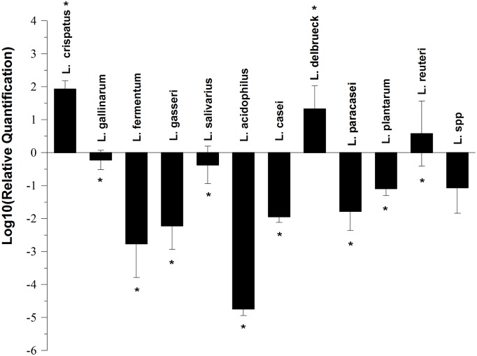
The results of quantitative analysis of the 11 Lactobacillus species in UC patients and healthy controls are shown in Fig 1. Compared with the healthy controls, the number of L. gallinarum, L. fermentum, L. gasseri, L. salivarius, L. acidophilus, L. casei, L. paracasei, and L. plantarum decreased significantly in UC patients (P < 0.05), while the number of L. crispatus, L. delbrueckii, and L. reuteri increased significantly (P < 0.05).
Fig 1. The relative quantification of Lactobacillus species in UC patients.
The fold change was calculated using log10 RQ where RQ is 2−ΔΔCt. Statistical significance was determined by a t-test comparing ΔCt of healthy controls to ΔCt of UC patients. *P < 0.05.
Effect of different Lactobacillus strains on weight loss in DSS-induced colitis in BALB/c mice
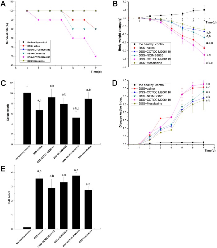
Four days after inducing colitis, deaths began to occur due to gastrointestinal bleeding. The survival statuses of the animals at the end of the experiments are shown in Fig 2A. All groups exhibited significant weight loss during DSS treatment compared to the healthy control group, which was given water and a standard diet (P < 0.05), especially the negative control group. The positive control group had significantly less weight loss compared with the negative control group (-0.71 ± 0.28 g vs. -1.84 ± 0.72 g, P < 0.05). The weight loss in the L. fermentum CCTCC M206110 group (-0.82 ± 0.39 g vs. -1.84 ± 0.72 g, P < 0.05) and the L. plantarum NCIMB8826 group (-1.19 ± 0.55 g vs. -1.84 ± 0.72 g, P < 0.05) was also significantly less than the negative control group; however, the weight loss was significantly higher in the L. crispatus CCTCC M206119 group than in the negative control group (-2.26 ± 0.51 g vs. -1.84 ± 0.72 g, P < 0.05; Fig 2B).
Fig 2. Effects of Lactobacillus strains NCIMB8826, CCTCC M206119, and CCTCC M206110 in dextran sulfate sodium-colitis mice.
(A) Survival rate of six BALB/c mouse groups (n = 10) by the end of the experiment. (B) Effects of Lactobacillus strains treatments on colitis-induced body weight loss. The x-axis shows the mean weight (mean ± SD) recorded for each group on each day of the 7-day colitis induction. (C) Effects of Lactobacillus strain treatments on colitis-induced changes in colonic length. (D) Effects of Lactobacillus strains on colitis-induced changes in DAI scores. The daily mean DAI score ± SD is plotted for each group; E: Mean ± SD DAI scores on day 9. aP < 0.05 vs. the healthy control group; bP < 0.05 vs. the negative (saline) control group; cP < 0.05 vs. the positive (mesalazine) control group.
Effect of different Lactobacillus strains on colon length in DSS-induced colitis in BALB/c mice
All groups exhibited significant colon length reduction during DSS treatment compared to the healthy control group, which was given water and a standard diet (P < 0.05). The mesalazine group had significantly improved colon length shortening compared with the negative control group (9.18 ± 1.39 cm vs. 6.71 ± 1.47 cm, P < 0.05). In addition, the L. fermentum (9.22 ± 1.69 cm vs. 6.71 ± 1.47 cm, P < 0.05) and L. plantarum groups (7.95 ± 1.19 cm vs. 6.71 ± 1.47 cm, P < 0.05) both had significantly reduced shortening of the colon length compared with the negative control group. Furthermore, the L. fermentum group had more improvement in colon length shortening than the L. plantarum group (9.22 ± 1.69 cm vs. 7.95 ± 1.19 cm, P < 0.05); however, the results indicated that L. crispatus exacerbated the colon length shortening in comparison with the negative control group (5.25 ± 1.19 cm vs. 6.71 ± 1.47 cm, P < 0.05; Fig 2C).
Effect of different Lactobacillus strains on DAI in DSS-induced colitis in BALB/c mice
All groups exhibited a significantly higher DAI score during DSS treatment compared to the healthy control group, which was given water and a standard diet (P < 0.05; Fig 2D). Compared with the negative control group, mesalazine treatment resulted in a significant decrease in the DAI score (2.77 ± 0.63 vs. 3.57 ± 0.32, P < 0.05). Moreover, the DAI scores of animals in the L. fermentum group were also significantly less than the negative control group (2.90 ± 0.22 vs. 3.57 ± 0.32, P < 0.05); however, the results indicated no significant difference in the DAI in the L. plantarum group or L. crispatus group compared with the negative control group (P > 0.05; Fig 2E).
Effect of different Lactobacillus strains on histologic changes in DSS-induced colitis in BALB/c mice
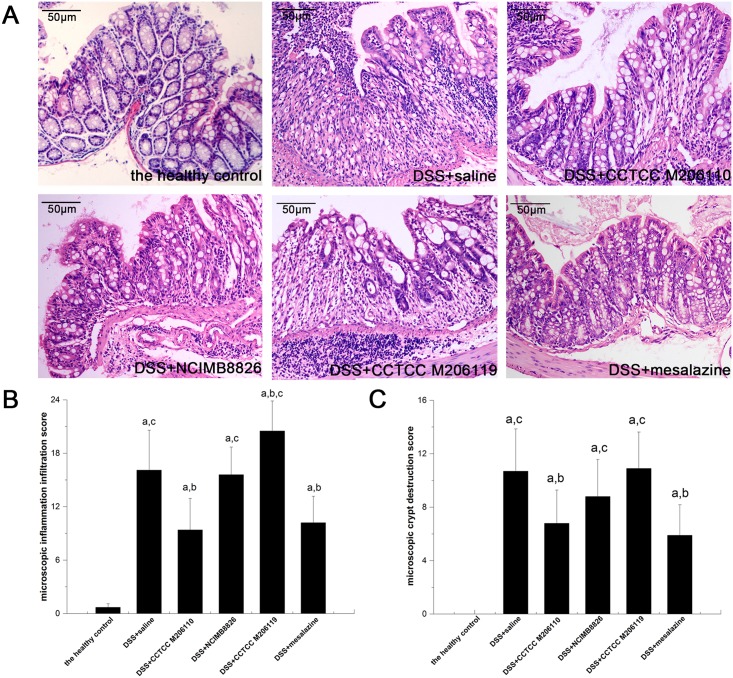
The colon sections of the healthy control group showed intact mucosae with glands secreting abundant mucin. The colon sections of DSS-treated animals showed loss of the epithelial layer, goblet cell depletion, neutrophil infiltration, and distortion/destruction of the crypt architecture compared with the healthy controls; however, when the mice were co-treated with L. fermentum, the level of inflammatory infiltration was significantly lower compared with the negative control (9.40 ± 3.52 vs. 16.1 ± 4.48, P < 0.05), as was the extent of crypt structure destruction (6.80 ± 2.49 vs. 10.70 ± 3.16, P < 0.05). The results indicated no significant difference in the severity of inflammatory infiltration or crypt destruction between the L. plantarum group and the negative group (P > 0.05). Notably, more severe inflammatory infiltration was observed in the L. crispatus group compared with the negative controls (20.50 ± 3.37 vs. 16.1 ± 4.48, P < 0.05), with no significant difference in the severity of crypt destruction (Fig 3).
Fig 3. Effects of Lactobacillus strain treatments on colitis-induced changes in intestinal histopathologic features.
(A) Histologic analysis of representative colons from the mice (original magnification, hematoxylin and eosin staining, 200×). (B) Microscopic inflammatory infiltration score; (C) Microscopic crypt destruction score. aP < 0.05 vs. the healthy control group; bP < 0.05 vs. the negative (saline) control group; cP < 0.05 vs. the positive (mesalazine) control group.
Discussion
Therapies aimed at modifying and manipulating the gut flora have been implicated in the pathogenesis of UC through the use of probiotics and have received increased attention [19].
The application of Lactobacillus to UC is based on the evidence indicating the reduction of the fecal Lactobacillus count in UC patients [20–22]. In the current study, we demonstrated that the change in Lactobacillus in UC patients was more complicated at the species level. Not all Lactobacillus species were decreased in UC patients. The results of quantitative analysis of the 11 Lactobacillus species indicated that some Lactobacillus species increased significantly in UC patients compared with healthy controls, such as L. crispatus. Furthermore, the therapeutic effects of Lactobacillus strains were correlated with changes in the concentrations in the distal colon[23,24]. Lactobacillus strains of different species could exert completely opposite effects in UC.
Based on our study, L. crispatus numbers increased significantly in UC patients compared with healthy controls. The animal experiments further revealed that L. crispatus CCTCC M206119 aggravated weight loss and colonic histologic damage in the mouse colitis model. Previous studies on the effects of L. crispatus in induced colitis have reported conflicting results. Castagliuolo et al. [25] have demonstrated that L. crispatus M247 reduced the severity of DSS colitis in a dose-dependent fashion, while Ulinski and Aoun [26] revealed that the L. crispatus CCTCC M206119 strain is involved in the exacerbation of intestinal inflammation in DSS-colitis mice. Thus, we should be more cautious in administering Lactobacillus to UC patients in clinical practice.
The results obtained in the present study reveal that L. fermentum CCTCC M206110 appeared to be effective at attenuating DSS-induced colitis in BALB/c mice, given that the important clinical parameters, such as mortality, weight loss, DAI scores, and colonic histologic damage were improved in mice receiving L. fermentum compared with the controls. A previous study demonstrated that mice with colitis treated with L. fermentum had an improved survival rate, DAI score, and colonic mucosa histologic scoring [27]. The study further indicated that L. fermentum attenuated trinitrobenzenesulfonic acid-induced colitis, which was associated with an increase in intestinal superoxide dismutase activity and a reduction in oxidative stress, nuclear factor κB (NF-κB) activity, and cytokine production [28,29]. Our study tested the effect of L. fermentum CCTCC M206110 in DSS-induced colitis because of a previously noted reduction in L. fermentum species in UC patient feces. The present study indicates that L. fermentum exhibited beneficial anti-oxidative and anti-inflammatory properties in the mouse colitis model. Thus, a L. fermentum probiotic could be recommended as potential adjuvant therapy in combination with olsalazine to achieve a more efficacious treatment for UC. Nevertheless, because the associated clinical data are rather limited and far from convincing, the concomitant use of L. fermentum warrants well-designed, large, randomized, placebo-controlled trials to investigate the unresolved issues related to efficacy, dose, duration of use, and single or multistrain formulation.
In the current study, we failed to observe an obvious attenuation effect of L. plantarum NCIMB8826 in DSS-induced colitis in BALB/c mice. The results showed that L. plantarum NCIMB8826 improved weight loss and colon length, but with no significant influence on the DAI or colonic histologic damage in the mouse colitis model. During the last decade, the therapeutic effect of L. plantarum strains on ameliorating experimental colitis in mouse models has been reported [30–32]. Additionally, the anti-inflammatory and immunomodulatory activities of L. plantarum were evaluated, including reducing the production of pro-inflammatory cytokines (tumor necrosis factor (TNF)-α, interleukin (IL)-1, and IL-6) and inhibiting the Toll-like receptor (TLR)4-linked NF-κB and mitogen-activated protein kinase (MAPK) signaling pathways [31]. Furthermore, L. plantarum exhibited antioxidant properties by significantly decreasing lipid peroxidation (TBARS) and nitric oxide production and increasing the glutathione concentration [30,33]. It is arbitrary and inexact to draw the conclusion that L. plantarum is ineffective in DSS-induced colitis based on the present study, given the strong possibility of a false-negative result due to an insufficient sample size. Thus, the potential probiotic effect of L. plantarum on UC must be further assessed.
Hence, it is of great importance to establish the mechanism(s) underlying the beneficial interaction between the colonic epithelium, intestinal immunity, and the microbiota for the extensive but evidence-based application of probiotics in UC. Based on the previous studies, the gut microbiota plays a role in shaping the mucosal immune system [34]. Clostridia and Bacteroides have been proven to attenuate intestinal inflammation in mice through the induction of CD4+FOXP3+ regulatory T lymphocytes [35–37]. Other members of the microbiota can attenuate mucosal inflammation by antagonizing the activation of NF-κB or modifying the cytokine status [37].
In conclusion, the change in Lactobacillus in UC patients was more complicated at the species level. Not all Lactobacillus species are beneficial for UC patients. Administration of L. crispatus CCTCC M206119 supplement aggravated DSS-induced colitis, while L. fermentum CCTCC M206110 proved to be effective at attenuating DSS-induced colitis. L. plantarum NCIMB8826 showed no obvious attenuation effect in DSS-induced colitis. Thus, the potential probiotic effect of L. plantarum species on UC has yet to be assessed. Future studies should aim to determine the mechanisms underlying the interactions between Lactobacillus, the colonic epithelium and intestinal immunity, which may be essential for the extensive but evidence-based application of probiotics in UC.
Supporting Information
(DOCX)
Data Availability
All relevant data are within the paper and its Supporting Information files.
Funding Statement
National Natural Science Foundation of China (NO. 81470801), received by F. Lu.
References
- 1.Molodecky NA, Soon IS, Rabi DM, Ghali WA, Ferris M, Chernoff G, et al. Increasing incidence and prevalence of the inflammatory bowel diseases with time, based on systematic review. Gastroenterology. 2012;142(1):46–54 e42; quiz e30. 10.1053/j.gastro.2011.10.001 [DOI] [PubMed] [Google Scholar]
- 2.Ng SC, Bernstein CN, Vatn MH, Lakatos PL, Loftus EV Jr. Tysk C, et al. Geographical variability and environmental risk factors in inflammatory bowel disease. Gut. 2013;62(4):630–49. 10.1136/gutjnl-2012-303661 [DOI] [PubMed] [Google Scholar]
- 3.Hanauer SB. Inflammatory bowel disease: epidemiology, pathogenesis, and therapeutic opportunities. Inflammatory bowel diseases. 2006;12 Suppl 1:S3–9. . [DOI] [PubMed] [Google Scholar]
- 4.Sartor RB. Microbial influences in inflammatory bowel diseases. Gastroenterology. 2008;134(2):577–94. 10.1053/j.gastro.2007.11.059 [DOI] [PubMed] [Google Scholar]
- 5.Sartor RB. Therapeutic manipulation of the enteric microflora in inflammatory bowel diseases: antibiotics, probiotics, and prebiotics. Gastroenterology. 2004;126(6):1620–33. . [DOI] [PubMed] [Google Scholar]
- 6.Miele E, Pascarella F, Giannetti E, Quaglietta L, Baldassano RN, Staiano A. Effect of a probiotic preparation (VSL#3) on induction and maintenance of remission in children with ulcerative colitis. The American journal of gastroenterology. 2009;104(2):437–43. 10.1038/ajg.2008.118 [DOI] [PubMed] [Google Scholar]
- 7.Sood A, Midha V, Makharia GK, Ahuja V, Singal D, Goswami P, et al. The probiotic preparation, VSL#3 induces remission in patients with mild-to-moderately active ulcerative colitis. Clinical gastroenterology and hepatology: the official clinical practice journal of the American Gastroenterological Association. 2009;7(11):1202–9, 9 e1. . [DOI] [PubMed] [Google Scholar]
- 8.Herias MV, Koninkx JF, Vos JG, Huis in't Veld JH, van Dijk JE. Probiotic effects of Lactobacillus casei on DSS-induced ulcerative colitis in mice. International journal of food microbiology. 2005;103(2):143–55. . [DOI] [PubMed] [Google Scholar]
- 9.Chen LL, Zou YY, Lu FG, Li FJ, Lian GH. Efficacy profiles for different concentrations of Lactobacillus acidophilus in experimental colitis. World journal of gastroenterology: WJG. 2013;19(32):5347–56. PubMed Central PMCID: 3752571. 10.3748/wjg.v19.i32.5347 [DOI] [PMC free article] [PubMed] [Google Scholar]
- 10.Oliva S, Di Nardo G, Ferrari F, Mallardo S, Rossi P, Patrizi G, et al. Randomised clinical trial: the effectiveness of Lactobacillus reuteri ATCC 55730 rectal enema in children with active distal ulcerative colitis. Alimentary pharmacology & therapeutics. 2012;35(3):327–34. . [DOI] [PubMed] [Google Scholar]
- 11.Zocco MA, dal Verme LZ, Cremonini F, Piscaglia AC, Nista EC, Candelli M, et al. Efficacy of Lactobacillus GG in maintaining remission of ulcerative colitis. Alimentary pharmacology & therapeutics. 2006;23(11):1567–74. . [DOI] [PubMed] [Google Scholar]
- 12.Schultz M, Veltkamp C, Dieleman LA, Grenther WB, Wyrick PB, Tonkonogy SL, et al. Lactobacillus plantarum 299 V in the treatment and prevention of spontaneous colitis in interleukin-10-deficient mice. Inflammatory bowel diseases. 2002;8(2):71–80. . [DOI] [PubMed] [Google Scholar]
- 13.Navidshad B, Liang JB, Jahromi MF. Correlation coefficients between different methods of expressing bacterial quantification using real time PCR. International journal of molecular sciences. 2012;13(2):2119–32. PubMed Central PMCID: 3292011. 10.3390/ijms13022119 [DOI] [PMC free article] [PubMed] [Google Scholar]
- 14.Feng Y, Gong J, Yu H, Jin Y, Zhu J, Han Y. Identification of changes in the composition of ileal bacterial microbiota of broiler chickens infected with Clostridium perfringens. Veterinary microbiology. 2010. January 6;140(1–2):116–21. 10.1016/j.vetmic.2009.07.001 [DOI] [PubMed] [Google Scholar]
- 15.Cooper HS, Murthy SN, Shah RS, Sedergran DJ. Clinicopathologic study of dextran sulfate sodium experimental murine colitis. Laboratory investigation; a journal of technical methods and pathology. 1993;69(2):238–49. . [PubMed] [Google Scholar]
- 16.Murthy SN, Cooper HS, Shim H, Shah RS, Ibrahim SA, Sedergran DJ. Treatment of dextran sulfate sodium-induced murine colitis by intracolonic cyclosporin. Digestive diseases and sciences. 1993;38(9):1722–34. . [DOI] [PubMed] [Google Scholar]
- 17.Kokesova A, Frolova L, Kverka M, Sokol D, Rossmann P, Bartova J, et al. Oral administration of probiotic bacteria (E. coli Nissle, E. coli O83, Lactobacillus casei) influences the severity of dextran sodium sulfate-induced colitis in BALB/c mice. Folia microbiologica. 2006;51(5):478–84. . [DOI] [PubMed] [Google Scholar]
- 18.Sanchez-Fidalgo S, Villegas I, Aparicio-Soto M, Cardeno A, Rosillo MA, Gonzalez-Benjumea A, et al. Effects of dietary virgin olive oil polyphenols: hydroxytyrosyl acetate and 3, 4-dihydroxyphenylglycol on DSS-induced acute colitis in mice. The Journal of nutritional biochemistry. 2015;26(5):513–20. 10.1016/j.jnutbio.2014.12.001 [DOI] [PubMed] [Google Scholar]
- 19.Koido S, Ohkusa T, Kajiura T, Shinozaki J, Suzuki M, Saito K, et al. Long-term alteration of intestinal microbiota in patients with ulcerative colitis by antibiotic combination therapy. PloS one. 2014;9(1):e86702 PubMed Central PMCID: 3906066. 10.1371/journal.pone.0086702 [DOI] [PMC free article] [PubMed] [Google Scholar]
- 20.Cammarota G, Ianiro G, Cianci R, Bibbo S, Gasbarrini A, Curro D. The involvement of gut microbiota in inflammatory bowel disease pathogenesis: potential for therapy. Pharmacology & therapeutics. 2015;149:191–212. . [DOI] [PubMed] [Google Scholar]
- 21.Macfarlane GT, Blackett KL, Nakayama T, Steed H, Macfarlane S. The gut microbiota in inflammatory bowel disease. Current pharmaceutical design. 2009;15(13):1528–36. . [DOI] [PubMed] [Google Scholar]
- 22.Ott SJ, Musfeldt M, Wenderoth DF, Hampe J, Brant O, Folsch UR, et al. Reduction in diversity of the colonic mucosa associated bacterial microflora in patients with active inflammatory bowel disease. Gut. 2004;53(5):685–93. . PubMed Central PMCID: 1774050. [DOI] [PMC free article] [PubMed] [Google Scholar]
- 23.Sartor RB. Therapeutic manipulation of the enteric microflora in inflammatory bowel diseases: antibiotics, probiotics, and prebiotics. Gastroenterology. 2004. May;126(6):1620–33. . [DOI] [PubMed] [Google Scholar]
- 24.Chen LL, Zou YY, Lu FG, Li FJ, Lian GH. Efficacy profiles for different concentrations of Lactobacillus acidophilus in experimental colitis. World J Gastroenterol.2013; 19: 5347–5356. 10.3748/wjg.v19.i32.5347 [DOI] [PMC free article] [PubMed] [Google Scholar]
- 25.Castagliuolo I, Galeazzi F, Ferrari S, Elli M, Brun P, Cavaggioni A, et al. Beneficial effect of auto-aggregating Lactobacillus crispatus on experimentally induced colitis in mice. FEMS immunology and medical microbiology. 2005;43(2):197–204. . [DOI] [PubMed] [Google Scholar]
- 26.Ulinski T, Aoun B. Pediatric idiopathic nephrotic syndrome: treatment strategies in steroid dependent and steroid resistant forms. Current medicinal chemistry. 2010;17(9):847–53. . [DOI] [PubMed] [Google Scholar]
- 27.Geier MS, Butler RN, Giffard PM, Howarth GS. Lactobacillus fermentum BR11, a potential new probiotic, alleviates symptoms of colitis induced by dextran sulfate sodium (DSS) in rats. International journal of food microbiology. 2007;114(3):267–74. . [DOI] [PubMed] [Google Scholar]
- 28.Zhang J, Liu H, Wang Q, Hou C, Thacker P, Qiao S. Expression of catalase in Lactobacillus fermentum and evaluation of its anti-oxidative properties in a dextran sodium sulfate induced mouse colitis model. World journal of microbiology & biotechnology. 2013;29(12):2293–301. . [DOI] [PubMed] [Google Scholar]
- 29.Zuo F, Yu R, Feng X, Khaskheli GB, Chen L, Ma H, et al. Combination of heterogeneous catalase and superoxide dismutase protects Bifidobacterium longum strain NCC2705 from oxidative stress. Applied microbiology and biotechnology. 2014;98(17):7523–34. 10.1007/s00253-014-5851-z [DOI] [PubMed] [Google Scholar]
- 30.Satish Kumar CS, Kondal Reddy K, Reddy AG, Vinoth A, Ch SR, Boobalan G, et al. Protective effect of Lactobacillus plantarum 21, a probiotic on trinitrobenzenesulfonic acid-induced ulcerative colitis in rats. International immunopharmacology. 2015;25(2):504–10. 10.1016/j.intimp.2015.02.026 [DOI] [PubMed] [Google Scholar]
- 31.Liu YW, Su YW, Ong WK, Cheng TH, Tsai YC. Oral administration of Lactobacillus plantarum K68 ameliorates DSS-induced ulcerative colitis in BALB/c mice via the anti-inflammatory and immunomodulatory activities. International immunopharmacology. 2011;11(12):2159–66. 10.1016/j.intimp.2011.09.013 [DOI] [PubMed] [Google Scholar]
- 32.Jang SE, Han MJ, Kim SY, Kim DH. Lactobacillus plantarum CLP-0611 ameliorates colitis in mice by polarizing M1 to M2-like macrophages. International immunopharmacology. 2014;21(1):186–92. 10.1016/j.intimp.2014.04.021 [DOI] [PubMed] [Google Scholar]
- 33.Dijkstra G, Moshage H, Jansen PL. Blockade of NF-kappaB activation and donation of nitric oxide: new treatment options in inflammatory bowel disease? Scandinavian journal of gastroenterology Supplement. 2002. (236):37–41. . [DOI] [PubMed] [Google Scholar]
- 34.Wasilewski A, Zielinska M, Storr M, Fichna J. Beneficial Effects of Probiotics, Prebiotics, Synbiotics, and Psychobiotics in Inflammatory Bowel Disease. Inflammatory bowel diseases. 2015;21(7):1674–82. . [DOI] [PubMed] [Google Scholar]
- 35.Atarashi K, Tanoue T, Oshima K, Suda W, Nagano Y, Nishikawa H, et al. Treg induction by a rationally selected mixture of Clostridia strains from the human microbiota. Nature. 2013;500(7461):232–6. 10.1038/nature12331 [DOI] [PubMed] [Google Scholar]
- 36.Martin R, Miquel S, Chain F, Natividad JM, Jury J, Lu J, et al. Faecalibacterium prausnitzii prevents physiological damages in a chronic low-grade inflammation murine model. BMC microbiology. 2015;15:67 PubMed Central PMCID: 4391109. 10.1186/s12866-015-0400-1 [DOI] [PMC free article] [PubMed] [Google Scholar]
- 37.Sanchez B, Gueimonde M, Pena AS, Bernardo D. Intestinal microbiota as modulators of the immune system. Journal of immunology research. 2015;2015:159094 PubMed Central PMCID: 4363913. 10.1155/2015/159094 [DOI] [PMC free article] [PubMed] [Google Scholar]
Associated Data
This section collects any data citations, data availability statements, or supplementary materials included in this article.
Supplementary Materials
(DOCX)
Data Availability Statement
All relevant data are within the paper and its Supporting Information files.


