Abstract
Carcinosarcomas of the bile ducts are very rare tumors consisting of both epithelial and mesenchymal elements. We report a case of bile duct carcinosarcoma and its clinical, radiological and pathological features and a brief review on this rare condition.
Keywords: Carcinosarcoma, sarcomatoid carcinoma, bile duct
Introduction
Carcinosarcomas, also known as sarcomatoid carcinomas, are very rare tumors consisting of both epithelial and mesenchymal elements. These tumors have been reported in many different organs, including the pancreas, lung, uterus, ovary, esophagus, stomach and kidney (1-5). However, carcinosarcoma of the biliary tree is rare, with only a handful of cases reported in the bile ducts (6-11). We report an interesting case of a bile duct carcinosarcoma and a brief literature review.
Case presentation
A 91-year-old female patient was referred to the hepato-pancreato-biliary (HPB) service for further management of an intrabiliary mass near the biliary confluence detected on her CT scan (Figure 1). She had elevated liver enzymes [aspartate aminotransferase (AST): 133 U/L; alanine aminotransferase (ALT): 141 U/L and alkaline phosphate (ALP): 728 U/L], but bilirubin was within the normal range. Tumor markers were abnormal; alpha-fetoprotein (αFP), carbohydrate antigen 19-9 (CA 19-9) and carcinoembryonic antigen (CEA) were all elevated at 6.7 ng/mL, 5.8 ng/mL and 147 U/mL, respectively.
Figure 1.
Computed tomography cross-sectional and coronal images. White arrows illustrate the hilar location and the intraductal papillary—like features of the bile duct carcinosarcoma distending the biliary confluence and causing proximal intrahepatic biliary dilatation.
The consensus at a multi-disciplinary HPB tumor conference was an intraluminal soft tissue mass at the confluence of the left and right main bile ducts resulting in marked bilobar intrahepatic biliary dilatation, suspicious for a papillary hilar cholangiocarcinoma. A decision was made for limited surgical resection of the hilar cholangiocarcinoma, peri-hilar lymphadenectomy and a hepatojejunostomy without a liver resection due to patient’s age and personal preference to avoid a major hepatectomy. Additionally, given the difficulties of palliating jaundice in the setting of a large intraductal tumor, even a limited resection was considered preferable to non-operative decompression.
Intraoperatively, after exclusion of peritoneal metastases, a large papillary intrabiliary duct tumor arising from the biliary confluence was found and a complete gross resection was achieved by removing the biliary confluence and the tumor in-situ, and hepatojejunostomy was fashioned to the right, left and caudate ducts separately. The patient recovered uneventfully but the disease recurred after 2 months and she passed away soon after.
Histological features
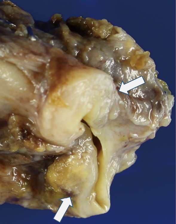
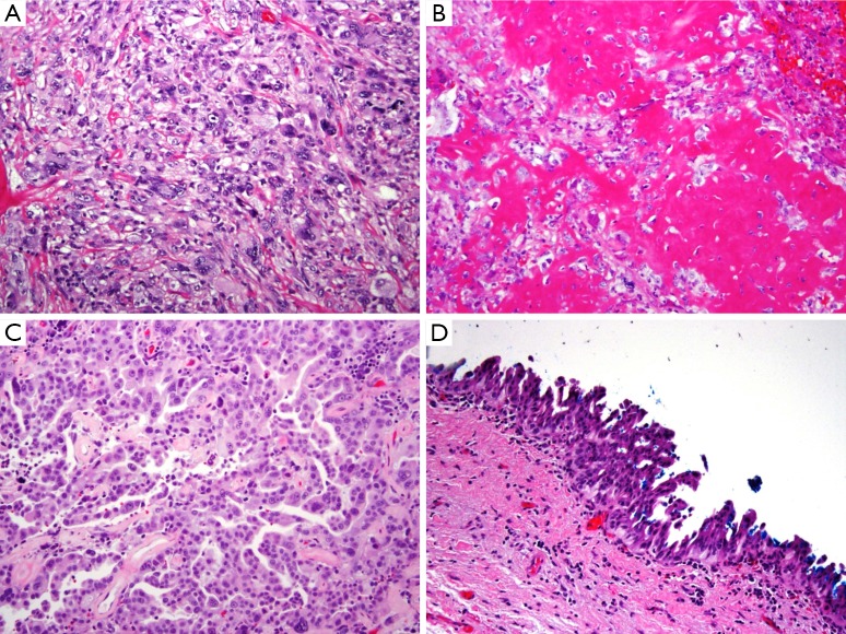
On gross examination, the tumor was primarily exophytic with masses seen within both the right and left hepatic ducts. An area of mural invasion was noted involving the right hepatic duct wall (Figure 2). Histologically, both the exophytic and murally invasive components were composed primarily of sarcomatoid elements, mostly in the form of spindle and pleomorphic tumor cells with prominent mitotic activity (Figure 3A). A minor component of the tumor showed osteogenic differentiation with formation of malignant osteoid (Figure 3B). The carcinoma component was minor, constituting about 10% of the entire tumor with morphological features of biliary adenocarcinoma and the overall architectural pattern being papillary (Figure 3C). Notably, the underlying bile duct epithelium showed foci of low and high grade dysplasia (Figure 3D).
Figure 2.

Gross specimen. Arrows illustrate the areas of mural invasion involving the right hepatic duct wall near the resection margins when the tumor specimen was bivalved.
Figure 3.
Histological features illustrating the mixed components of the tumor. (A) Both the exophytic and morally invasive components contained prominent sarcomatoid elements, in the form of spindle and pleomorphic tumor cells with prominent mitotic activity; (B) minor component of the tumor showed osteogenic differentiation with formation of malignant osteoid; (C) the carcinoma component, constituting about 10% of the entire tumor, had morphological features characteristic of biliary adenocarcinoma with tumor cells being cuboidal and the overall architectural pattern being papillary; (D) the underlying bile duct epithelium showed foci of low and high grade dysplasia. Hematoxylin and Eosin stain. Magnification: (A-C) is 100× and (D) is 40×.
Discussion
Carcinosarcomas are pathologically characterized by the presence of both epithelial and mesenchymal components within the same tumor. The most common site for carcinosarcoma of the biliary tract is the gallbladder with fewer than 100 cases reported in the literature; comparatively, there are only 7 cases of carcinosarcoma of the bile ducts reported in the English literature to date (Table 1) (6-13).
Table 1. Summary of patients’ characteristics in current series and in literature.
| Author/year | Gender/age | Presentation | Investigation/tumor markers | Location | Gross/size (mm)* | Surgery | Pathology | IHC makers (carcinomatous component) | IHC makers (sarcomatous component) | Prognosis |
|---|---|---|---|---|---|---|---|---|---|---|
| Loudet al.1997 (8) | F/35 | Obstructive jaundice | ERCP, CT, PTC | Hilar | Polypoidal/Size-NR | BD resection | Biphasic pattern of carcinomatous and sarcomatoid components | NR | NR | NR |
| Yoon et al. 2003 (9) | M/78 | Abdominal pain and obstructive jaundice | CT, PTC/tumor markers not done | Mid CBD | Infiltrative/40 | PD | Biphasic pattern of carcinomatous and sarcomatoid components merging into another,2/22 LN + ve for sarcomatoid component | Cytokeratin++; CEA+; P53+; PCNA+ | Cytokeratin++; Vimentin++; α-SMA+; NSEfocal +; P53++; PCNA++ | Died on5POD due to cardiac complications |
| Kadonoet al.2005 (7) | F/75 | Acute obstructive suppurative cholangitis | ERCP with cytology(-), PTC, CT/CEA ↑, CA19-9 ↑ | Mid CBD | Infiltrative/Size-NR | PD | Biphasic pattern of carcinomatous and sarcomatoid components with chondroid tissue, osteoid formation with focal ossification and a squamous differentiation | EMA+; Mucin-1 antigen+; Keratin+ | Vimentin+; EMA+; thrombomodulin+; α-1 antichymotrypsin+; S100+ | 2 years; died from local recurrence |
| Jang et al. 2005 (10) | F/68 | Abdominal pain, anorexia and weight loss | CT, EUS/AFP-ve, CEA-ve, CA19-9 ↑ | Distal CBD | Polypoidal/35 | PD | Biphasic pattern of predominantly sarcomatous and focally carcinomatous components, no heterologous sarcomatous components | Cytokeratin ++; CEA++ | Cytokeratin+; vimentin+ (diffuse) | 1 year disease free |
| Sodergren et al.2005 (6) | F/64 | Ill health and anorexia | US, ERCP, CT/tumor markers not done | Hilar | Polypoidal/20 | BD resection | Biphasic pattern with epithelial and mesenchymal elements with foci that resemble smooth muscle and other foci with a chondroid appearance | Cytokeratin +; EMA+; CEA + | Vimentin+; SMA+; desmin+; myoglobin+; S100+ (focal) | 5-yeardisease free |
| Aurelloet al.2008 (11) | F/73 | Obstructive jaundice | US, ERCP with cytology(+), PTC, CT/CEA ↑, CA19-9 ↑ | Distal CBD | Polypoidal/26 | PD | Biphasic pattern with heterologous differentiation ki-67 YMIB-1 (80%, uniformly distributed) | Cytokeratin AE1/AE3+; EMA+; CAM 5.2+ | Cytokeratin AE1/AE3+ (focal);EMA+ (focal); CAM 5.2+ (focal); vimentin+ (diffuse) | Alive,7 months; disease-free |
| Tanakaet al.2012 (12) | M/71 | Recurrent cholangitis | CT, ERCP | Distal CBD | Polypoidal/75 | PD | Biphasic pattern with predominant sarcomatous and a few carcinomatous components in the ductal wall and invasive front; ki-67 index (40%) | NR | Negative for epithelial andnon-epithelial markers (not specified) | 3 months; disease-free, died at 10 months |
| Lee et al. (present case) | F/91 | Fatigue and abnormal LFT | CT/AFP ↑, CEA ↑, CA19-9 ↑ | Hilar | Polypoidal/20 | BD resection | Biphasic pattern of papillary adenocarcinoma, poorly; differentiated carcinoma, spindle cell and pleomorphic sarcoma, with a heterologous element of osteoid sarcoma | NR | NR | Died at 2 months |
*, largest dimension; +/++, positive staining/strongly positive staining. PD, pancreaticoduodenectomy; CT, computed tomography; US, ultrasound; ERCP, endoscopic retrograde cholangiopancreatogram; PTC, percutaneous transhepatic cholangiogram; EUS, endoscopic ultrasound; LFT, liver function tests; CA 19-9, carbohydrate antigen 19-9; CEA, carcinoembryonic antigen; αFP, alpha-fetoprotein; BD, bile duct; CBD, common bile duct; NR, not reported; IHC, immunohistochemistry; α-SMA, α-smooth muscle actin; NSE, neuron specific enolase; PCNA, proliferating cell nuclear antigen; EMA, epithelial membrane antigen.
The pathogenesis of these rare tumors is uncertain but several hypotheses exist. It has been theorized that carcinosarcoma arises from totipotent stromal stem cells that are capable of divergent differentiation. Another postulation is the collision tumor theory that suggests the distinct and concurrent malignant proliferation of both the epithelial and mesenchymal components within the same tissue. It has also been suggested that a carcinoma can transform into a sarcoma by metaplastic transformation (14-16). A distinguishing feature of a true carcinosarcoma is the biphasic nature of the tumor with a lack of transition between the two epithelial and sarcomatous components as opposed to a poorly differentiated carcinoma with spindle cell pattern (6,14). The sarcomatous element commonly consists of undifferentiated spindle cells and a variety of heterogeneous components such as, chondro-, osteo-, leiomyo-, rhabdomyosarcoma cells (Table 1). The epithelial element usually consists of adenocarcinoma and occasionally, components such as squamous-, small cell- and undifferentiated carcinomas (17-20). Genetic analysis has been utilized to evaluate the pathogenesis of these tumors (21). They have also been sub-classified into two sub-groups: one with predominant sarcomatous differentiation or predominantly carcinomatous differentiation.
The demographics of biliary carcinosarcoma are similar to that of gallbladder adenocarcinoma, with the majority occurring in elderly women and strongly associated with cholelithiasis (22). The prognosis is dismal, largely extrapolated from the experience of gallbladder carcinosarcomas. The reported median survival time of patients with carcinosarcomas of the gallbladder after surgical resection was only 7 months with a 1-, 2-, and 3-year survival rates of 37.2%, 31.0%, and 31.0% respectively (23). The median time to recurrence for patients who died was less than 2 months (23). The TNM staging system is a valuable prognostic tool in gallbladder carcinoma but has little role in gallbladder carcinosarcoma; a recent review demonstrated that the survival time is in matter of months regardless of their tumor stage (24-29).
In general, carcinosarcomas are locally aggressive tumors with a propensity to metastasize systemically even in early stages (7,30). It been suggested that the aggressiveness of the tumor depends on the sarcomatoid component, as they metastasize to the lymph nodes and distant organs more readily. Interestingly, it is also the sarcomatoid component that forms the bulk of polypoidal element that leads to an earlier presentation as it obstructs the bile ducts, in contrast to gallbladder carcinosarcomas, where the carcinomatoid element forms the infiltrative component of the tumor (6,7,9,11,17). Kubota et al. (31) studied ki-67 in a case of gallbladder carcinosarcoma and compared it to a cohort (n=11) of patients with ordinary gallbladder adenocarcinoma: the ki-67 index in the spindle-cell component was higher than that in the epithelial component, which may account for the more aggressive biological behavior of the carcinosarcoma. This observation is consistent with other studies reporting that Ki-67 has prognostic value in various types of carcinosarcoma and other malignant tumors as well (31,32). Due to the rarity of bile duct carcinosarcoma, its true prognosis and clinicopathological features are not well established. Based on the reported cases, the survival time can vary widely, from 2 months to 5 years of disease-free survival (Table 1).
Management of all biliary malignancy, including carcinosarcoma, remains challenging, and this is particularly true for those arising from the proximal biliary tree. The strategy has largely been extrapolated from the experience in treating cholangiocarcinoma, with resection as the mainstay of treatment. As is also true for the more common biliary adenocarcinoma, there is no clear evidence that chemotherapy or radiotherapy confers any survival benefit, either as adjuvant treatment or in the palliative setting (20,25). However, judging from the poor results, it is clear that surgery alone is inadequate. Adjuvant chemotherapy has been used in carcinosarcomas of the female genital tract but with disappointing results and the role of radiation is currently unclear as well (6,7,20,30). Lumsden et al. reported improved survival with the use of intracavitary radiotherapy via a T-Tube for a case of a gallbladder carcinosarcoma (28).
This case report includes interesting radiologic and histologic features of carcinosarcoma of the bile duct. Carcinosarcomas often demonstrate unusual gross and radiographic features. Polypoid growth, which was also observed in this patient, was reported to be the characteristic gross feature of carcinosarcomas (5,7,8,33). The features of intraductal expansion, distension of the biliary confluence by the polypoidal tumor and subsequent proximal biliary tree obstruction were noted in our patient and are illustrated in Figure 1. This growth pattern is also seen in papillary cholangiocarcinoma, which is often a more indolent disease. As the present cases illustrates, the radiographic features of carcinosarcoma and papillary cholangiocarcinoma may be indistinguishable.
Conclusions
We report a case of carcinosarcoma of the bile duct with biphasic areas of papillary adenocarcinoma, poorly differentiated carcinoma intermingled with spindle cell and pleomorphic sarcoma and a heterologous element of osteoid sarcoma. To the best of our knowledge, this is the eighth case of biliary carcinosarcoma, it’s pathological and immunohistochemical characteristics define and distinguish it from a hilar cholangiocarcinoma. Complete resection is the optimal mode of treatment when possible, but the prognosis for most patients is poor; roles for chemotherapy and/or radiation therapy are undefined.
Acknowledgements
None.
Informed Consent: Written informed consent was obtained from the patient for publication of this case report and any accompanying images. A copy of the written consent is available for review by the Editor-in-Chief of this journal.
Footnotes
Conflicts of Interest: The authors have no conflicts of interest to declare.
References
- 1.Shimada K, Iwase K, Aono T, et al. Carcinosarcoma of the gallbladder producing alpha-fetoprotein and manifesting as leukocytosis with elevated serum granulocyte colony-stimulating factor: report of a case. Surg Today 2009;39:241-6. [DOI] [PubMed] [Google Scholar]
- 2.Jonson AL, Bliss RL, Truskinovsky A, et al. Clinical features and outcomes of uterine and ovarian carcinosarcoma. Gynecol Oncol 2006;100:561-4. [DOI] [PubMed] [Google Scholar]
- 3.Batsakis JG, Suarez P. Sarcomatoid carcinomas of the upper aerodigestive tracts. Adv Anat Pathol 2000;7:282-93. [DOI] [PubMed] [Google Scholar]
- 4.Zarbo RJ, Crissman JD, Venkat H, et al. Spindle-cell carcinoma of the upper aerodigestive tract mucosa. An immunohistologic and ultrastructural study of 18 biphasic tumors and comparison with seven monophasic spindle-cell tumors. Am J Surg Pathol 1986;10:741-53. [DOI] [PubMed] [Google Scholar]
- 5.Iyomasa S, Kato H, Tachimori Y, et al. Carcinosarcoma of the esophagus: a twenty-case study. Jpn J Clin Oncol 1990;20:99-106. [PubMed] [Google Scholar]
- 6.Sodergren MH, Silva MA, Read-Jones SL, et al. Carcinosarcoma of the biliary tract: two case reports and a review of the literature. Eur J Gastroenterol Hepatol 2005;17:683-5. [DOI] [PubMed] [Google Scholar]
- 7.Kadono J, Hamada N, Higashi M, et al. Carcinosarcoma of the extrahepatic bile duct. J Hepatobiliary Pancreat Surg 2005;12:328-31. [DOI] [PubMed] [Google Scholar]
- 8.Loud PA, Warshauer DM, Woosley JT, et al. Carcinosarcoma of the extrahepatic bile ducts: cholangiographic and CT appearance. Abdom Imaging 1997;22:85-6. [DOI] [PubMed] [Google Scholar]
- 9.Yoon GS, Choi DL. Sarcomatoid carcinoma of common bile duct: a case report. Hepatogastroenterology 2004;51:106-9. [PubMed] [Google Scholar]
- 10.Jang KS, Jang SH, Oh YH, et al. Sarcomatoid Carcinoma of the Distal Common Bile Duct- A Case Report. Korean J Pathol 2005;39:360-3. [Google Scholar]
- 11.Aurello P, Milione M, Dente M, et al. Synchronous carcinosarcoma of the intrapancreatic bile duct and carcinoma in situ of wirsung duct: a case report. Pancreas 2008;36:95-7. [DOI] [PubMed] [Google Scholar]
- 12.Tanaka M, Ajiki T, Matsumoto I, et al. Duodenal protrusion by carcinosarcoma of the extrahepatic bile duct. Dig Endosc 2012;24:484. [DOI] [PubMed] [Google Scholar]
- 13.Wick MR, Swanson PE. Carcinosarcomas: current perspectives and an historical review of nosological concepts. Semin Diagn Pathol 1993;10:118-27. [PubMed] [Google Scholar]
- 14.Nomura K, Aizawa S, Ushigome S. Carcinosarcoma of the liver. Arch Pathol Lab Med 2000;124:888-90. [DOI] [PubMed] [Google Scholar]
- 15.de Brito PA, Silverberg SG, Orenstein JM. Carcinosarcoma (malignant mixed müllerian (mesodermal) tumor) of the female genital tract: immunohistochemical and ultrastructural analysis of 28 cases. Hum Pathol 1993;24:132-42. [DOI] [PubMed] [Google Scholar]
- 16.Nappi O, Glasner SD, Swanson PE, et al. Biphasic and monophasic sarcomatoid carcinomas of the lung. A reappraisal of ‘carcinosarcomas’ and ‘spindle-cell carcinomas’. Am J Clin Pathol 1994;102:331-40. [DOI] [PubMed] [Google Scholar]
- 17.Sadamori H, Fujiwara H, Tanaka T, et al. Carcinosarcoma of the gallbladder manifesting as cholangitis due to hemobilia. J Gastrointest Surg 2012;16:1278-81. [DOI] [PubMed] [Google Scholar]
- 18.Kim MJ, Yu E, Ro JY, et al. Sarcomatoid carcinoma of the gallbladder with a rhabdoid tumor component. Arch Pathol Lab Med 2003;127:e406-8. [DOI] [PubMed] [Google Scholar]
- 19.Takahashi Y, Fukushima J, Fukusato T, et al. Sarcomatoid carcinoma with components of small cell carcinoma and undifferentiated carcinoma of the gallbladder. Pathol Int 2004;54:866-71. [DOI] [PubMed] [Google Scholar]
- 20.Ajiki T, Nakamura T, Fujino Y, et al. Carcinosarcoma of the gallbladder with chondroid differentiation. J Gastroenterol 2002;37:966-71. [DOI] [PubMed] [Google Scholar]
- 21.Iwaya T, Maesawa C, Tamura G, et al. Esophageal carcinosarcoma: a genetic analysis. Gastroenterology 1997;113:973-7. [DOI] [PubMed] [Google Scholar]
- 22.Born MW, Ramey WG, Ryan SF, et al. Carcinosarcoma and carcinoma of the gallbladder. Cancer 1984;53:2171-7. [DOI] [PubMed] [Google Scholar]
- 23.Okabayashi T, Sun ZL, Montgomey RA, et al. Surgical outcome of carcinosarcoma of the gall bladder: a review. World J Gastroenterol 2009;15:4877-82. [DOI] [PMC free article] [PubMed] [Google Scholar]
- 24.Pu JJ, Wu W. Gallbladder carcinosarcoma. BMJ Case Rep 2011;2011:bcr0520103009. [DOI] [PMC free article] [PubMed]
- 25.Liu KH, Yeh TS, Hwang TL, et al. Surgical management of gallbladder sarcomatoid carcinoma. World J Gastroenterol 2009;15:1876-9. [DOI] [PMC free article] [PubMed] [Google Scholar]
- 26.Fong Y, Jarnagin W, Blumgart LH. Gallbladder cancer: comparison of patients presenting initially for definitive operation with those presenting after prior noncurative intervention. Ann Surg 2000;232:557-69. [DOI] [PMC free article] [PubMed] [Google Scholar]
- 27.Oh TG, Chung MJ, Bang S, et al. Comparison of the sixth and seventh editions of the AJCC TNM classification for gallbladder cancer. J Gastrointest Surg 2013;17:925-30. [DOI] [PubMed] [Google Scholar]
- 28.Lumsden AB, Mitchell WE, Vohman MD. Carcinosarcoma of the gallbladder: a case report and review of the literature. Am Surg 1988;54:492-4. [PubMed] [Google Scholar]
- 29.Fagot H, Fabre JM, Ramos J, et al. Carcinosarcoma of the gallbladder. A case report and review of the literature. J Clin Gastroenterol 1994;18:314-6. [DOI] [PubMed] [Google Scholar]
- 30.Harris MA, Delap LM, Sengupta PS, et al. Carcinosarcoma of the ovary. Br J Cancer 2003;88:654-7. [DOI] [PMC free article] [PubMed] [Google Scholar]
- 31.Kubota K, Kakuta Y, Kawamura S, et al. Undifferentiated spindle-cell carcinoma of the gallbladder: an immunohistochemical study. J Hepatobiliary Pancreat Surg 2006;13:468-71. [DOI] [PubMed] [Google Scholar]
- 32.Ariyoshi K, Kawauchi S, Kaku T, et al. Prognostic factors in ovarian carcinosarcoma: a clinicopathological and immunohistochemical analysis of 23 cases. Histopathology 2000;37:427-36. [DOI] [PubMed] [Google Scholar]
- 33.Inoshita S, Iwashita A, Enjoji M. Carcinosarcoma of the gallbladder. Report of a case and review of the literature. Acta Pathol Jpn 1986;36:913-20. [DOI] [PubMed] [Google Scholar]


