Abstract
We demonstrate that a poly(lactide-co-glycolide) (PLG) cancer vaccine can be used in combination with immune checkpoint antibodies, anti–CTLA-4 or anti–PD-1, to enhance cytotoxic T cell (CTL) activity and induce the regression of solid B16 tumors in mice. Combination therapy obviated the need for vaccine boosting and significantly skewed intratumoral reactions toward CTL activity, resulting in the regression of B16 tumors up to 50mm2 in size and 75% survival rates. These data suggests that combining material-based cancer vaccines with checkpoint antibodies has the potential to mediate tumor regression in humans.
INTRODUCTION
Cancer accounts for almost 15% of all deaths, worldwide, and has produced consistent death rates for over 50 years1. However, recent advances in immunotherapy generate anti-tumor, cytotoxic T cells (CTLs) that may dramatically impact patient outcomes2,3. The T cell receptor complex (TCR) recognizes tumor-derived peptides bound to surface, major histocompatibility complexes (MHC) to produce specific and systemic tumor cell targeting. Cancer vaccine protocols load antigen presenting cells (APCs) with tumor-associated antigens and activate them to co-stimulate and expand anti-tumor, CTL immunity3. Clinically, vaccines have demonstrated the ability to augment T-cell reactions, and in 2010 the U.S. Food and Drug Administration approved the first, solid tumor vaccine, Provenge, for the treatment of advanced prostate cancer3,4. Importantly, immunizations typically do not produce tumor shrinkage, which limits survival benefits4. This may be due, in part, to tumor-mediated signaling of CTLA-4 and PD-1, which dampens T-cell activity2. CTLA-4 suppresses T-cell activation by blocking T-cell costimulation during TCR ligation, whereas PD-1 engagement by PD-L1 and PD-L2 (expressed by tumor cells or co-opted resident cells) limits T-cell activity within tumors by promoting anergy, death, or exhaustion2,5,6. In addition, both CTLA-4 and PD-1 are expressed by T regulatory cells (Tregs), which may reside within tumors or lymphoid tissues to further suppress T cell activation2,6,7. This knowledge of tumor and T-cell biology is the basis for antibody therapies that specifically block these immunosuppressive checkpoints. Recently, the anti–CTLA-4 antibody ipilimumab has demonstrated clinical efficacy, being the first agent to significantly prolong the overall survival of inoperable stage III/IV melanoma patients8,9. Antibodies targeting the PD-1 pathway have recently entered the clinic, and are showing dramatic effects in subsets of patients in a variety of cancer types6. Despite these successes, most treated subjects still succumb to progressive disease, indicating that vaccines or antibody therapies alone are insufficient to effect complete tumor cell killing3,4,6,9.
Approaches that utilize therapeutic vaccines to produce CTLs, while inhibiting their suppression via checkpoint antibodies, should be synergistic in amplifying T-cell immunity and effecting tumor shrinkage. We recently developed a biomaterial-based, therapeutic vaccine that has unprecedented effectiveness in maintaining sustained T-cell responses and produces tumor regression in preclinical models of melanoma and other cancers10,11. To test the effects of immune checkpoint blockade antibodies (anti–CTLA-4 and anti–PD-1) and cancer vaccines in combination, we utilized a therapeutic B16-F10 melanoma model.
MATERIALS & METHODS
Cell Lines
B16-F10 melanoma cells were obtained from American Type Culture Collection (catalog: ATCC CRL-6475) in 2010 and 2012. Upon receipt, the cells were cultured to passage 3, aliquoted and frozen in liquid nitrogen. For tumor experiments, B16-F10 cells were thawed and cultured in DMEM (Life Technologies, Inc.), containing 10% fetal bovine serum (Life Technologies, Inc.), 100 units/ml penicillin, and 100 μg/ml streptomycin. The cells were maintained at 37°C in a humidified 5% CO2/95% air atmosphere and early passage cells (between 4 and 9) were utilized for experiments.
Mice
C57BL/6 mice (6–8-week-old female; Jackson Laboratories), were cared for in accordance with the American Association for the Accreditation of Laboratory Animal Care International regulations. Experiments were all approved by the Harvard University Institutional Animal Care and Use Committee.
PLG Vaccine Fabrication
A 85:15, 120 kD copolymer of D,L-lactide and glycolide (PLG) (Alkermes, Cambridge, MA) was utilized in a gas-foaming process to form porous PLG matrices10. In brief, PLG microspheres encapsulating GM-CSF were first made using standard double emulsion10. To incorporate tumor lysates into PLG scaffolds, biopsies of B16-F10 tumors that had grown subcutaneously in the backs of C57BL/6J mice (Jackson Laboratory, Bar Harbor Maine), were digested in collagenase (250 U/ml) (Worthington, Lakewood, NJ) and suspended at a concentration equivalent to 107 cells per ml after filtration through 40 μm cell strainers. The tumor cell suspension was subjected to 4 cycles of rapid freeze in liquid nitrogen and thaw (37°C) and then centrifuged at 400 rpm for 10 min. The supernatant (1ml) containing tumor lysates was collected and lyophilized. To incorporate CpG-oligodeoxynucleotides (ODNs) into PLG scaffolds, CpG-ODN 1826, 5′-tcc atg acg ttc ctg acg tt-3′, (Invivogen, San Diego, CA) was first condensed with poly(ethylenimine) (PEI, Mn ~60,000, Sigma Aldrich) by dropping CpG-ODN 1826 solutions into a PEI solution, while vortexing the mixture11. The charge ratio between PEI and CpG-ODN (NH3+:PO4−) was kept constant at 7 during condensation. The condensate solutions were then vortexed with 60 μl of 50% (wt/vol) sucrose solution, lyophilized and mixed with dry sucrose to a final weight of 150 mg.
PLG microspheres were then mixed with the sucrose containing PEI-CpG-ODN condensate and tumor lysate and compression molded. The resulting disc was allowed to equilibrate within a high-pressure CO2 environment, and a rapid reduction in pressure causes the polymer particles to expand and fuse into an interconnected structure42. The sucrose was leached from the scaffolds by immersion in water, yielding scaffolds that were 80–90% porous.
Vaccine Assays
For therapeutic vaccination, animals were challenged with a subcutaneous injection of 105 B16-F10 melanoma cells (ATCC, Manassas, NJ) in the back of the neck. At day 3 a subset of mice were treated with 100 μg of either anti-CTLA-4 (9D9), or anti-PD-1 (RMP1-14) (Bioxcell, West Lebabnon, NH), or both antibodies and treatment was repeated every three days for thirty days. At day 9 after tumor challenge, PLG vaccines with melanoma tumor lysates and GM-CSF in combination with CpG-ODN were implanted subcutaneously into the lower left flank of C57BL/6J mice. Animals were monitored for the onset of tumor growth (approximately 1mm3) and sacrificed for humane reasons when tumors grew to 20–25 mm (longest diameter).
T cell infiltration and activation
PLG vaccines were excised at day 14 and the ingrown tissue was digested into single cell suspensions using a collagenase solution (Worthington, 250 U/ml) that was agitated at 37°C for 45 minutes. CTLA-4 and PD-1 antibody therapy for these experiments were administered at the same timepoints as the vaccination experiments and terminated with vaccine explantation. The cell suspensions were then poured through a 40μm cell strainer to isolate cells from scaffold particles and the cells were pelleted and washed with cold PBS and counted using a Z2 coulter counter (Beckman Coulter). The inguinal lymph nodes draining the vaccine sites were also excised and processed into single cell suspensions. On the indicated days, B16-F10 tumors were also removed from mice, and digested in 1 mg/mL collagenase II (250 U/ml) (Worthington, Lakewood, NJ) and 0.1 mg/mL DNase for 1 hour at 37°C, and dissociated cells were filtered through a 40-μm filter. Negative T-cell separation was performed using a murine, pan T cell separation kit (Miltenyi Biotec, San Diego, CA), which primarily removes innate immune cells and APCs along with debris and necrotic cells from suspension.
To assess T cells isolated from the vaccine site, draining lymph node, and tumors, isolated cells were directly stained with antibodies for phenotype characterization by fluorescence-activated cell sorting (FACS) analysis. PE-Cy7 conjugated CD3 stains were performed in conjunction with APC-conjugated CD8a (CD8 T cells), FITC-conjugated CD4 (CD4 Tcells) and PE-conjugated FoxP3 (Tregs) and analyzed with flow cytometry. Tumor infiltrating leukocytes were also costained FITC-anti-IFNγ, and PE-anti-CD107a for analysis of these activation markers. All antibodies and the FoxP3 antibody staining kit were obtained from eBioscience, San Diego, CA. Cells were gated according to single positive FITC, APC and PE stainings, using isotype controls. The percentage of cells staining positive for each surface antigen was recorded.
Statistical analysis
All values in the present study were expressed as mean ± S.D. Statistical significance of differences between the groups were analyzed by a two-tailed, Student’s t test and a P value of less than 0.05 was considered significant.
RESULTS
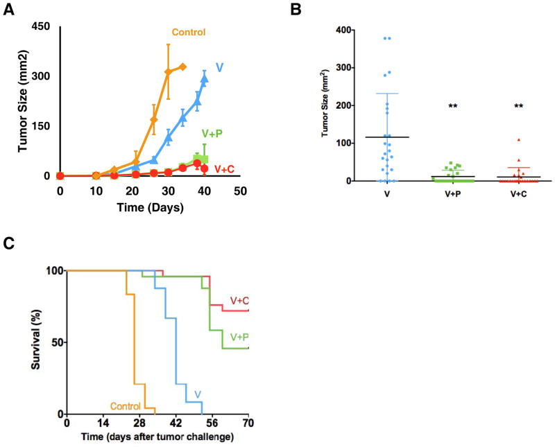
PLG matrices were fabricated, as described10, to coordinate the recruitment and anti-tumor programming of dendritic cells via the controlled presentation of tumor lysates with GM-CSF and CpG-rich ODN (PLG vaccines). In mice bearing 3-day-old B16 melanoma tumors (5×10⁵ cells), treatment with CTLA-4 and PD-1 antibodies alone have no effect on tumor progression and survival outcomes in these animals (Supplementary Fig. S1). Moreover, a single PLG vaccination modestly suppressed tumor progression but did not effect long-term survival in any mice bearing B16 melanoma tumors (Fig. 1). Strikingly, treatment with CTLA-4 and PD-1 antibodies at day 3 combined with a single PLG vaccination at day 9 significantly reduced the rate of tumor progression and resulted in long-term survival rates of 75% and 40%, respectively, with complete tumor regression in the surviving mice (Fig. 1A–C). Tumors were pretreated with antibody blockade prior to vaccination because this sequence likely reflects the clinical setting where these antibodies are becoming standards of care. If antibody administration is ceased after vaccination, the effects on tumor inhibition are lost (Supplementary Fig. S2), suggesting that blockade treatment significantly augments the subsequent T cell responses induced by vaccination.
Figure 1. Tumor protection induced by therapeutic PLG vaccination in combination with blockade antibodies.
(A) A comparison of the tumor growth of untreated mice (Control) bearing established melanoma tumors (inoculated with 5×10⁵ B16-F10 cells and allowed to develop for 9 days) and mice treated after tumor challenge with PLG vaccines (V) or PLG vaccines in combination with antibodies to PD-1 (V+P) or CTLA-4 (V+C). (B) Dot plot of the tumor size in untreated mice (control), and mice treated with PLG vaccines (V) or PLG vaccines in combination with antibodies to PD-1 (V+P) or CTLA-4 (V+C) at 30 days after tumor challenge. (C) A comparison of the survival of untreated mice (Control) and mice treated with PLG vaccines (V) or PLG vaccines in combination with antibodies to PD-1 (V+P) or CTLA-4 (V+C) after tumor challenge. The antibody treatments were initiated on day 3 and injected i.p. every 3 days for 30 days after tumor challenge. Values in A (n=24) represent mean and SEM. ** P<0.01 as the vaccine compared to other experimental conditions (V vs Control, V vs V+P, V vs V+C) unless otherwise noted. This experiment was performed three times (n=8; N=24 in total) and results were pooled together.
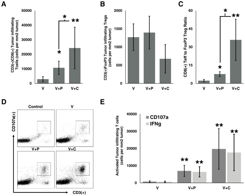
Indeed, combining blockade antibodies with PLG vaccination significantly skewed the tumor infiltrating leukocyte (TIL) response toward active, cytotoxic T cells, relative to suppressive Tregs (Fig. 2 & S2). This is consistent with the finding of tumor regression, as higher CD8/Treg ratios within tumors are indicative of effective vaccination3,9. PLG vaccination at day 9 after tumor challenge induced significant CD3+CD8+ T cell infiltration into 20-day-old B16 tumors, resulting in approximately 3,000 cytotoxic T cells per mm2 of tumor (Fig. 2A). The addition of anti-PD-1 treatment to vaccination provided a 3.7-fold increase in tumor infiltrating CD3+CD8+ T cells, whereas the addition of anti-CTLA-4 therapy produced over 24,000 CD3+CD8+ cytotoxic T cells per mm2 of tumor (Fig. 2A). In contrast, these treatment groups had no effect on the numbers of tumor-resident CD4+FoxP3+ Tregs (Fig. 2B). The intratumoral ratio of CD8+ effectors to Tregs at day 18 tripled with PD-1 antibody administration compared to vaccination alone (Fig. 2C). Combining anti-CTLA-4 with vaccination resulted in a 15-fold increase in the Teff/Treg ratio compared to vaccination alone at day 18 (35.9 to 2.3; Fig. 2C). The same analysis was conducted at day 30 after tumor challenge, and only immunizations combined with anti-CTLA-4 were able to generate significant CD8+/Treg ratios (approximately 6-fold increase; Supplementary Fig. S3) consistent with the long-term survival data. In addition, supplementing vaccination with PD-1 or CTLA-4 antibody therapy resulted in 3-fold and 8-fold increases in intratumoral, cytotoxic T cell activation, respectively, as determined by CD107a and IFN-γ co-expression (Fig. 2, D and E). The addition of checkpoint blockade enhanced not only the density of activated, CD8+ TILs but also the percentage of total CD8+ T cells that were activated (Fig 2, D and E), indicating that these treatments promoted T-cell cytotoxicity locally, within tumors.
Figure 2. Engineered PLG vaccine in combination with blockade antibodies enhances intratumoral T effector cell activity.
(A) The total number of CD3+CD8+ T cells and (B) CD3+FoxP3+ T regulatory cells isolated from the B16 tumors of untreated mice (Control) and mice treated with PLG vaccines alone (Vax) or in combination with a antibodies to PD-1 (+PD-1) and CTLA-4 (+CTLA4). (C) The ratio of CD3+CD8+ T cells to CD3+FoxP3+ T regulatory cells isolated from the B16 tumors of untreated mice (Control) and mice treated with PLG vaccines alone (Vax) or in combination with antibodies to PD-1 (+PD-1) and CTLA-4 (+CTLA-4). (D) FACS plots representing tumor infiltrating leukocytes in tumors of untreated mice (Control) and mice treated with PLG vaccines alone (Vax) or in combination with antibodies to PD-1 (+PD-1) and CTLA-4 (+CTLA4). Single cell suspensions were prepared from tumors at day 18 and stained for activated cytotoxic T-cell markers, CD8 and CD107a. Numbers in FACS plots indicate the percentage of the cell population positive for both markers. (E) The numbers of CD8+, tumor-infiltrating T cells positive for either IFNγ or CD107a in blank matrices (Control), PLG vaccines alone or vaccines in combination with antibodies to PD-1 and CTLA-4. The antibody treatments were administered i.p. as described for vaccination experiments until tumor excision at day 18 for Tcell infiltration analysis. Vaccination was initiated 9 days after tumor challenge. All cellular staining was performed following MACS cell separation of total cells suspensions extracted from tumors. Values in A, B, C, and D (n=10) represent mean and standard deviation. * P<0.05 ** P<0.01 as compared to the vaccine alone (V vs V+P; V vs V+C) unless otherwise noted. This experiment was performed twice.
Combining vaccination with both CTLA-4 and PD-1 antibodies initially suppressed tumor growth over single antibody combinations with PLG vaccines (Supplemental Fig. S4A). Tumor inhibition coincided with increased cytotoxic T cell-activity relative to vaccine augmentation with a single antibody (Supplementary Fig. S4B–D). However, this combined checkpoint inhibition with vaccination did not significantly affect long-term survival in comparison to the addition of one blockade (data not shown). These data suggest that combining PLG vaccines with either CTLA-4 or PD-1 antibodies can dramatically enhance the numbers of activated, effector T cells within tumors, which likely perform tumor cell killing to enhance survival in these animals.
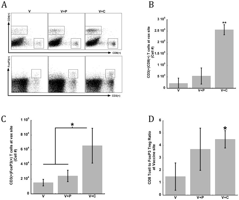
CTLA-4 and PD-1 antibody treatment also enriched the vaccine site with CD4+ and CD8+ T cells at day 14 after implantation (Fig. 3A). CTLA-4 antibody treatment combined with vaccination significantly increased the local numbers of CD8+ cytotoxic T cells by 5 and 10 fold over PD-1 addition, or vaccines alone (Fig. 3B). CTLA-4 blockade also enhanced T regulatory cell activity within vaccines relative to PD-1 combination or vaccination alone (Fig. 3C). Importantly, the cytotoxic T cell to T reg ratio was approximately 3-fold greater with antibody addition in comparison to vaccine treatments alone (Fig. 3D). In addition, increased CD8+ T cell numbers and reduced FoxP3+ T reg numbers were found within the vaccine-draining inguinal lymph nodes with combination treatment of CTLA-4 and vaccines, in contrast to PD-1 addition or vaccines alone (Supplementary Fig. S5).
Figure 3. Engineered PLG vaccine in combination with CTLA-4 maintains local T cell activity at vaccine site.
The T cell infiltration into PLG vaccines implanted in mice for 14 days. (A) FACs histograms of CD4+, CD8+ and CD4+FoxP3+ T cell infiltrates isolated from PLG implants in mice treated with PLG vaccines alone (V) or in combination with PD-1 antibody (V+P) or anti-CTLA-4 antibody (V+C). The numbers of (B) CD3+CD8+ T cells, (C) CD4+FoxP3+ Tregs and (D) The ratio of CD3+CD8+ effector T cells to CD4+FoxP3+ T cells isolated from the vaccine site in mice treated with PLG vaccines alone (V) or in combination with PD-1 antibody (V+P) or anti-CTLA-4 antibody (V+C). The antibody treatments were administered i.p. as described for vaccination experiments until vaccine explantation. CD8/Treg ratio was calculated from cell numbers. Values represent mean and standard deviation (n=5). * P<0.05 ** P<0.01 as vaccine alone compared to to all other experimental conditions (V vs V+P; V vs V+C) unless otherwise noted. This experiment was performed once.
DISCUSSION
In summary, this study indicates that combination therapy of antibodies and PLG vaccines can enhance anti-tumor CTL activation and promote significant tumor regression. Single PLG vaccination is able to produce significant numbers of activated TILs, but these cells fail to shrink tumors unless this response is amplified by either CTLA-4 or PD-1 signaling. Combination therapy produced effector TIL numbers that comprised up to 25% of all tumor cells.
In contrast to previous studies utilizing checkpoint blockade treatment alone or in combination with vaccines12,13,14, antibody therapy administered with vaccination in this study had no effect on Treg populations at the vaccine site or in tumors. In contrast, a recent study demonstrated that combining anti-CTLA-4 with Gvax can deplete Treg numbers in a tumor microenvironment and this is dependent on the presence of tumor infiltrating, Fc receptor–expressing macrophages14. Thus, observations of Treg depletion is associated with cellular and molecular changes in the tumor microenvironment that may be modulated by the vaccine strategy. Notably, the PLG vaccine used in this study has been optimized to control the presentation of GM-CSF and adjuvants, relative to other vaccine formulations (eg. Gvax), in order to enhance T effector activity and downregulate Tregs and other immunosuppressive mechanisms induced by GM-CSF11,14. Therefore, the PLG vaccine likely creates a tumor and vaccine microenvironment that responds to anti-CTLA-4 by preferentially enhancing effector T cell generation and expansion over Tregs. Further, in vitro studies investigating the functional capacity and antigen specificity of effector and regulatory T cells should be performed to clarify their activation state and the mechanisms by which inhibitors synergize with PLG vaccination. Experiments in other, more immunosuppressive, tumor models may further elucidate the effects of blockade inhibitors on PLG vaccine-induced Tregs.
Interestingly, vaccination with anti-CTLA-4 and not anti-PD-1 increased CD8+ effector cell activity at the vaccine sites and within lymph nodes, consistent with enhanced costimulation by resident APCs. Additionally, immunization supplemented with anti-PD-1 conferred significant effector T cell activation only within tumors, and not at the scaffold site, suggesting that PD-1 activity predominates within the B16 tumor environment relative to the vaccine site. Further work, including an assessment of PD-1+ and CTLA-4+ T cells at the sites of vaccination and tumors should be performed to elucidate the superior effect of CTLA-4 inhibition over PD-1. These data have significant clinical relevance, since the PLG system, designated WDvax, is currently in phase I testing for the treatment of metastatic melanoma (clinicaltrials.gov ID-NCT01753089), and suggest that vaccinations be utilized in combination with blockade antibodies.
Supplementary Material
Acknowledgments
GRANT SUPPORT
This work was supported by grant funding from the National Institutes of Health and the National Institute of Biomedical Imaging and Bioengineering (R01 EB015498) and internal funding from the Wyss Institute for Bioinspired Engineering.
References
- 1.Global Cancer: Facts and Figures. 2. American Cancer Society Report; 2011. pp. 1–60. [Google Scholar]
- 2.Pardoll DM. The blockade of immune checkpoints in cancer immunotherapy. Nat Review Cancer. 2012:1–13. doi: 10.1038/nrc3239. [DOI] [PMC free article] [PubMed] [Google Scholar]
- 3.Dougan M, Dranoff G. Immune Therapy for Cancer. Annu Rev Immunol. 2009;27:83–117. doi: 10.1146/annurev.immunol.021908.132544. [DOI] [PubMed] [Google Scholar]
- 4.Kantoff PW, Higano CS, Shore ND, Berger ER, Small EJ, Penson DF, et al. Sipuleucel-T immunotherapy for castration-resistant prostate cancer. N Engl J Med. 2010;363:411–422. doi: 10.1056/NEJMoa1001294. [DOI] [PubMed] [Google Scholar]
- 5.Korman AJ, Peggs KS, Allison JP. Checkpoint blockade in cancer immunotherapy. Adv Immunol. 2006;90:297–339. doi: 10.1016/S0065-2776(06)90008-X. [DOI] [PMC free article] [PubMed] [Google Scholar]
- 6.Ferris R. PD-1 targeting in cancer immunotherapy. Cancer. 2013;119:E1–3. doi: 10.1002/cncr.27832. [DOI] [PMC free article] [PubMed] [Google Scholar]
- 7.Peggs KS, Quezada SA, Chambers CA, Korman AJ, Allison JP. Blockade of CTLA-4 on both effector and regulatory T cell compartments contributes to the antitumor activity of anti-CTLA-4 antibodies. J of Exp Med. 2009;206:1717–1725. doi: 10.1084/jem.20082492. [DOI] [PMC free article] [PubMed] [Google Scholar]
- 8.Wolchok JD, Hodi FS, Weber JS, Allison JP, Urba WJ, Robert C, et al. Development of ipilimumab: a novel immunotherapeutic approach for the treatment of advanced melanoma. Ann NY Acad Sci. 2013;1291:1–13. doi: 10.1111/nyas.12180. [DOI] [PMC free article] [PubMed] [Google Scholar]
- 9.Hodi FS, O’Day SJ, McDermott DF, Weber RW, Sosman JA, Haanen JB, et al. Improved survival with ipilimumab in patients with metastatic melanoma. N Engl J Med. 2010;363:711–723. doi: 10.1056/NEJMoa1003466. [DOI] [PMC free article] [PubMed] [Google Scholar]
- 10.Ali OA, Huebsch N, Cao L, Dranoff G, Mooney DJ. Infection-mimicking materials to program dendritic cells in situ. Nat Mat. 2009;2009:1–8. doi: 10.1038/nmat2357. [DOI] [PMC free article] [PubMed] [Google Scholar]
- 11.Ali OA, Emerich D, Dranoff G, Mooney DJ. In Situ Regulation of DC Subsets and T Cells Mediates Tumor Regression in Mice. Science Trans Med. 2009;1:8ra19–8ra19. doi: 10.1126/scitranslmed.3000359. [DOI] [PMC free article] [PubMed] [Google Scholar]
- 12.Duraiswamy J, Kaluza KM, Freeman GJ, Coukos G. Dual blockade of PD-1 and CTLA-4 combined with tumor vaccine effectively restores T-cell rejection function in tumors--response. Cancer Res. 2013;73(12):3591–603. doi: 10.1158/0008-5472.CAN-12-4100. [DOI] [PMC free article] [PubMed] [Google Scholar]
- 13.Curran MA, Montalvo W, Yagita H, Allison JP. PD-1 and CTLA-4 combination blockade expands infiltrating T cells and reduces regulatory T and myeloid cells within B16 melanoma tumors. PNAS. 2010;107:4275–4280. doi: 10.1073/pnas.0915174107. [DOI] [PMC free article] [PubMed] [Google Scholar]
- 14.Simpson TR, Montaivo-Ortiz W, Sepilveda MA, Bergerhoff K, Arce F, Roddie C, et al. Fc-dependent depletion of tumor-infiltrating regulatory T cells co-defines the efficacy of anti-CTLA-4 therapy against melanoma. J Exp Med. 2013;210(9):1695–710. doi: 10.1084/jem.20130579. [DOI] [PMC free article] [PubMed] [Google Scholar]
- 15.Ali OA, Verbeke C, Johnson C, Sands RW, Lewin SA, White D, et al. Identification of immune factors regulating anti-tumor immunity using polymeric vaccines with multiple adjuvants. Cancer Res. 74(6):1670–81. doi: 10.1158/0008-5472.CAN-13-0777. [DOI] [PMC free article] [PubMed] [Google Scholar]
Associated Data
This section collects any data citations, data availability statements, or supplementary materials included in this article.


