Abstract
Background
Crimean-Congo hemorrhagic fever virus (CCHFV) is a member of the nairovirus, a genus in the Bunyaviridae family, which causes a life threatening disease in human. Currently, there is no vaccine against CCHFV and detailed structural analysis of CCHFV proteins remains undefined. The CCHFV M RNA segment encodes two viral surface glycoproteins known as Gn and Gc. Viral glycoproteins can be considered as key targets for vaccine development.
Objectives
The current study aimed to investigate structural bioinformatics of CCHFV Gn protein and design a construct to make a recombinant bacmid to express by baculovirus system.
Materials and Methods
To express the Gn protein in insect cells that can be used as antigen in animal model vaccine studies. Bioinformatic analysis of CCHFV Gn protein was performed and designed a construct and cloned into pFastBacHTb vector and a recombinant Gn-bacmid was generated by Bac to Bac system.
Results
Primary, secondary, and 3D structure of CCHFV Gn were obtained and PCR reaction with M13 forward and reverse primers confirmed the generation of recombinant bacmid DNA harboring Gn coding region under polyhedron promoter.
Conclusions
Characterization of the detailed structure of CCHFV Gn by bioinformatics software provides the basis for development of new experiments and construction of a recombinant bacmid harboring CCHFV Gn, which is valuable for designing a recombinant vaccine against deadly pathogens like CCHFV.
Keywords: Hemorrhagic Fever Virus, Crimean-Congo, Glycoprotein, Viral Proteins, Bioinformatics, Baculovirus
1. Background
Crimean-Congo hemorrhagic fever virus (CCHFV) causes severe hemorrhagic symptoms in human with high mortality rate, approximately 30% (1, 2). The virus can be transmitted to human through the bites of infected ticks, direct contact with virus contaminated tissues or blood, or nosocomial infections. Incubation period is between 2 to 7 days, and patients show flu-like symptoms and then hemorrhagic state starts with bleeding from the mucous membranes and development of petechiae, associated with thrombocytopenia and leucopenia (3, 4). The ability of virus to cause nosocomial infections, make CCHFV a major public health concern (5). CCHFV is an arthropod virus that belongs to the family Bunyaviridae. Family Bunyaviridae comprises five genera: Orthobunyavirus, Hantavirus, Phlebovirus, Nairovirus, and Tospovirus. CCHFV, like all viruses in this family, has segmented negative-strand RNA genome. Small RNA segment (S) encodes a nucleocapsid (N) protein; medium RNA segment (M) encodes 2 membrane glycoproteins named Gn and Gc; and the large RNA segment (L) encodes an RNA dependent RNA polymerase. The glycoproteins are derived from a polyprotein by a proteolysis cleavage that leads to 2 structural glycoproteins known as Gn and Gc (6).
CCHFV has been reported in more than 30 countries in Europe, Asia, and Africa. In Iran, CCHFV is an important health problem that is endemic in southeast region, Sistan-Baluchistan Province but sporadic cases occur in other provinces such as Fars, Isfahan, Kerman, Yazd and etc. Many people are infected with the virus through different routes of transmission and unfortunately some of them die (7-9). Members of the genus Hyalloma tick have been linked to the natural infection cycle of CCHFV. Ticks population survey in Iran has been shown that Hyalloma spp. are distributed in all parts of Iran and CCHFV has been isolated from ticks (10-12). Since mortality rate of CCHFV disease is very high, infection with this virus is a major threat for public health, especially when a safe vaccine is not available yet and antiviral chemotherapy (ribavirin) could not efficiently treat this illness (8, 13). So any effort to develop a vaccine against CCHFV has high importance.
Baculovirus is an enveloped virus with double stranded circular DNA of 88 - 180kb as viral genome that packaged within rod-shaped nucleocapsids. Safety, high level expression of protein and posttranslational modification of expressed protein are advantages of baculovirus as an eukaryotic expression system. Recently engineered baculovirus vectors are developed which allows the propagation of virus genomes in Escherichia coli as a bacmid. Several research groups have expressed many microorganism genes in baculovirus expression system (14-16). Since interaction of CCHFV with its receptor is through its surface glycoproteins (Gn and Gc) and neutralizing antibodies are stimulated against these proteins, these surface proteins are important candidates for developing a potential vaccine (17). Prior to cloning and production of recombinant protein, the sequence, structure, and similarity of target protein must be identified. Bioinformatics analysis has opened new insights into protein sequence and structural features.
2. Objectives
We decided to study the structure of CCHFV Gn protein and generate a recombinant DNA bacmid harboring CCHFV Gn gene that can be expressed in insect cells by baculovirus expression system. Following expression, the recombinant Gn protein of CCHFV could be used as a vaccine against CCHFV or for laboratory diagnosis of this viral agent.
3. Materials and Methods
3.1. Analysis of the Nucleotide Sequence of CCHFV M Segment
We retrieved the sequence of M segment of CCHFV from national center for biotechnology institute (NCBI) database, and BLASTN analysis was performed based on that.
3.2. Phylogenetic Analysis
To study CCHFV genetic diversity, we performed phylogenetic analysis by using MEGA software (version 6). A set of different strains of complete CCHFV M segment sequences were retrieved from NCBI and were aligned using CLUSTAL algorithm at the amino acid levels. Nucleotide phylogenetic trees were also investigated using neighbor-joining method.
3.3. Primary Structural Analysis
The amino acid sequence of Gn protein of CCHFV was retrieved from the NCBI database. ExPASy ProtParam server has been applied for the study of physicochemical characterization like theoretical isoelectric point (PI), molecular weight, and molecular formula, total number of positive and negative residues, instability index, extinction coefficient, aliphatic index, and grand average of hydropathy (GRAVY).
3.4. Secondary Structural Analysis
We applied a new highly accurate secondary structure prediction method, PSIPRED, (available at http://bioinf.cs.ucl.ac.uk/psipred) for computation of secondary structural features of CCHFV Gn protein sequences.
3.5. Protein 3D Structure
I-TASSER is a hierarchical protein structure modeling approach based on the second-structure enhanced profile-profile threading alignment, not homology modeling. 3D model of Gn protein of CCHFV strain accession number: DQ446216.1 was generated using I-TASSER (http://zhanglab.ccmb.med.umich.edu/I-TASSER), a web based server. High C-score and correlation between C-score and TM-score of model determine the best model. The C-score is a confidence score for estimating the quality of predicted models by I-TASSER. It is calculated based on the significance of threading template alignments and the convergence parameters of the structure assembly simulations. The C-score is typically in the range of -5 to 2, where a C-score of higher value signifies a model with a high confidence and vice versa.
3.6. Designing the Construct and Addition of V5 tag to Gn
After selection of Gn coding sequence, the sequence was evaluated by on line RestrictionMapper software version 3 (available at http://www.restrictionmapper.org), to select appropriate restriction enzymes. For directional cloning, we designed 2 enzyme recognition sites for Bam HI and Xho I (Fermentas, Lithuania) on the 5’ and 3’ end of the construct, respectively. Owning to the lack of monoclonal antibody against Gn, we added V5 tag to C-terminal of Gn for western blot analysis.
3.7. Codon Optimization and Synthesis of the Construct
To obtain high level of protein expression, the codons of Gn sequence were optimized for expression in insect cells (Sf9 cells). The construct was synthesized by Bioneer (Seoul, South Korea) and delivered in pGEM-B2 vector (Bioneer, South Korea), named pGEM-B2-Gn.
3.8. Amplification of pGEM-B2-Gn
To amplify the construct harboring our interest gene, we transformed E. coli strain TOP10 (Invitrogen, Carlsbad, CA, USA) with pGEM-B2-Gn plasmid by calcium chloride method. Briefly, E. coli TOP10 was cultivated in Lysogeny broth (LB), containing 10 mg/mL ampicillin at 37°C in a shaker incubator for 18 hours, to transform competent cells. Two microliter of pGEM-B2-Gn was added to 100 µL of competent cells and following a brief vortex, the mixture was placed on ice for 40 minutes after vortex and spin. Then, it was heat shocked at 42°C for 90 seconds and immediately placed on ice for 5 minutes. Next, 1 mL of LB, antibiotic free medium, was added to the transformed cells incubated at 37°C for 2 hours. After incubation, the transformed cells harboring pGEM-B2-Gn plasmid were plated and incubated at 37°C on LB agar plates, containing 10 mg/mL ampicillin and tetracycline overnight.
3.9. Plasmid Purification
Cells harboring the recombinant plasmid were cultured in antibiotic containing LB medium for 18 hours at 37°C in a shaker incubator. Miniprep kit (Bioneer, South Korea) was used to purify plasmid from E. coli TOP10, according to the manufacturer’s instructions. Briefly, 5 mL of bacterial culture was harvested and lysed. Then, the lysate was cleared by centrifugation and poured onto the silica column. The DNA molecules selectively bind to silica particle of the column. The plasmid DNA was eluted in 20 μL of distilled water. Plasmid DNA concentrations were determined by absorbance at 260 nm using picodrop spectrophotometer (Picodrop, UK).
3.10. Enzyme Digestion of pGEM-B2-Gn
With regard to the presence of Bam HI and Xho I restriction sites on plasmids extracted from transformed colonies, the digestion reaction was carried out to release the fragment of interest. Each 20 μL digestion reaction contained 10 μL of plasmid, 1 µL of each restriction enzyme, 2 μL of 10X buffer, and 6 μL of dH2O. Digestion was performed by incubation at 37°C for 2 hours and digestion products were analyzed by electrophoresis on 1% agarose gel.
3.11. Gel extraction of Digested Products
The specific fragment containing desired gene were purified from the agarose gel by accuprep gel purification kit (Bioneer, South Korea) based on manufacturers’ recommendations. Pure DNA was eluted with a small volume of low salt buffer provided by the kit.
3.12. Ligation Into pFastBacHTb Vector
Purified fragments from agarose gel were ligated into pFastBacHTb vector (Invitrogen, Carlsbad, CA, USA) according to manufacturer’s instructions. The molar ratio of vector to insert was 1:3. Ligation reaction was set up with 10 μL volume containing 1 μL pFastBacHTb plasmid, 3 μL of purified fragment, 1 μL of T4 DNA ligase enzyme, 2 μL of 5X buffer, and 3 μL of nuclease free distilled water. After gentle mix and a brief centrifugation, the ligation reaction mixture was incubated at 8°C overnight. The resulting plasmid was named pFastBac-Gn.
3.13. Enzyme Digestion and Sequencing of the pFastBacHTb-Gn
To confirm and determine the presence and size of inserts in pFastBacHTb-Gn, recombinant plasmid was simultaneously digested with 2 enzymes BamH I and Xho I, then the digestion products were loaded on 1% agarose gel and electrophoresed for about an hour. Also to verify authenticity of the fragments and correct in-frame insertion of construct in pFastBacHTb vector, sequencing in the forward and reverse directions was performed. Gene runner v3.05 (Hastings Software, Inc. USA) was used for sequencing data assembly and analysis. Recombinant vectors were stored at -20°C until transformation.
3.14. Generation of Recombinant Bacmid DNA Blue-White Screening
Preparation of competent cells from E. coli strain DH10 Bac was performed by calcium chloride method (18). E. coli DH10 Bac was cultivated in LB, containing 10 mg/mL tetracycline and 10 mg/mL kanamycin in a shaker incubator for 18 hours. Cells harboring pFastBac-Gn plasmid were plated and grown at 37°C on LB agar plates (10 g NaCl, 5 g yeast extract, 10 g bacto-tryptone) with kanamycin, tetracycline both containing 10 mg/mL, 7 mg/mL gentamicin, 100 mM IPTG (Fermentas, Lithuania), and 20 mg/mL X-Gal (Fermentas, Lithuania) for 48 hours. For blue-white screening after overnight incubation, plates were placed at 4°C for 2 hours and cells from white colonies were harvested and cultured on antibiotic containing LB agar plates. After 16 hours incubation at 37°C, cells harboring the recombinant plasmids grew up.
3.15. Bacmid Purification and PCR Amplification
White colonies in LB agar were transferred to LB broth, containing 10 mg/mL kanamycin, tetracycline, and 7 mg/mL gentamicin and incubated for 18 hours in shaker incubator, and then high molecular weight bacmid was extracted with laboratory-made solutions based on Invitrogen protocol (tools.lifetechnologies.com/content/sfs/manuals/bactobac_man.pdf). Successful recombination was verified by PCR analysis using M13 forward 5’-GTTTTCCCAGTCACGAC-3’ and M13 reverse 5’-CAGGAAACAGCTATGAC-3’. PCR reaction was carried out with M13 forward and reveres primers in a total volume of 25 μL mixture, containing 5 μL of extracted bacmid, 5 μL of 10X PCR buffer with MgCl2, 1 μL of 20 μM of each primer, 2 μL of 10 mM dNTPs mix, and 1 U/μL Taq DNA polymerase (Fermentas, Lithuania). PCR program comprised an initial denaturation at 93°C for 3 minutes; 94°C for 45 seconds, 55°C for 45 seconds, 72°C for 5 minutes (35 cycles), and finally the reaction was held at 4˚C. PCR products were electrophoresed on 1% agarose gel. To ensure of the intact structure of bacmid, it was electrophoresed on 0.7% agarose for 18 hours.
4. Results
4.1. Blast Search and Multiple Sequence Alignments
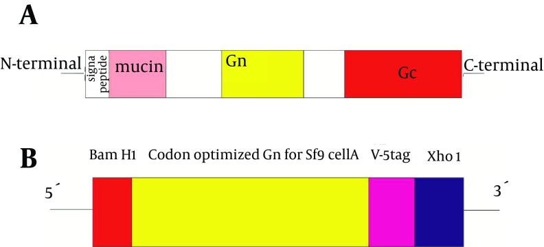
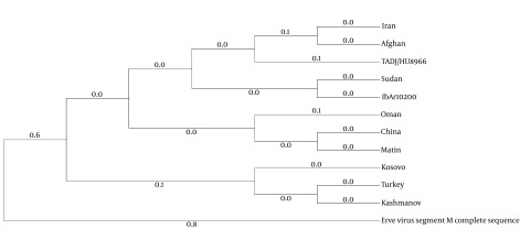
Sequence analysis of CCHFV M segment was performed and the region related to Gn was selected and used for designing the construct (Figure 1). The result of phylogenetic analysis among M RNA genome of the CCHFV strains is shown in Figure 2. Multiple Sequence alignments were built with CLUSTAL algorithm (as implemented in the MEGA package version 6) at the amino acid level for the M segment and neighbor-joining phylogenetic tree was constructed.
Figure 1. Schematic Representation of CCHFV Full Length Glycoprotein Precursor.
A, Our synthetic construct and its cloning sites and tags B, Figures not to scale.
Figure 2. Phylogenetic Tree Based on CCHFV M Segment Alignment.
CCHFV strains were presented with regard to the country of origin.
4.2. Structural Description of CCHFV Gn
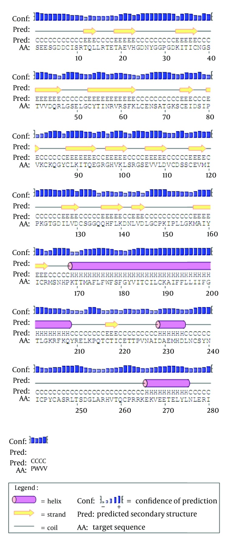
Physicochemical parameters of the CCHFV Gn protein were shown in Table 1. Secondary structural features are shown in Figure 3. The results showed that all residues lie under coil, strand, and alpha helix. Tertiary structure of CCHFV Gn was predicted using I-TASSER Server as shown in Figure 4. I-TASSER result provided the best model with high C-score and high correlation between C-score and TM-score of model 1 (0.91). Different C-scores, cluster density, and number of decoys are shown in Table 2. Predicted model 1 was chosen because of low correlation between C-score and TM-score of other models; TM-score RMDS were not reported by I-TASSER.
Table 1. Physicochemical Properties of CCHFV Gn Protein Derived From ProtParama.
| Property | Values |
|---|---|
| Number of amino acids | 285 |
| Molecular weight | 33587.8 |
| Theoretical pI | 6.65 |
| Negatively charged residues (Asp + Glu) | 35 |
| Positively charged residues (Arg + Lys) | 34 |
| Formula | C 1469 H 2355 N 401 O 445 S 26 |
| Total number of atoms | 4696 |
| Extinction coefficients | 25660 |
| Estimated half-life | 30 hours (mammalian reticulocytes, in vitro) > 20 hours (yeast, in vivo) > 10 hours (Escherichia coli, in vivo) |
| Instability index | 32.97 |
| Aliphatic index | 87.13 |
| GRAVY | -0.122 |
aAbbreviations: GRAVY, Grand Average of Hydropathy; pI, Theoretical Isoelectric Point.
Figure 3. Secondary Structural Features of CCHFV Gn Computed by PSIPRED.
Figure 4. 3D Structure of the CCHFV Gn Protein Predicted Using I-TASSER.
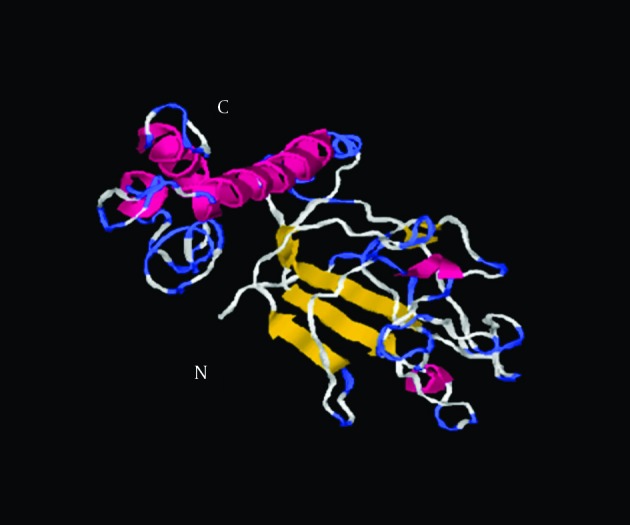
This model was selected as the best model because of the high correlation between C-score and TM-score.
Table 2. I-TASSER Parameters for Predicted 3D Models.
| S. No | Model | C-scorea | TM-scoreb | RMSDb | No. of Decoysc | Cluster Densityd |
|---|---|---|---|---|---|---|
| 1 | Model 1 | -3.29 | 0.35 ± 0.12 | 14 ± 3.9 | 813 | 0.0129 |
aC-score is a confidence score for estimating the quality of predicted models by I-TASSER. C-score is typically in the range of -5 to 2, whereas a C-score of higher value signifies a model with a high confidence and vice versa.
bTM-score and RMSD are known standards for measuring structural similarity between two structures which are usually used to measure the accuracy of structure modeling when the native structure is known. TM-score has the value in (0, 1), where 1 indicates a perfect match between two structures.
cNumber of decoys represents the number of structural decoys that used in generating each model.
dCluster density represents the density of cluster.
4.3. Amplification of pGEM-B2-Gn
The pGEM-B2-Gn plasmid was successfully amplified in E. coli TOP10. Transformed E. coli TOP10 produced colonies in LB agar, containing 2 antibiotics of ampicillin and tetracycline. Digestion and agarose gel electrophoresis of recombinant pGEM-B2-Gn with 2 enzymes Bam HI /Xho I produced a 912 bp fragment that was similar to the expected construct size.
4.4. Subcloning of the Construct Into pFastBacHTb Vector
The purified fragment from agarose gel was successfully subcloned into pFastBacHTb vector. Transformed E. coli TOP10 with pFastHTb-Gn was grown on LB agar, containing ampicillin and tetracycline.
4.5. Enzyme Digestion and Sequencing
To confirm and determine the presence and size of inserts in pFastBacHTb-Gn, recombinant plasmid was simultaneously digested with 2 enzymes, Bam HI/Xho I. After electrophoresis of digestion reaction on 1% agarose gel, 2 bands were detected in each lane and attributed to pFastBacHTb band (4857bp) and insert band (912bp for construct). As shown in Figure 5 inserts of expected length were detected. The nucleotide sequencing confirmed that the gene of interest was inserted properly in-frame with His-tag under the control of polyhedron promoter.
Figure 5. Digestion of Recombinant pFastHTb-Gn With Restriction Enzymes.
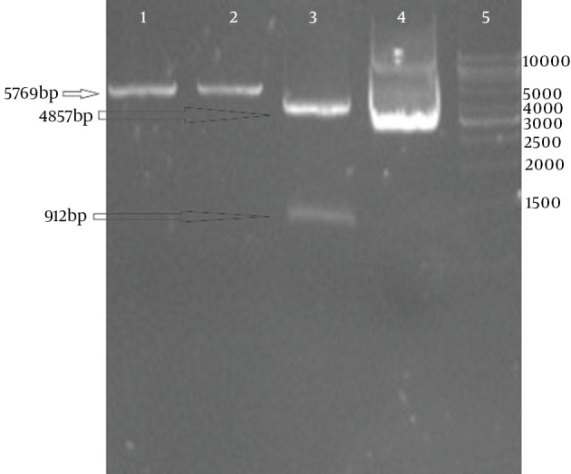
Lane 1, single digestion with Bam HI; lane 2, Single digestion with Xho I; lane 3, Double digestion; lane 4, Un cut vector; lane 5, 1kb marker (Fermentas, Lithuania).
4.6. Generation of Recombinant Bacmid and Blue-White Screening
The recombinant bacmid was generated successfully as described in Materials and Methods. Transformed E. coli DH10 Bac produced white colonies while untransformed bacteria formed blue colonies in LB agar, containing IPTG and X-gal.
4.7. Polymerase Chain Reaction Amplification
Polymerase Chain Reaction (PCR) was used to confirm the recombinants. PCR was performed using M13 forward and reverse primers, which generated 3342 bp fragment. Selected white colonies generated strong bands after PCR that showed execution of recombination process as expected. As shown in Figure 6, inserts of expected length were detected. Bacmid DNA was extracted from overnight culture of white colonies in LB agar and electrophoresised on 0.7% agarose (Figure 7).
Figure 6. PCR With M13 Primers on Bacmid.
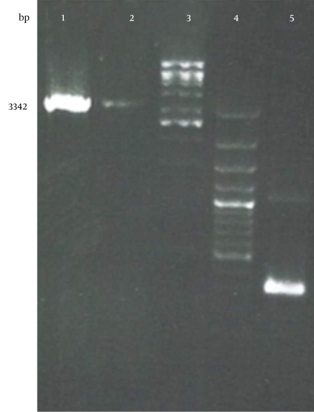
Lane 1, 2; bacmid with insert, lane 3; 1kbp marker (Fermentas, Lithuania), lane4; 100bp marker, lane 5; bacmid without insert.
Figure 7. Electrophoresis of Bacmid in 0.7% Agarose Gel for 18 Hours.
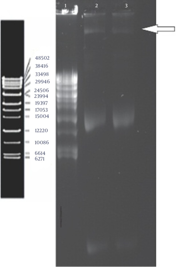
Lane 1, ladder lambda (Fermentas, Lithuania); Lane 2 and 3, bacmid.
5. Discussion
Crimean-Congo hemorrhagic fever virus causes severe hemorrhage in human with mortality rate of approximately 30%. It is reported in many countries of Africa, Asia, Middle East, and southeast Europe. The geographic distribution of CCHFV is the most extensive in comparison with other medically tick-borne viruses and has expanded in recent years as an increasing number of epidemics and sporadic cases have been reported in many regions of the world. Global climate change may be a reason for increasing the number of cases in recent years (1-4). Despite the fact that CCHFV is a member of Bunyaviridae, it has some unusual properties compared to members of other genera: first, CCHFV genome M segment encodes an unusually large polyprotein, which is a large precursor with 1684 amino acids in length; second, viral glycoprotein precursors usually undergo only one proteolysis processing step but (in the case of CCHFV) M polyprotein needs a complex series of proteolysis processing before generating mature glycoproteins; third, CCHFV glycoproteins (Gn and Gc) contain cysteine residues, suggesting the presence of disulfide bonds and a complex secondary structure (1, 13, 17, 19).
Currently, there is no available vaccine against CCHFV and detailed structural analysis of CCHFV proteins remains undefined. In this study, first we used bioinformatic software to determine the structure of CCHFV Gn protein, and then design and clone a construct recombinant bacmid to express Gn for studying its immunogenic potentials in future. The primary structure of CCHFV Gn was speculated with ExPASy ProtParam (20). Physicochemical parameters of the CCHFV Gn protein showed that it has 285-aa long sequences and a molecular weight of 33587.8 Daltons with theoretical isoelectric point (PI) of 6.65. The instability index is computed to be 32.67; the instability index provides an estimate of the stability of protein of interest in a test tube. Values greater than 40 indicate that the protein may be unstable in vitro. Based on ProtParam analysis results, Gn is classified as a stable protein. The secondary structure of CCHFV Gn was predicted and analyzed by PSIPRED (21). The tertiary structure prediction was performed by I-TASSER server using the best align template (22). Out of 5 generated similar models of the target sequence, the best one was chosen to employ the criteria of good C-score, TM Score. The predicted 3D structure will provide more insight in understanding the structure and function of the protein.
Neighbor-joining analysis had separated CCHFV into 4 broad groups, which predominantly contained viruses from Europe/Turkey, Africa, Middle East, and West Asia. M segment sequence alignment of Iran strain compared to other strains showed a close relationship between Iran strain and Afghanistan strain with 99% similarity. Both Gn and Gc are modified by N-linked glycosylation so eukaryotic expression system is necessary for proper folding of glycoproteins. Erickson and collogues in their study indicated that 2 glycosylation sites (557N and 755N) are present in mature Gn glycoprotein of CCHFV. Of them, 557N site is important and mutational blocking of Gn glycosylation at 557N results in mislocalization and retention of Gn and other proteins synthesized from the virus M segment ORF (GP160, GP85, GP38, and Gc) in the endoplasmic reticulum (19, 23).
Gn and Gc glycoproteins of CCHFV may involve in the host range, cell tropism, and pathogenicity of this vertebrate and tick virus, and are the targets for neutralizing antibodies. So, cloning, expression, and determination of neutralizing epitopes may have significant diagnostic and therapeutic applications (18, 19). Currently, diverse prokaryotic and eukaryotic expression systems have been developed for cloning and expression of recombinant proteins. E. coli is one of the most widely-used prokaryotic expression systems because of its easy manipulation, low cost, and high production of recombinant protein. Lack of posttranslational modification mechanisms in bacterial cells such as glycosylation, proteolysis protein maturation, or limited capacity for formation of disulfide bonds are disadvantages of this expression system (24, 25). In the case of enveloped viruses, proteolysis cleavage and glycosylation are very important, which can modulate biological characteristics of the virus. Expression of Gn in E. coli might yield large amounts of protein; however, Gn is a glycosylated protein with disulfide bonds so its expression in E. coli cannot produce an active protein.
Among eukaryotic systems, the baculovirus expression system has many advantages such as high protein production, correct folding and safety and most importantly, posttranslational modification (like glycosylation) in insect cells which is critical for Gn antigen. Many of researchers have used baculovirus expression systems in various viral agents (26-28). The role of the glycoprotein encoded by M segment in neutralizing and protecting antibody in animal and human body infected by Hantavirus has already been demonstrated. In the case of CCHFV, several studies have shown that passive transfer of convalescent serum of patients infected with CCHFV to acutely infected patients can confer some protection to the patients (19). Reports about the production of neutralizing MAbs against CCHFV glycoproteins are limited. In one study, Bertolotti-Ciarlet and collogues demonstrated that monoclonal antibodies (MAbs) obtained from mice against CCHFV Gn were more effective on protecting mice from a lethal CCHFV challenge than MAbs against Gc when administrated either 24 hours before or after the infection (17).
Many researchers have used Gn glycoprotein of other members of family Bunyaviridae as a candidate vaccine component in different expression systems and concluded that this gene can be considered as a potent immunogen in vaccine development (29-33). Currently, there are no specific treatments or licensed vaccine for CCHFV. Reports on detailed bioinformatic study and also developing a vaccine against CCHFV are limited. On the other hand, the production of native antigens for CCHFV has several limitations such as high risk of contamination by handling the virus; high level bio-containment facilities required for working with the virus (not available in many laboratories) and low number of cases (2). Therefore, producing CCHFV surface glycoproteins, including Gn by recombinant DNA technology and characterizing their immunogenicity’s will be important in vaccine development as well as in research and diagnostic laboratories. Because CCHFV is a health problem in our country, any attempt on developing a candidate vaccine for this virus is extremely important. Cloning and production of recombinant protein is the first step in designing a safe vaccine against a highly pathogenic organism like CCHFV. To the best of our knowledge, our project was the first attempt to clone CCHFV glycoprotein in baculovirus expression system in Iran and an indispensable step in future studies for CCHFV vaccine development.
Acknowledgments
We would like to thank our colleagues who assist us in conducting this study. This paper was part of the project of Virus PhD student Mr Mehdi Rahpeima performed in Health Research Institute, Infectious and Tropical Diseases Research Center, Ahvaz Jundishapur University of Medical Sciences, Ahvaz, Iran.
Footnotes
Authors’ Contribution:Mehdi Rahpeyma and Alireza Samarbaf-Zadeh developed the concept and designed the study. Manoochehr Makvandi and Ata Ghadiri advised the study. Mehdi Rahpeyma performed the experiments, conducted bioinformatics analysis and wrote the paper. Alireza Samarbaf-Zadeh and Fatemeh Fotouhi interpreted the results and revised the manuscript.
Funding/Support:This study was supported by the grant No. 91119 approved in Health Research Institute, Infectious and Tropical Diseases, Ahvaz Jundishapur University of Medical Sciences, Iran.
References
- 1.Sanchez AJ, Vincent MJ, Nichol ST. Characterization of the glycoproteins of Crimean-Congo hemorrhagic fever virus. J Virol. 2002;76(14):7263–75. doi: 10.1128/JVI.76.14.7263-7275.2002. [DOI] [PMC free article] [PubMed] [Google Scholar]
- 2.Canakoglu N, Berber E, Ertek M, Yoruk MD, Tonbak S, Bolat Y, et al. Pseudo-plaque reduction neutralization test (PPRNT) for the measurement of neutralizing antibodies to Crimean-Congo hemorrhagic fever virus. Virol J. 2013;10:6. doi: 10.1186/1743-422X-10-6. [DOI] [PMC free article] [PubMed] [Google Scholar]
- 3.Flick R, Flick K, Feldmann H, Elgh F. Reverse genetics for crimean-congo hemorrhagic fever virus. J Virol. 2003;77(10):5997–6006. doi: 10.1128/JVI.77.10.5997-6006.2003. [DOI] [PMC free article] [PubMed] [Google Scholar]
- 4.Mardani M, Keshtkar-Jahromi M, Ataie B, Adibi P. Crimean-Congo hemorrhagic fever virus as a nosocomial pathogen in Iran. Am J Trop Med Hyg. 2009;81(4):675–8. doi: 10.4269/ajtmh.2009.09-0051. [DOI] [PubMed] [Google Scholar]
- 5.Bente DA, Alimonti JB, Shieh WJ, Camus G, Stroher U, Zaki S, et al. Pathogenesis and immune response of Crimean-Congo hemorrhagic fever virus in a STAT-1 knockout mouse model. J Virol. 2010;84(21):11089–100. doi: 10.1128/JVI.01383-10. [DOI] [PMC free article] [PubMed] [Google Scholar]
- 6.Garrison AR, Radoshitzky SR, Kota KP, Pegoraro G, Ruthel G, Kuhn JH, et al. Crimean-Congo hemorrhagic fever virus utilizes a clathrin- and early endosome-dependent entry pathway. Virology. 2013;444(1-2):45–54. doi: 10.1016/j.virol.2013.05.030. [DOI] [PubMed] [Google Scholar]
- 7.Izadi S, Naieni KH, Madjdzadeh SR, Nadim A. Crimean-Congo hemorrhagic fever in Sistan and Baluchestan Province of Iran, a case-control study on epidemiological characteristics. Int J Infect Dis. 2004;8(5):299–306. doi: 10.1016/j.ijid.2003.10.008. [DOI] [PubMed] [Google Scholar]
- 8.Mardani M, Jahromi MK, Naieni KH, Zeinali M. The efficacy of oral ribavirin in the treatment of crimean-congo hemorrhagic fever in Iran. Clin Infect Dis. 2003;36(12):1613–8. doi: 10.1086/375058. [DOI] [PubMed] [Google Scholar]
- 9.Sharifi-Mood B, Metanat M, Hashemi-Shahri SM, Mardani M, Hashemi SA, Fayyaz-Jahani F. Crimean-Congo hemorrhagic fever following consumption of uncooked liver: case series study. Iran J Clin Infect Dis. 2011;6(3):128–30. [Google Scholar]
- 10.Rahbari S, Nabian S, Shayan P. Primary report on distribution of tick fauna in Iran. Parasitol Res. 2007;101 Suppl 2:S175–7. doi: 10.1007/s00436-007-0692-7. [DOI] [PubMed] [Google Scholar]
- 11.Lak SS, Vatandoost H, Telmadarraiy Z, Mahdi RE, Kia E. Seasonal activity of ticks and their importance in tick-borne infectious diseases in West Azerbaijan, Iran. J Arthropod-Borne Dis. 2008;2(2):28–34. [Google Scholar]
- 12.Nabian S, Rahbari S. Occurrence of soft and hard ticks on ruminants in Zagros mountainous areas of Iran. J Arthropod-Borne Dis. 2008;2(1):16–20. [Google Scholar]
- 13.Carter SD, Surtees R, Walter CT, Ariza A, Bergeron E, Nichol ST, et al. Structure, function, and evolution of the Crimean-Congo hemorrhagic fever virus nucleocapsid protein. J Virol. 2012;86(20):10914–23. doi: 10.1128/JVI.01555-12. [DOI] [PMC free article] [PubMed] [Google Scholar]
- 14.Kato T, Thompson JR, Park EY. Construction of new ligation-independent cloning vectors for the expression and purification of recombinant proteins in silkworms using BmNPV bacmid system. PLoS One. 2013;8(5):e25502. doi: 10.1371/journal.pone.0064007. [DOI] [PMC free article] [PubMed] [Google Scholar]
- 15.Roy P, Noad R. Use of bacterial artificial chromosomes in baculovirus research and recombinant protein expression: current trends and future perspectives. ISRN microbiol. 2012;2012:11. doi: 10.5402/2012/628797. [DOI] [PMC free article] [PubMed] [Google Scholar]
- 16.Abdel-Moneim AS, Giesow K, Keil GM. High-level protein expression following single and dual gene cloning of infectious bronchitis virus N and S genes using baculovirus systems. Viral Immunol. 2014;27(2):75–81. doi: 10.1089/vim.2013.0114. [DOI] [PubMed] [Google Scholar]
- 17.Bertolotti-Ciarlet A, Smith J, Strecker K, Paragas J, Altamura LA, McFalls JM, et al. Cellular localization and antigenic characterization of crimean-congo hemorrhagic fever virus glycoproteins. J Virol. 2005;79(10):6152–61. doi: 10.1128/JVI.79.10.6152-6161.2005. [DOI] [PMC free article] [PubMed] [Google Scholar]
- 18.Bergeron E, Albarino CG, Khristova ML, Nichol ST. Crimean-Congo hemorrhagic fever virus-encoded ovarian tumor protease activity is dispensable for virus RNA polymerase function. J virology. 2010;84(1):216–26. doi: 10.1128/JVI.01859-09. [DOI] [PMC free article] [PubMed] [Google Scholar]
- 19.Altamura LA, Bertolotti-Ciarlet A, Teigler J, Paragas J, Schmaljohn CS, Doms RW. Identification of a novel C-terminal cleavage of Crimean-Congo hemorrhagic fever virus PreGN that leads to generation of an NSM protein. J Virol. 2007;81(12):6632–42. doi: 10.1128/JVI.02730-06. [DOI] [PMC free article] [PubMed] [Google Scholar]
- 20.Sato Y, Takaya A, Yamamoto T. Meta-analytic approach to the accurate prediction of secreted virulence effectors in gram-negative bacteria. BMC Bioinformatics. 2011;12:442. doi: 10.1186/1471-2105-12-442. [DOI] [PMC free article] [PubMed] [Google Scholar]
- 21.McGuffin LJ, Bryson K, Jones DT. The PSIPRED protein structure prediction server. Bioinformatics. 2000;16(4):404–5. doi: 10.1093/bioinformatics/16.4.404. [DOI] [PubMed] [Google Scholar]
- 22.Zhang Y. I-TASSER server for protein 3D structure prediction. BMC Bioinformatics. 2008;9:40. doi: 10.1186/1471-2105-9-40. [DOI] [PMC free article] [PubMed] [Google Scholar]
- 23.Erickson BR, Deyde V, Sanchez AJ, Vincent MJ, Nichol ST. N-linked glycosylation of Gn (but not Gc) is important for Crimean Congo hemorrhagic fever virus glycoprotein localization and transport. Virology. 2007;361(2):348–55. doi: 10.1016/j.virol.2006.11.023. [DOI] [PubMed] [Google Scholar]
- 24.Carneiro S, Ferreira EC, Rocha I. Metabolic responses to recombinant bioprocesses in Escherichia coli. J Biotechnol. 2013;164(3):396–408. doi: 10.1016/j.jbiotec.2012.08.026. [DOI] [PubMed] [Google Scholar]
- 25.Nallamsetty S, Waugh DS. A generic protocol for the expression and purification of recombinant proteins in Escherichia coli using a combinatorial His6-maltose binding protein fusion tag. Nat Protoc. 2007;2(2):383–91. doi: 10.1038/nprot.2007.50. [DOI] [PubMed] [Google Scholar]
- 26.Jarvis DL. Developing baculovirus-insect cell expression systems for humanized recombinant glycoprotein production. Virology. 2003;310(1):1–7. doi: 10.1016/s0042-6822(03)00120-x. [DOI] [PMC free article] [PubMed] [Google Scholar]
- 27.Harrison RL, Jarvis DL. Protein N‐Glycosylation in the Baculovirus–Insect Cell Expression System and Engineering of Insect Cells to Produce “Mammalianized” Recombinant Glycoproteins. Adv virus res. 2006;68:159–91. doi: 10.1016/s0065-3527(06)68005-6. [DOI] [PubMed] [Google Scholar]
- 28.Ramya R, Mohana Subramanian B, Sivakumar V, Senthilkumar RL, Sambasiva Rao KR, Srinivasan VA. Expression and solubilization of insect cell-based rabies virus glycoprotein and assessment of its immunogenicity and protective efficacy in mice. Clin Vaccine Immunol. 2011;18(10):1673–9. doi: 10.1128/CVI.05258-11. [DOI] [PMC free article] [PubMed] [Google Scholar]
- 29.Metz SW, Geertsema C, Martina BE, Andrade P, Heldens JG, van Oers MM, et al. Functional processing and secretion of Chikungunya virus E1 and E2 glycoproteins in insect cells. Virol J. 2011;8:353. doi: 10.1186/1743-422X-8-353. [DOI] [PMC free article] [PubMed] [Google Scholar]
- 30.Barros MC, Galasso TG, Chaib AJ, Degallier N, Nagata T, Ribeiro BM. Yellow fever virus envelope protein expressed in insect cells is capable of syncytium formation in lepidopteran cells and could be used for immunodetection of YFV in human sera. Virol J. 2011;8:261. doi: 10.1186/1743-422X-8-261. [DOI] [PMC free article] [PubMed] [Google Scholar]
- 31.Kim S, Chang J. Baculovirus-based Vaccine Displaying Respiratory Syncytial Virus Glycoprotein Induces Protective Immunity against RSV Infection without Vaccine-Enhanced Disease. Immune Netw. 2012;12(1):8–17. doi: 10.4110/in.2012.12.1.8. [DOI] [PMC free article] [PubMed] [Google Scholar]
- 32.Zoth SC, Gomez E, Carballeda JM, Carrillo E, Berinstein A. Expression of a secreted version of the hemagglutinin-neuraminidase glycoprotein of Newcastle disease virus: its evaluation as a diagnostic reagent. J Vet Diagn Invest. 2011;23(3):519–23. doi: 10.1177/1040638711404153. [DOI] [PubMed] [Google Scholar]
- 33.Shi X, Jarvis DL. Protein N-glycosylation in the baculovirus-insect cell system. Curr Drug Targets. 2007;8(10):1116–25. doi: 10.2174/138945007782151360. [DOI] [PMC free article] [PubMed] [Google Scholar]


