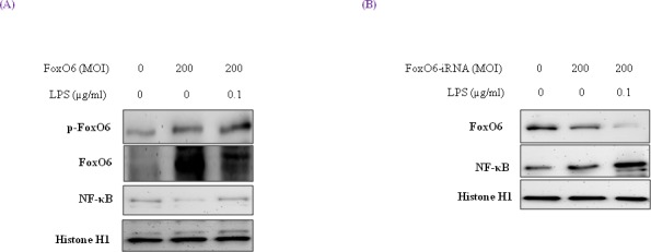
Figure 2. Activation of NF-κB through FoxO6 by LPS.

HepG2 cells were grown to 80% confluence in 100 mm dishes in DMEM, pre-treated (1 day) with or without FoxO6 (100 MOI), and then stimulated with 100 ng/ml LPS. A. HepG2 cells were pretransduced with 200 MOI of FoxO6 vector in the absence or presence of LPS. Cells were analyzed by Western blotting using p-FoxO6, FoxO6, NF-κB, and Histone H1 antibody. B. After stimulating HepG2 cells with LPS (100 ng/ml) in the absence or presence of FoxO6-siRNA (200 MOI), levels of NF-κB were determined in cell extracts.
