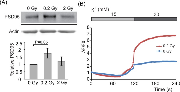
Figure 3. PSD95 expression levels in response to radiation.
A. E18 hippocampal neurons were irradiated with 0, 0.2, or 2 Gy radiation on DIV 7. After 5 days, cell lysates were collected and analyzed by western blotting using anti-PSD95 or actin antibody. Level of PSD95 was normalized to actin. Quantified results are shown as mean ± S.E.M from three independent experiments. B. E18 hippocampal neurons were irradiated with 0 or 0.2 Gy radiation on DIV 7. Five days after radiation, neurons were incubated with FM® 4-64FX dye followed by increasing concentrations of K+ and images were taken. The intensity of fluorescence was quantified as described in material and methods. Blue line: 0 Gy radiation (n = 8) and red line: 0.2 Gy radiation (n = 22).

