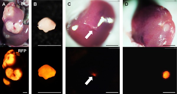
Figure 5. Establishment of orthotopic liver metastasis mouse models.

Upper panels show bright-field images and lower are RFP images. A. Multiple liver metastases were initially established after spleen injection of HT-29-RFP cells in the donor mouse. B. The metastasis were resected and cut into small fragments. C. Single fragments were then orthotopically implanted in the left lobe of the liver in the experimental mice through an incision (arrow). D. Four weeks after implantation, an orthotopic liver metastasis mouse model was established. Scale bars: 3 mm.
