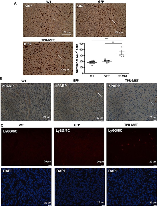
Figure 5. Activation of MET signaling in SMS-CTR ERMS cells enhances tumor proliferation, decreases tumor apoptosis and induces infiltration of neutrophils in vivo.

A. Representative images of the staining for Ki67 in tumor sections show areas of tumor cells proliferation. Number of Ki67 positive cells was calculated in non-necrotic areas of tumor specimens, n = 5. Arrows indicate Ki67 positive cells. B. Representative images of the staining for cleaved PARP demonstrate decreased apoptosis in TPR-MET SMS-CTR tumors. C. Representative images of the staining for neutrophils (Ly6G/6C) show infiltration of murine neutrophils to TPR-MET SMS-CTR tumors. *p < 0.05, **p < 0.01, ***p < 0.001. Data in graphs are represented as mean +/− SEM. Arrows indicate cPARP positive cells.
