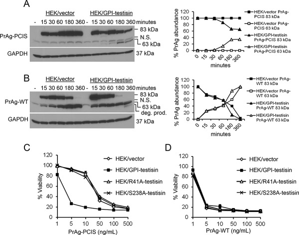
Figure 4. Expression of GPI-anchored testisin in HEK293T cells increases PrAg-PCIS processing and PrAg-PCIS toxin-induced tumor cell killing.

A. Cell-expressed testisin increases processing of PrAg-PCIS. HEK293T cells stably expressing wild-type testisin (HEK/GPI-testisin) or vector alone (HEK/vector) were incubated for up to 6 hours with 500 ng/mL PrAg-PCIS in growth media. At each time point, cells were washed in PBS to remove non-bound proteins and immunoblotted using anti-PrAg antibodies to investigate PrAg cleavage. The blot was reprobed with anti-GAPDH antibody to assess protein loading and is representative of two independent experiments. Densitometric analysis shows cleavage activation of PrAg-PCIS, as indicated by the appearance of the PrAg-PCIS 63-kDa and loss of PrAg-PCIS 83-kDa, in HEK/GPI-testisin cells. B. Cell-expressed testisin increases processing of PrAg-WT. HEK/GPI-testisin or HEK/vector cells were treated as in A) and analyzed for PrAg cleavage. The blot was reprobed with anti-GAPDH antibody to assess protein loading and is representative of two independent experiments. Densitometric analysis shows efficient processing of PrAg-WT to the 63-kDa form in both cell lines. In HEK/GPI-testisin cells, an additional band was detected, likely an in vitro degradation product. C. Active testisin increases PrAg-PCIS toxin-induced cytotoxicity. The indicated cell lines were incubated for 6 hours in growth media with PrAg-PCIS (0–500 ng/mL) and FP59 (50 ng/mL), and then media was replaced with fresh media. Cell viability was assayed 48 hours later by MTT assay. D. PrAg-WT toxin-induced cytotoxicity is not dependent on active testisin. The indicated cell lines were treated with PrAg-WT and FP59 and viability measured as in C). MTT assays represent the mean of a total of 6 experiments (3 separate experiments, with triplicate samples, for each of two independent pools of stably-transfected cells).
