
Figure 4. TAM-derived HO-1 reverted EMT in other words triggered mesenchymal to epithelial transition MET in vivo.
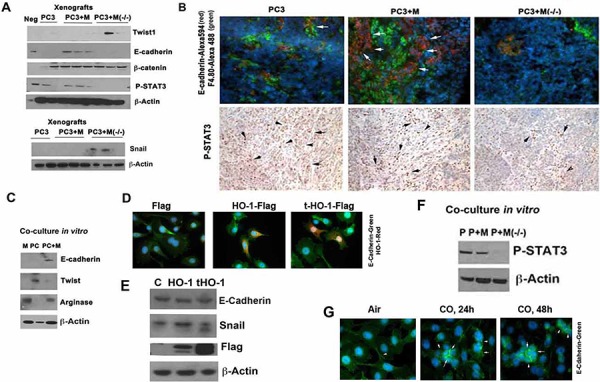
A. PC3 cells alone or PC3 mixed with BMDM from Hmox1fl/fl (M) and LyzM-Cre:Hmox1fl/fl (M−/−) mice were inoculated into the flanks of nude mice as described in Figure 3 and xenografts were harvested on day 26. Immunoblotting with antibody against twist1, E-cadherin, Snail and β-catenin was performed. Neg: normal skin. B. Immunostaining and immunohistochemistry with antibody against E-cadherin (Alexa 594-red) and F4.80, a marker of macrophages (Alexa488-green) were applied in the xenografts as above. C. PC3 (PC) cells were co-cultured with BMDM (M) and harvested after 24 h. Immunoblotting with antibody against E-cadherin, twist1 and arginase was performed in the lysates of the co-cultures. Immunoblots correspond to the mixture of proteins isolated from both cell types. D–E. PC-3 cells were transfected with HO-1 (cytoplasmic) and truncated HO-1 (cytoplasmic and nuclear) and the levels of E-cadherin and HO-1 were correlated by immunostaining (HO-1-Alexa549, E-cadherin-Alexa488) or immunoblotting. Data are representative for 3 independent experiments. F. PC3 cells co-cultured in vitro with BMDM from Hmox1fl/fl (M) and LyzM-Cre:Hmox1fl/fl (M−/−) mice were harvested after 24 h and lysates were immunoblotted for HO-1 (m-mouse, h-human) and P-STAT3. Data are representative for 3 independent experiments. G. PC3 cells were treated with exogenous CO (250 ppm) for 24 or 48 h and the levels of E-cadherin were measured by immunofluorescence staining. Data are representative for 3 independent experiments. Arrows indicate positive staining for E-cadherin.