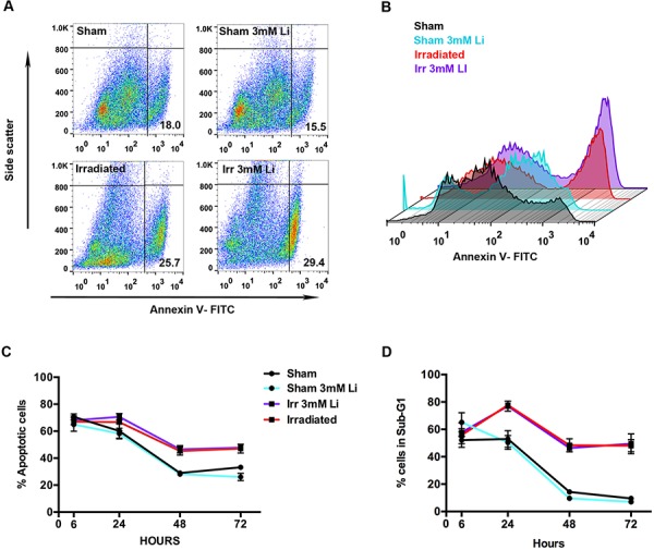
Figure 4. Lithium at 3 mM did not halt irradiation-induced apoptosis in young neural stem cells.

A. Representative scatter plot of the distribution of the cells stained with FITC for annexin V (x-axis) plotted against the side scatter (y-axis) analysed by flow cytometry showing the percentages (%) of apoptotic cell in the lower right quadrant in each group: sham, sham+3 mM LiCl, irradiated and irradiated+3 mM LiCl. B. Representative picture illustrating the peaks of intensity of annexin V-FITC (x-axis) plotted against the count of the events (y-axis) in sham (black), sham+3 mM LiCl (light blue), irradiated (red) and irradiated+3 mM LiCl (purple). C. Quantification of the percentage of apoptotic cells, positive for annexin V, at different times showing that irradiation strongly induces apoptosis in this cell type at 48 and 72 hours ***p48 hours = 0.0004, ***p72 hours = 0.0002. Lithium at 3 mM did not rescue this cell type from apoptosis. D. Time course of the percentage of cells in sub-G1. Irradiation displays an effect on cell death at 24, 48 and 72 hours, **p24 hours = 0.0040, ****p48 hours < 0.0001, ****p72 hours < 0.0001. Lithium at 3 mM did not reduce the increased percentage of cells found in sub-G1. Data are presented as mean ± SEM, n = 3–6.
