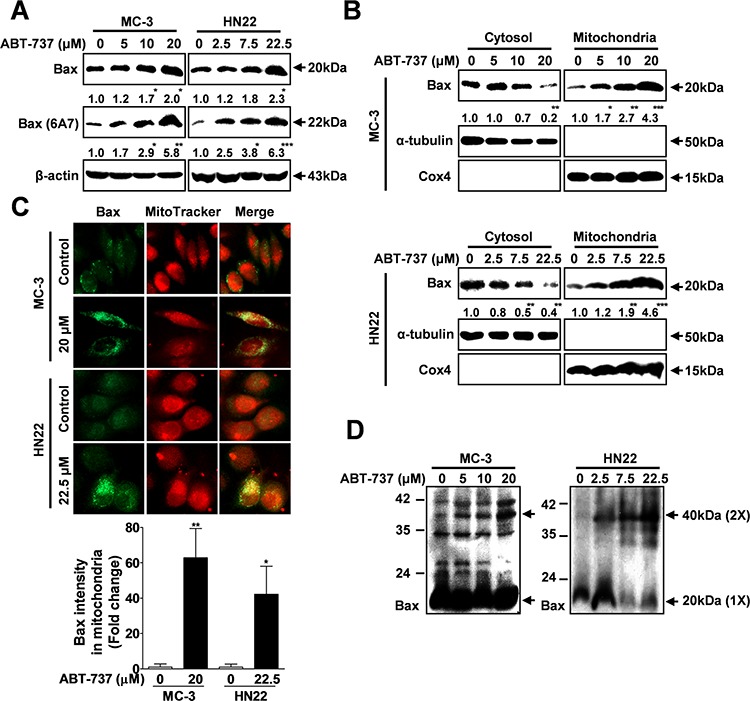
Figure 3. ABT-737 induces mitochondrial-dependent apoptosis through Bax.

MC-3 and HN22 cells were treated with or without ABT-737 for 24 hr, total cellular protein was prepared, and the protein levels of total and active Bax (6A7) were evaluated by Western blot analysis A. MC-3 and HN22 cells were treated with or without ABT-737 for 24 hr; afterwards, cytosolic and mitochondrial fractions were prepared and the protein levels of total and active Bax (6A7) were evaluated by Western blot analysis B. Immunocytochemistry revealed the mitochondrial translocation of Bax from the cytoplasm after ABT-737 treatment (20 μM in MC-3 cells and 22.5 μM in HN22 cells) for 24 hr. MitoTracker (red) staining labels mitochondria within each field C. To assess whether ABT-737 treatment has an effect on the dimerization of Bax, the levels of dimeric Bax were measured with or without ABT-737 treatment in MC-3 and HN22 cells D. β-actin was used as an internal control. The Cox4 exclusive mitochondria marker was used as a control for the separation of mitochondrial and cytosolic fractions. The results are shown as the mean ± SD from three independent experiments. *P < 0.05, **P < 0.01, and ***P < 0.001.
