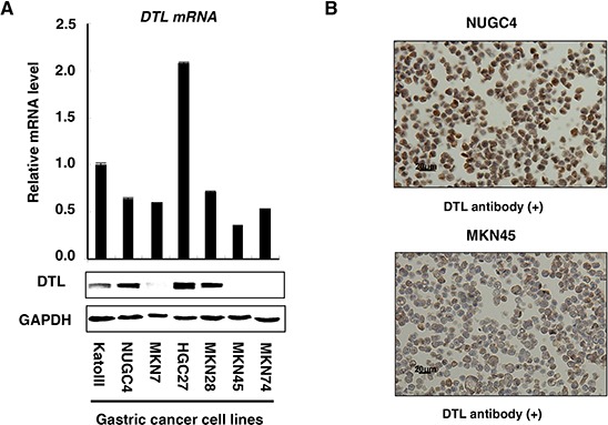
Figure 1. Expression profiles of DTL in 7 GC cell lines.

A. Quantitative real-time RT–PCR and western blotting analysis were performed using a DTL-specific antibody to determine DTL mRNA (top) and protein (bottom) expression in the gastric cancer cell lines KatoIII, NUGC4, MKN7, HGC27, MKN28, MKN45, and MKN74. DTL overexpression was observed in the KatoIII, NUGC4, HGC27, and MKN28 cells (4/7 lines, 57%). B. A formalin-fixed gastric cancer NUGC4 cell line that overexpresses DTL, in which > 50% of cells stained positively, was used as a positive control; an MKN45 cell line with a low expression of DTL was included as a negative control.
