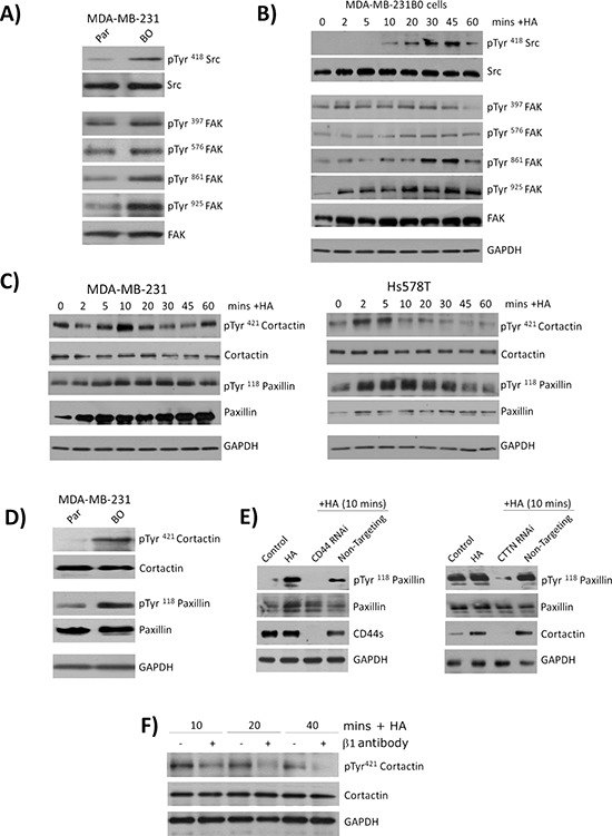
Figure 4. CD44 signaling promotes the phosphorylation of cytoskeletal proteins cortactin and paxillin.

A. Panel of immunoblots comparing the expression and phosphorylation status of the kinases Src and FAK in the parental and bone-tropic MDA-MB-231 cell lines. B. Immunoblots comparing the expression and phosphorylation status of Src and FAK following stimulation of MDA-MB-231BO cells with HA. C. Panel of immunoblots comparing the expression and phosphorylation status of the cytoskeletal proteins cortactin and paxillin in MDA-MB-231 (left panel) and Hs578T (right panel) breast cancer cell lines. D. Immunoblot comparing the basal expression and phosphorylation status of cortactin and paxillin in parental MDA-MB-231 and bone-tropic MDA-MB-231BO cells. E. Panel of immunoblots illustrating the levels of phosphorylation detected in paxillin in response to HA stimulation of MDA-MB-231 cells in the absence or presence of CD44 (left panel) or the absence and presence of cortactin (CTTN). F. Immunoblots measuring the levels of cortactin phospgorylation in HA-stimulated MDA-MB-2331 cells in the absence or presence of a neutralizing antibody to β1-integrin receptors. Equal protein loading in all immunoblots is confirmed by reanalysis of GAPDH expression. Blots shown in A-F are representative of at least three independent experiments.
