

Abstract
Background Fractures of the distal radius with small volar ulnar marginal fracture fragments are difficult to stabilize with standard volar locking plates. The purpose of this study is to describe alternative techniques available to stabilize these injuries.
Materials and Methods Five patients were identified retrospectively with unstable volar lunate facet fracture fragments treated with supplemental fixation techniques. The demographic data, pre- and postoperative radiographic parameters, and early outcomes data were analyzed. The AO classification, preoperative and final postoperative ulnar variance, articular step-off, volar tilt, radial inclination, and teardrop angle were measured. The lunate subsidence and length of the volar cortex available for fixation were measured from the initial injury films.
Description of Technique Lunate facet fixation was based on the morphology of the fragment, and stabilization was achieved with headless compression screws in three patients, a tension band wire construct in one, and two cortical screws in another.
Results Five patients with a mean age of 58 years (range: 41–82) were included. There were two AO C3.2 and three B3.3 fractures. Preoperative radiographic measurements including radial inclination, tilt, and ulnar variance all improved after surgery and were maintained within normal limits at 3-month follow-up. There was no change in the teardrop angle at final follow-up (70–64 degrees; p = 0.14). None of the patients had loss of fixation or volar carpal subluxation. The mean visual analog scale pain score at 3 months was 1 (range: 0–2).
Conclusions The morphology of volar lunate facet fracture fragments is variable, and fixation must be customized to the particular pattern. Small fragments may preclude the use of plates and screws for fixation. These fractures can be managed successfully with tension band wire constructs and headless screws. These low-profile implants may decrease the risk of tendon irritation that might accompany distally placed plates.
Keywords: distal radius, fracture, lunate facet, fixation
Fractures of the distal radius with small marginal fragments are unstable and can be difficult to manage. The volar lunate facet fragment has received attention over the last decade due to the potential for loss of fixation and carpal subluxation.1 The volar lunate facet may fracture in isolation or more commonly is associated with volar shearing fractures as well as severe comminuted fractures of the distal radius. Stable fixation of the volar lunate facet is critical because failure to do so results in volar carpal subluxation or dislocation and subsequent impaired function, pain, and posttraumatic arthritis.1 2 3 4 Despite the fact that surgeons are aware of this potential pitfall, loss of fixation continues to occur.2
Jupiter et al observed that the vast majority of volar shearing fractures of the distal radius have two or more small marginal articular components.5 The scaphoid and lunate portions of the volar articular surface shear off of the distal radius as a unit but can fragment into separate pieces in the sagittal plane. The volar lunate facet fragment is the most difficult to stabilize because it projects volar to the axis of the palmer surface of the radius, making it inherently unstable and difficult to capture with a standard volar locking plate.2 3 6 7 8 9 10 11 12
Beck et al found that the size of the volar lunate facet fragment and the amount of initial displacement determined whether a volar plate alone can maintain fracture reduction.2 More than 5 mm of initial lunate facet subsidence and lunate facets less than 15 mm in length were more likely to displace after adequate fixation with a volar plate.
The treatment of volar shearing fractures of the distal radius with comminution of the volar rim is challenging but loss of fixation and malunion can be avoided by the use of a fragment-specific fixation in addition to volar locking plates. Techniques that have been described for stabilization of the lunate facet include tension band wire constructs,13 Kirschner wires with external fixator,10 14 pin plates,15 Kirschner wire constructs,11 arthroscopic reduction and pinning,16 screw and washer fixation,3 2.4-mm T- and L-plates,17 and more recently volar hook plate fixation.9 18 The purpose of this study is to describe alternative techniques to stabilize small volar ulnar marginal fracture fragments with a low-profile implant to minimize the potential for tendon irritation.
Methods
The operative log of a single surgeon (N.G.H.) was reviewed between January 2014 and October 2015 for patients who underwent open reduction and internal fixation of the distal radius. A total of 52 patients underwent operative fixation of the distal radius. Each patient had been entered into a distal radius fracture registry that is used by our institution to track adult patients who undergo surgical management of distal radius fractures. Radiographs were classified at the time of surgery by the treating hand surgeon (N.G.H.) using the AO classification for distal radius fractures. Patients at least 18 years of age with surgically treated AO B3.3 or C type fractures were included. Patients with AO type A, B1, B2, B3.1, and B3.2 fractures were excluded. An AO B3.3 fracture has comminution of the volar rim of the distal radius with separate scaphoid and lunate facets. Fractures with an associated lunate facet fracture that were stable with volar plate fixation alone were excluded. Stability was defined as the ability of a volar plate to capture the volar lunate facet fragment with a least two locking screws. Fractures treated with a volar plate with less than one screw capturing the volar lunate facet fragment were considered unstable. All radiographs were reviewed to confirm maintenance of reduction of the lunate facet in those patients that had fixation with a volar plate and two screws to further verify stability. Five patients met criteria for entry into the study.
Patients are evaluated at 2 and 6 weeks and 3 months after surgical intervention per our distal radius fracture registry protocol. The registry was reviewed to determine sex, age, date of injury, hand dominance, medical comorbidities (diabetes, rheumatoid arthritis, use of systemic steroids, or renal failure requiring dialysis), history of smoking, mechanism of injury, occupation, associated injuries, pain level, and postoperative wrist range of motion. The implant manufacturer, type, size, and number of implants used for fixation were recorded from the medical record and radiographs. The AO classification, preoperative and final postoperative ulnar variance, articular step-off, volar tilt, radial inclination, and teardrop angle (TDA) were measured from radiographs using a goniometer by a single observer (N.G.H.). The radiographs were taken in a uniform fashion with the shoulder abducted 90 degrees, elbow flexed 90 degrees, and the wrist in neutral alignment for posteroanterior radiographs, and with the shoulder adducted to the side, elbow flexed 90 degrees, and the forearm and wrist in neutral alignment for lateral radiographs. The volar TDA was measured using the method previously described by Medoff.19 The lunate subsidence distance (LSD) and the length of the volar cortex available for fixation (LVC) were measured from the initial injury films using the method previously described by Beck et al.2
Surgical Technique
All fractures were fixed with a Stryker VariAx (Kalamazoo, MI) distal radius volar locking plate. In each case, fixation of the lunate facet fragment was attempted with the volar plate but additional implants were added to achieve full stability. The two patients with C-type fractures required additional fixation of the radial column with a fragment-specific Stryker radial column pin plate. One of these two patients also required an l-shaped Stryker pin plate to support the dorsal lunate facet.
The lunate facet fracture fragment varied in morphology between cases. When visualized from a volar approach, the facet was triangular in three cases, boomerang shaped in one, and rectangular in one. The rectangular fragment was entirely distal to the watershed line. The triangular-shaped fragments were fixed with a tension band construct (case 1) or headless screws (cases 2, 3, and 4). The tension band construct was made with two 0.035-inch Kirschner wires and 26-gauge dental wire. A single Stryker twin fix screw was used in case 4 and two SBi (Small Bone Innovations, Morrisville, PA) 2.0-mm headless screws were used in cases 2 and 5. The boomerang-shaped lunate facet fragment was fixed with two 2.0-mm Synthes (West Chester, PA) titanium cortical screws. There were no cases of subluxation of the volar lunate facet fragment or joint dislocation. All fractures healed, and there were no infections, hardware-related issues, or complex regional pain syndrome.
Case 1
An 82-year-old right-handed man fell from a standing height and had an AO B3.3 fracture. He was diagnosed with osteoporosis with a T-score of −3.0. There was comminution of the radial column as well as separate scaphoid and lunate facet fragments that were identified on plain radiograph. (Fig. 1A, B) A computed tomography (CT) scan was not obtained. A standard volar approach between the flexor carpi radialis and radial artery was utilized. The volar marginal fracture fragments included a radial, central, and lunate facet portion. The radial and central fragments were stabilized with a Stryker volar plate; however, the lunate facet required additional fixation. Only one screw in the ulnar most aspect of the plate could be placed into the lunate facet fragment. Supplemental fixation of the facet was performed with a tension band technique. Two 0.035-inch Kirschner wires were drilled into the most distal margin of the volar lunate facet, directed proximally to avoid the articular surface. The wires were advanced to engage the dorsal cortex. A 26-gauge dental wire was wrapped in a figure of 8 around the distal margin of the wires and then around the most ulnar screw head in the distal row of screws in the plate. The ulnar screw, which had been engaged in the locking hole of the plate, was backed out to allow the wire to be wrapped around it. Once the wire was tightened, the screw was subsequently advanced until it engaged the wire against the plate, securing the fixation of the volar lunate facet. Alternatively, the wire can be placed around the shaft of the screw, beneath the plate prior to final tightening of the screw. The wires were then backed out slightly, cut short, and then bent back over the wire, similar to the technique used to fix an olecranon fracture. (Fig. 2A, B) The tension band helps to prevent volar subluxation of the fragment but also creates a compressive force on the fracture fragment when the wrist is extended.
Fig. 1.

(A, B) Preoperative anteroposterior and lateral radiographs (patient 1).
Fig. 2.
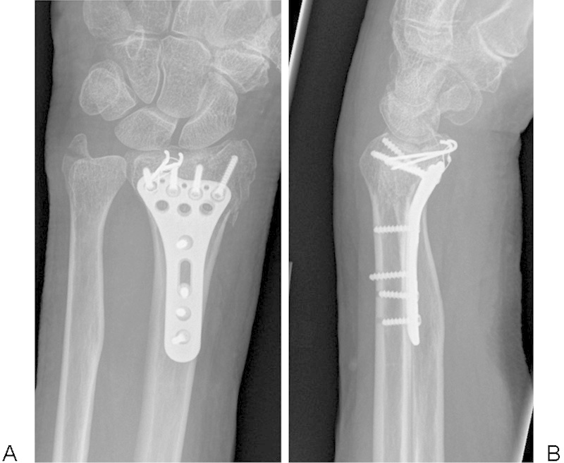
(A, B) Postoperative anteroposterior and lateral radiographs (patient 1).
Case 2
A 49-year-old right-handed woman fell from a standing height and fractured the right wrist. The fracture was classified as an AO C3.2 with a radial styloid and separate dorsal and volar lunate facet fragments (Fig. 3A, B). The small size of the lunate facet fragment was better delineated on two- and three-dimensional CT reconstructions (Figs. 4 and 5). The Stryker VariAx volar plate did not extend distally enough to allow the ulnar most screws to capture the volar lunate facet fragment (Fig. 6). The intraoperative measurement of the LVC was 10 mm (Fig. 7). Stable fixation of the lunate facet fracture fragment was achieved with two 2.0-mm SBi (Small Bone Innovations) headless screws (Fig. 8A, B).
Fig. 3.

(A, B) Preoperative anteroposterior and lateral radiographs of the distal radius (patient 2).
Fig. 4.

A two-dimensional CT scan in the sagittal plane showing the small size of the volar lunate facet fragment. A black arrow is pointing to the volar lunate facet.
Fig. 5.

A three-dimensional CT scan showing the volar lunate facet fragment.
Fig. 6.

An intraoperative view of the volar lunate facet fragment with a black arrow pointing toward the fragment. Note the soft tissues limiting access to the volar ulnar corner from a standard volar approach.
Fig. 7.

The intraoperative view of the volar lunate facet fragment measuring less than 10 mm in length. The short white arrow is pointing to the proximal margin and the long white arrow to the distal margin. This view is from an anteromedial approach between the ulnar neurovascular bundle and the flexor tendons.
Fig. 8.

(A, B) Anteroposterior and lateral postoperative views of the distal radius demonstrate fragment-specific fixation with the addition of two headless screws to secure the volar lunate facet fragment.
The most common operative approach to these fractures is through the distal limb of the Henry approach,20 which may limit adequate exposure to the volar lunate facet of distal radius.1 This fracture was approached through a central incision that allowed mobilization of the flexor tendons utilizing windows to the volar surface of the distal radius on the radial and ulnar sides. The ulnar window is approached between the flexor tendons and the ulnar artery and nerve. The most distal aspect of the pronator quadratus must be elevated to visualize the facet fragment. The guidewires for a tension band wire construct or headless screws may be directed away from the distal radioulnar and radiocarpal joints from this more ulnar-based approach. When attempting to place guidewires through the more radially based approach, the flexor tendons limit the ability to place wires distally and force the wire to be directed in a more ulnar direction.
The remainder of the cases required additional fixation using various constructs. Case 3 was stabilized with two 2.0-mm Synthes titanium screws placed along the most ulnar arm of the boomerang-shaped lunate facet fragment (Figs. 9 10 to 11A–C). Case 4 required a single Stryker twin fix headless screw to maintain stability of the lunate facet. Case 5 had a triangular-shaped lunate facet stabilized by two 2.0-mm SBi (Small Bone Innovations) headless compression screws. All patients were treated with a volar plaster splint for 10 to 14 days and transitioned to a Velcro wrist brace after suture removal. They were all sent for formal occupational hand therapy with active, active-assisted, and passive range of motion allowed at 2 weeks after surgery. Strengthening was allowed at 6 weeks after surgery.
Fig. 9.

Preoperative anteroposterior view of the volar lunate facet fracture with white arrow pointing to the fracture line (patient 3).
Fig. 10.

Preoperative lateral view (patient 3).
Fig. 11.

(A–C) Anteroposterior, lateral, and oblique views of the fixation of the volar lunate facet fracture with two cortical screws. Note the trajectory from ulnar to radial provided by the anteromedial approach.
Results
Between January 2014 and October 2015, five patients underwent operative fixation of a distal radius fracture with an unstable lunate facet fracture fragment. Patients' ages ranged from 41 to 82 years with a mean of 58 years. There were three men and two women. All of the patients were right-handed; the injured wrist was left in three cases and right in two. The mechanism of injury was a fall from standing height in three and high-energy motorcycle accidents in two. There were two retirees, two light laborers, and one heavy laborer. None of the patients used tobacco. None of the patients were diabetic, had rheumatoid arthritis, used systemic steroids, or had renal failure requiring dialysis. Based on the AO classification of fractures, there were two AO C3.2 and three B3.3 fractures.
All radiographic parameters improved after surgical intervention to a normal range as measured at the 3-month post-op. The mean preoperative and 3-month postoperative radial inclination, volar tilt, ulnar variance, TDA, and step-off are listed in Tables 1 and 2. There was no change in the mean TDA when comparing the preoperative to postoperative alignment (70–64 degrees; p = 0.14). The mean preoperative LSD was 4 mm (range: 0–11) and the mean LVC was 14 mm (range: 9–19). Wrist range of motion measurements and visual analog scale (VAS) pain scores were only available for three of the patients. The 3-month range of motion values are listed in Table 3. The mean VAS pain score at 3 months was 1 (range: 0–2).
Table 1. Preoperative radiographic parameters.
| Patient | RI injury | UV injury | Tilt injury | TDA injury | LSD | LVC | Step injury |
|---|---|---|---|---|---|---|---|
| 1 | 9 | 4 | 22 | 77 | 4 | 14 | 2 |
| 2 | 12 | 1 | 0 | 72 | 0 | 9 | 0 |
| 3 | 21 | 0 | 16 | 69 | 3 | 19 | 3 |
| 4 | 18 | 0 | 18 | 58 | 3 | 12 | 4 |
| 5 | 12 | 11 | 22 | 75 | 11 | 16 | 1 |
| Mean | 14 | 3 | 16 | 70 | 4 | 14 | 2 |
Abbreviations: LSD, lunate subsidence distance (mm); LVC, length of volar cortex (mm); RI, radial inclination (degrees); Step, articular step-off (mm); TDA, teardrop angle (degrees); UV, ulnar variance (mm).
Table 2. Final radiographic parameters.
| Patient | RI final | UV final | Tilt final | Step final | TDA final |
|---|---|---|---|---|---|
| 1 | 20 | 0 | 4 | 0 | 72 |
| 2 | 22 | 0 | 12 | 0 | 68 |
| 3 | 20 | 0 | 8 | 0 | 67 |
| 4 | 13 | 0 | 12 | 0 | 63 |
| 5 | 22 | −2 | 4 | 0 | 50 |
| Mean | 19.4 | −0.4 | 8 | 0 | 64 |
Abbreviations: RI, radial inclination (degrees); Step, step-off (mm); TDA, teardrop angle (degrees); Tilt, volar tilt (degrees); UV, ulnar variance (mm).
Table 3. Range of motion (degrees).
| Patient | WE | WF | P | S | RD | UD |
|---|---|---|---|---|---|---|
| 1 | 60 | 60 | 80 | 80 | 15 | 15 |
| 3 | 60 | 60 | 80 | 80 | 10 | 20 |
| 5 | 30 | 10 | 85 | 45 | 15 | 10 |
| Mean | 50 | 43 | 82 | 68 | 13 | 15 |
Abbreviations: P, pronation; RD, radial deviation; S, supination; UD, ulnar deviation; WE, wrist extension; WF, wrist flexion.
Discussion
Fractures of the distal radius with an associated volar lunate facet fragment are unstable and can be difficult to manage. Despite changes in the design of volar plates with an extension at the volar ulnar corner to buttress the volar lunate facet, fixation remains a challenge in fragments less than 15 mm in length.2 To capture these small marginal fragments, the plate must be placed distal to the watershed line, which may result in tendon irritation and/or rupture.21 22 Low-profile implants such as tension band wire constructs and headless screws offer the ability to achieve stability but with less risk for tendon complications.
In the current study, all fractures maintained alignment after operative fixation. No patients experienced tendon irritation or returned for hardware removal. The key to maintenance of stability was to achieve compression and prevent rotation. The Kirschner wire/tension band figure-of-8 technique or the use of screws (headless or cortical) in conjunction with a volar plate achieved stable fixation. The mean LVC was 14 mm (range: 9–19 mm) and LSD 4 mm (range: 0–11 mm). The likelihood of displacement was lower in patients 3 and 5 with LVC > 15 mm. Only one patient (#5) had an LSD > 5 mm, indicating that the risk of displacement of these fractures was relatively low. In each case, the decision for supplemental fixation was made based on measurements of the LSD and LVC preoperatively as well as direct testing of the fragment for stability intraoperatively. If there was motion of the lunate facet with direct probing or simulated wrist range of motion, supplemental fixation was utilized.
There are several limitations to the current study, including the small sample size as well as the short follow-up. Only three of the five patients had full follow-up range of motion and pain data. The fixation techniques were variable and were not compared with a standard method. The measurements of the fracture size were based on plain radiographs in four of the five patients and only one CT scan was used. The use of plain radiographs could have introduced error in the measurements of the LSD and LVC. The short follow-up time limits the ability to determine if there were any tendon-related complications. The patient records were reviewed, and none have returned for hardware removal or tendon issues.
The lunate facet is critical to the stability of the wrist as it accounts for 46% of the contact area across the radiocarpal joint and bears 53% of the total force transmission in wrist extension and ulnar deviation.23 Furthermore, it is the attachment point for critical ligaments that support the radiocarpal articulation. Berger and Landsmeer performed anatomic studies that documented the origin of the short radiolunate ligament from the volar margin of the lunate facet and its attachment on the volar surface of the lunate.24 A small volar ulnar marginal fracture creates a functional radiolunate ligament avulsion.1 Since the volar lunate facet supports the carpus in power grip position and the volar ulnar corner is the attachment point for this critical stabilizing ligament, loss of fixation inevitably results in volar carpal subluxation.
In 2004, we reported a cohort of seven patients with volar shearing fractures of the distal end of the radius treated surgically with volar buttress plating who lost support of the lunate facet fragment with resultant volar carpal subluxation.1 Loss of fixation was due to the unique anatomy of the volar ulnar corner of the distal radius, which makes it difficult to adequately stabilize with a standard volar plate.1 3 6
The volar surface of the distal radius was traditionally felt to be flat but recent anatomic evidence showed that the volar lunate facet projects anterior to a plane defined by the volar surface of the distal radius.6 The lunate facet projects 3 ± 1 mm (range: 1–7 mm) anterior to the flat surface of the distal radius. The width of the lunate facet is 19 ± 4 mm (range: 10–26 mm). Volar plates for fixation of distal radius fractures were originally designed with a flat contour, which did not account for this unique anatomy.
Many techniques for fixation of the lunate facet have been described but each has its limitations. Chin and Jupiter originally described a wire loop fixation technique to stabilize small volar marginal osteochondral fragments of the distal radius.13 In their series of four patients, they achieved excellent stability and healing in each case; however, a single figure-of-8 wire provides limited rotational and sagittal plane stability. Moore and Dennison reported on nine patients treated with a spring wire fixation technique combined with a volar plate.11 Two Kirschner wires (0.035 inch) are drilled through the lunate facet fragment and subsequently bent to match the curvature of the volar distal radius cortex. A volar plate is then placed over the pins creating a spring plate affect. The authors reported excellent results but one must be cautious when using the plate to compresses the wires, as it is difficult to modulate the force and this might result in a flexed, malreduced lunate facet.
More recently, a volar hook plate (TriMed Orthopedics, Santa Clarita, CA) was designed to address the volar lunate facet. O'Shaughnessy et al reported on 26 wrists fixed with this implant and all went on to heal anatomically. Four patients required hardware removal due to tendon irritation. A third-generation design of the plate has attempted to correct a prominence that caused this irritation.9 Another plate design (Geminus plate, Skeletal Dynamics, Miami, FL) incorporates a separate hook plate that can be attached to the volar ulnar corner of the standard Geminus volar plate with a small screw, allowing the lunate facet to be captured. No reports on the outcomes of the plate have been published.
Ruch et al used palmar plating of the lunate facet combined with external fixation of high-energy compression fractures of the distal radius in 21 patients with good results.10 Arthroscopy with percutaneous pin placement has also been reported as an option for treatment of intra-articular fractures of the distal radius with an associated volar lunate facet fragment.16 The advent of volar locking plates and fragment-specific fixation has largely supplanted this as an option for most surgeons.
Kitay and Mudgal have used a single cannulated screw with a washer to secure the lunate facet.3 This provides a buttress effect but no rotational stability. Waters et al reported a single case where headless screws were used to fix concomitant fractures of the volar lunate facet of the distal radius and the capitate body. The lunate facet was stabilized with a countersunk 1.5-mm, noncannulated headless compression screw; however, the authors noted that a single headless compression screw might not provide rotational stability.25
The identification of the number of fracture fragments, their size, degree of displacement, and fragment morphology on standard radiographs is difficult in marginal fractures of the distal radius.3 In the current study, three volar lunate facet fracture patterns were identified requiring unique approaches to stabilization. Three of the fragments were triangular in shape. One of the fractures was boomerang shaped with a small, narrow proximal extension. One patient had a rectangular fracture distal to the watershed line. Because of the unique shapes of the lunate facet fracture fragment, it may be useful to obtain preoperative two- and three-dimensional CT imaging.26
To gain access to the lunate facet fracture fragment, a volar ulnar or anteromedial approach between the flexor tendons and the ulnar neurovascular bundle was used in four of the cases in this series.27 The use of this approach allows direct visualization of the lunate facet fragment, more accurate reduction, and a better trajectory for wire or screw fixation.
The treatment of volar lunate facet fractures is aided by accurate identification of the unique morphology of the fragment using plain radiographs or additional imaging with two- and three-dimensional CT scans, followed by an anteromedial approach to allow adequate visualization, and supplemental fragment-specific fixation techniques. Tension band wire constructs and headless screws are readily available and familiar to most surgeons. They provide stable fixation, are low profile, and, when used in combination with other implants, allow for early range of motion.
Conflict of Interest None.
Work performed at Kaiser Permanente Orange County
References
- 1.Harness N G, Jupiter J B, Orbay J L, Raskin K B, Fernandez D L. Loss of fixation of the volar lunate facet fragment in fractures of the distal part of the radius. J Bone Joint Surg Am. 2004;86-A(9):1900–1908. doi: 10.2106/00004623-200409000-00007. [DOI] [PubMed] [Google Scholar]
- 2.Beck J D, Harness N G, Spencer H T. Volar plate fixation failure for volar shearing distal radius fractures with small lunate facet fragments. J Hand Surg Am. 2014;39(4):670–678. doi: 10.1016/j.jhsa.2014.01.006. [DOI] [PubMed] [Google Scholar]
- 3.Kitay A, Mudgal C. Volar carpal subluxation following lunate facet fracture. J Hand Surg Am. 2014;39(11):2335–2341. doi: 10.1016/j.jhsa.2014.04.027. [DOI] [PubMed] [Google Scholar]
- 4.Knirk J L, Jupiter J B. Intra-articular fractures of the distal end of the radius in young adults. J Bone Joint Surg Am. 1986;68(5):647–659. [PubMed] [Google Scholar]
- 5.Jupiter J B, Fernandez D L, Toh C L, Fellman T, Ring D. Operative treatment of volar intra-articular fractures of the distal end of the radius. J Bone Joint Surg Am. 1996;78(12):1817–1828. doi: 10.2106/00004623-199612000-00004. [DOI] [PubMed] [Google Scholar]
- 6.Andermahr J, Lozano-Calderon S, Trafton T, Crisco J J, Ring D. The volar extension of the lunate facet of the distal radius: a quantitative anatomic study. J Hand Surg Am. 2006;31(6):892–895. doi: 10.1016/j.jhsa.2006.03.010. [DOI] [PubMed] [Google Scholar]
- 7.Thomsen S, Falstie-Jensen S. Palmar dislocation of the radiocarpal joint. J Hand Surg Am. 1989;14(4):627–630. doi: 10.1016/0363-5023(89)90179-2. [DOI] [PubMed] [Google Scholar]
- 8.Apergis E, Darmanis S, Theodoratos G, Maris J. Beware of the ulno-palmar distal radial fragment. J Hand Surg [Br] 2002;27(2):139–145. doi: 10.1054/jhsb.2001.0712. [DOI] [PubMed] [Google Scholar]
- 9.O'Shaughnessy M A, Shin A Y, Kakar S. Volar marginal rim fracture fixation with volar fragment-specific hook plate fixation. J Hand Surg Am. 2015;40(8):1563–1570. doi: 10.1016/j.jhsa.2015.04.021. [DOI] [PubMed] [Google Scholar]
- 10.Ruch D S, Yang C, Smith B P. Results of palmar plating of the lunate facet combined with external fixation for the treatment of high-energy compression fractures of the distal radius. J Orthop Trauma. 2004;18(1):28–33. doi: 10.1097/00005131-200401000-00006. [DOI] [PubMed] [Google Scholar]
- 11.Moore A M, Dennison D G. Distal radius fractures and the volar lunate facet fragment: Kirschner wire fixation in addition to volar-locked plating. Hand (NY) 2014;9(2):230–236. doi: 10.1007/s11552-013-9585-7. [DOI] [PMC free article] [PubMed] [Google Scholar]
- 12.Bellinghausen H W, Gilula L A, Young L V, Weeks P M. Post-traumatic palmar carpal subluxation. Report of two cases. J Bone Joint Surg Am. 1983;65(7):998–1006. [PubMed] [Google Scholar]
- 13.Chin K R, Jupiter J B. Wire-loop fixation of volar displaced osteochondral fractures of the distal radius. J Hand Surg Am. 1999;24(3):525–533. doi: 10.1053/jhsu.1999.0525. [DOI] [PubMed] [Google Scholar]
- 14.Axelrod T, Paley D, Green J, McMurtry R Y. Limited open reduction of the lunate facet in comminuted intra-articular fractures of the distal radius. J Hand Surg Am. 1988;13(3):372–377. doi: 10.1016/s0363-5023(88)80012-1. [DOI] [PubMed] [Google Scholar]
- 15.Schumer E D, Leslie B M. Fragment-specific fixation of distal radius fractures using the Trimed device. Tech Hand Up Extrem Surg. 2005;9(2):74–83. doi: 10.1097/01.bth.0000158974.15897.1b. [DOI] [PubMed] [Google Scholar]
- 16.Wiesler E R, Chloros G D, Lucas R M, Kuzma G R. Arthroscopic management of volar lunate facet fractures of the distal radius. Tech Hand Up Extrem Surg. 2006;10(3):139–144. doi: 10.1097/01.bth.0000221941.61060.10. [DOI] [PubMed] [Google Scholar]
- 17.Jupiter J B Marent-Huber M; LCP Study Group. Operative management of distal radial fractures with 2.4-millimeter locking plates: a multicenter prospective case series. Surgical technique J Bone Joint Surg Am 201092(Suppl 1 Pt 1):96–106. [DOI] [PubMed] [Google Scholar]
- 18.Bakker A J, Shin A Y. Fragment-specific volar hook plate for volar marginal rim fractures. Tech Hand Up Extrem Surg. 2014;18(1):56–60. doi: 10.1097/BTH.0000000000000038. [DOI] [PubMed] [Google Scholar]
- 19.Medoff R J. Essential radiographic evaluation for distal radius fractures. Hand Clin. 2005;21(3):279–288. doi: 10.1016/j.hcl.2005.02.008. [DOI] [PubMed] [Google Scholar]
- 20.Henry W. Edinburgh: Churchill Livingstone; 1973. Extensile Exposures. 2nd ed. [Google Scholar]
- 21.Soong M, Blazar P, Earp B. Further analysis of Soong grade and flexor tendon complications. J Hand Surg Am. 2015;40(7):1505. doi: 10.1016/j.jhsa.2015.03.036. [DOI] [PubMed] [Google Scholar]
- 22.Soong M, Earp B E, Bishop G, Leung A, Blazar P. Volar locking plate implant prominence and flexor tendon rupture. J Bone Joint Surg Am. 2011;93(4):328–335. doi: 10.2106/JBJS.J.00193. [DOI] [PubMed] [Google Scholar]
- 23.Paryavi E, Christian M W, Eglseder W A, Pensy R A. Sustentaculum lunatum: appreciation of the palmar lunate facet in management of complex intra-articular fractures of the distal radius. Am J Orthop. 2015;44(9):E303–E307. [PubMed] [Google Scholar]
- 24.Berger R A, Landsmeer J M. The palmar radiocarpal ligaments: a study of adult and fetal human wrist joints. J Hand Surg Am. 1990;15(6):847–854. doi: 10.1016/0363-5023(90)90002-9. [DOI] [PubMed] [Google Scholar]
- 25.Waters M J, Ruchelsman D E, Belsky M R, Leibman M I. Headless bone screw fixation for combined volar lunate facet distal radius fracture and capitate fracture: case report. J Hand Surg Am. 2014;39(8):1489–1493. doi: 10.1016/j.jhsa.2014.03.034. [DOI] [PubMed] [Google Scholar]
- 26.Harness N G, Ring D, Zurakowski D, Harris G J, Jupiter J B. The influence of three-dimensional computed tomography reconstructions on the characterization and treatment of distal radial fractures. J Bone Joint Surg Am. 2006;88(6):1315–1323. doi: 10.2106/JBJS.E.00686. [DOI] [PubMed] [Google Scholar]
- 27.Uzel A P, Bulla A, Laurent-Joye M, Caix P. Antero-medial approach to the wrist: anatomic basis and new application in cases of fracture of the lunate facet. Folia Morphol (Warsz) 2011;70(3):204–210. [PubMed] [Google Scholar]