Abstract
Purpose
To evaluate the efficacy and safety of customized orbital decompression surgery combined with eyelid surgery or strabismus surgery for mild to moderate thyroid-associated ophthalmopathy (TAO).
Methods
Twenty-seven consecutive subjects who were treated surgically for proptosis with disfigurement or diplopia after medical therapy from September 2009 to July 2012 were included in the analysis. Customized orbital decompression surgery with correction of eyelid retraction and extraocular movement disorders was simultaneously performed. The patients had a minimum preoperative period of 3 months of stable range of ocular motility and eyelid position. All patients had inactive TAO and were euthyroid at the time of operation. Preoperative and postoperative examinations, including vision, margin reflex distance, Hertel exophthalmometry, ocular motility, visual fields, Goldmann perimetry, and subject assessment of the procedure, were performed in all patients. Data were analyzed using paired t-test (PASW Statistics ver. 18.0).
Results
Forty-nine decompressions were performed on 27 subjects (16 females, 11 males; mean age, 36.6 ± 11.6 years). Twenty-two patients underwent bilateral operations; five required only unilateral orbital decompression. An average proptosis of 15.6 ± 2.2 mm (p = 0.00) was achieved, with a mean preoperative Hertel measurement of 17.6 ± 2.2 mm. Ocular motility was corrected through recession of the extraocular muscle in three cases, and no new-onset diplopia or aggravated diplopia was noted. The binocular single vision field increased in all patients. Eyelid retraction correction surgery was simultaneously performed in the same surgical session in 10 of 49 cases, and strabismus and eyelid retraction surgery were performed in the same surgical session in two cases. Margin reflex distance decreased from a preoperative average of 4.3 ± 0.8 to 3.8 ± 0.5 mm postoperatively.
Conclusions
The customized orbital decompression procedure decreased proptosis and improved diplopia, in a range comparable to those achieved through more stepwise techniques, and had favorable cosmetic results when combined with eyelid surgery or strabismus surgery for mild to moderate TAO.
Keywords: Diplopia, Exophthalmos, Eyelid retraction, Graves ophthalmopathy, Orbital decompression
The exact mechanism of thyroid-associated ophthalmopathy (TAO) is unknown; it is believed that fibroblasts in the orbital tissues express thyroid stimulating hormone (TSH) receptor. The resulting accumulation of T lymphocytes sensitized to antigens shared by both thyroid cells and orbital contents leads to immunological abnormalities causing infiltration of mucopolysaccharides, inflammatory cells, and immune complexes into the orbital fat and extraocular muscles [1]. This infiltration results in increases in extraocular orbital volume and pressure. Orbital soft tissue fibrosis persists in the stable phase of the disease. The signs and symptoms of ophthalmopathy are related to both fibrosis and compression of intraorbital content. Symptoms include deep orbital discomfort, tearing, photophobia, visual disturbances, and diplopia. In the most severe cases, compression at the orbital apex by enlarged extraocular muscles can produce optic neuropathy. Signs include disfiguring exophthalmos, corneal exposure from lid retraction, and motility disturbances. Many patients have disease that abates spontaneously, but clinically significant orbitopathy affects approximately 10% to 15% of patients, with about 5% to 6% developing severe orbitopathy [2].
Patients with mild disease (particularly in adolescent or young adult onset cases) might simply experience lid lag, lid retraction, lagophthalmos, and proptosis alone or in varying combinations. Some of these features regress in select patients with control of hyperthyroidism. Moderate disease consists of persistent lid retraction, lid lag, proptosis, and some manifestations of soft tissue change with swelling and intermittent myopathy that has an active course but eventually settles, usually within 6 months to 1 year. This end of the spectrum rarely leads to serious orbital or ocular problems and stabilizes relatively quickly. The imaging findings in this group consist of mild extraocular muscle enlargement with a disproportionate degree of proptosis that might reflect an increase in fat volume [3,4].
Surgery for TAO is traditionally conducted in a staged fashion [5,6,7]. In most cases, orbital decompression is performed first, and strabismus surgery is considered at the second stage if needed. One of the common complications associated with orbital decompression is development or worsening of diplopia, and primary or downgaze diplopia is the most cumbersome symptom [8,9,10,11]. The deep lateral wall decompression technique involves removal of thick areas of bone from the posterior lateral wall, specifically from the thickest area of the greater wing of the sphenoid bone (trigone); removal of this trigone posteriorly as far as the inner table of the cortical bone can achieve substantial expansion and reduction of proptosis [12,13]. Recent studies have shown that deep lateral wall decompression is associated with a much lower rate of new-onset diplopia (range, 2% to 15%) [14,15]. As a result, an eyelid operation is usually the last resort because both orbital decompression operation and strabismus operation affect eyelid position. Ben Simon et al. [16] compared the surgical outcomes of simultaneous orbital decompression and eyelid retraction surgeries and found that eyelid retraction surgery can be performed simultaneously with deep lateral wall orbital decompression without affecting functional or cosmetic outcome.
Patients who underwent several surgeries due to TAO complained about time required to complete surgical treatment, a long recovery period, and economic loss due to hospital bills for several admissions and hospital visits. To reduce patient pain, we chose to combine customized orbital decompression surgery, eyelid surgery, and strabismus surgery in TAO patients.
We assessed the clinical outcomes in our series, which was comprised of 49 eyes (27 subjects) that underwent customized orbital decompression for mild to moderate TAO with proptosis.
Materials and Methods
This retrospective study was conducted from September 2009 through July 2012 in the oculoplastic clinic of Bundang CHA Medical Center, Seongnam, Korea. The following inclusion criteria were used: (1) Graves' ophthalmopathy, (2) exophthalmic value of proptosis ≥17.0 mm or exophthalmic differences in both eyes ≥3 mm (asymmetric exophthalmos), and (3) stable thyroid function for at least 6 months, confirmed by an endocrinologist. Patients were excluded if they had active intraocular inflammation and/or infection. A total of 49 eyes from 27 patients with Graves' ophthalmopathy were enrolled. Orbital decompression surgery was performed in all eyes at the same time as eyelid retraction correction surgery and/or strabismus surgery. We examined history, thyroid function test, thyroid antibody test, and clinical manifestations of Graves' ophthalmopathy in order to plan customized orbital decompression surgery.
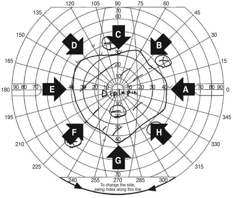
We asked patients about their history, subjective symptoms, symptom duration, and duration of thyroid disease at the first visit. Preoperative and postoperative evaluations included visual acuity, intraocular pressure, slit lamp exam, marginal reflex distance (MRD1) for lid height, Hertel exophthalmometry, presence of strabismus, and range of extraocular muscle motility using the sum of the angle in eight directions of a nondiplopic region in a binocular single vision field (BSV) (Fig. 1). Each patient underwent preoperative axial orbital computed tomography imaging with 2-mm cuts, and blood samples were obtained for thyroid function test and thyroid antibody test. The values of triiodothyronine, free thyroxine, TSH, thyroid stimulating antibody, TSH-receptor antibody, thyroglobulin antibody, and anti-thyroid peroxidase were obtained.
Fig. 1. Evaluation of diplopia using the sum of the angles in eight directions (0°, 45°, 90°, 135°, 180°, 225°, 270°, and 315° in binocular single vision field [BSV] charts) of a nondiplopic region in BSV. The sum of the angles in eight directions of a nondiplopic region in BSV was calculated using the following equation: A + B + C + D + E + F + G + H (°). The angle of the nondiplopic region in BSV at A = 0°, B = 45°, C = 90°, D = 135°, E = 180°, F = 225°, G = 270°, and H = 315°. The sum in this BSV chart was evaluated at 303° (42° + 35° + 30° + 38° + 38° + 38° + 45° + 37°).
Pictures of the patients' faces were taken before the operation, 1 week postoperatively, and 1 month, 3 months, and 6 months postoperatively. To analyze the change in lid height, particularly lid retraction, MRD1 was analyzed using Image J (National Institutes of Health, Bethesda, MD, USA) before and after surgery. Values of triiodothyronine, free thyroxine, and TSH were measured, and the thyroid antibody test was performed. All patients had a minimum preoperative period of 3 months of a stable range of ocular motility and eyelid position. All patients had inactive TAO and were euthyroid at the time of operation.
If patients showed only proptosis, we typically planned only orbital decompression; however, if patients showed proptosis with upper lid retraction (MRD1 >5 mm), we planned an additional surgery for lid correction. If patients showed proptosis with restrictive diplopia at the primary eye position, we also planned additional surgery for strabismus correction. If patients showed all symptoms of proptosis, eyelid retraction, and restrictive diplopia, we planned additional surgeries for lid correction and strabismus correction.
For customized orbital decompression, deep lateral decompression can reduce Hertel exophthalmometry by approximately 1.5 mm, and fat decompression can reduce Hertel exophthalmometry by approximately 1.0 mm per 1 mL. For patients with upper lid retraction, deep lateral decompression does not affect lid height after surgery. We planned transconjunctival levator disinsertion if the preoperative lid retraction value was less than 2 mm. If the preoperative lid retraction value was greater than 2 mm, we planned blepharotomy. If patients showed lower lid retraction, we planned englove lysis of lower lid retractors for correction [17]. If patients showed eyeball deviation as esotropia, we performed medial rectus muscle recession with an adjustment technique. If patients showed hypotropia, we performed inferior rectus muscle recession with an adjustment technique.
Surgical methods
1) Orbital decompression
All surgery was performed by one surgeon (HL) using general anesthesia. Epinephrine diluted with normal saline at 1 : 100,000 per 3 mL was injected for bleeding control before the incision was made. A transconjunctival approach using canthotomy was employed for orbital access. After conjunctival incision, the periosteum was dissected using an elevator. Then, using a high-speed drill and punch, cortical bone was removed from thick areas of the posterior lateral wall and the greater wing of the sphenoid bone (trigone). The extent of bone removal was individualized according to symptoms and proptosis severity. Orbital fat removal was performed from the infero-medial and infero-lateral intraconal spaces.
2) Eyelid retraction correction surgery
For eyelid retraction correction, Müller's muscle was removed through a transconjunctival approach, and trans-eyelid full-thickness blepharotomy was performed for severe cases. After disinfection, subcutaneous and subconjunctival local anesthesia was obtained using a total of 3 to 4 mL of 0.5% bupivacaine and 2% lidocaine with adrenaline (epinephrine) at a concentration of 1 : 200,000; following this, a transcutaneous approach was used at the level of the skin crease. For Müller's muscle excision, the surgeon inserted a traction suture into the upper lid and everted the lid over a Desmarres retractor. A short, full-thickness incision was created close to the upper border of the tarsal plate. The incision was then extended medially and laterally, parallel to the lid margin, to the full width of the tarsus. Then the Müller's muscle was disinserted and removed free from the upper fornix.
For trans-eyelid full-thickness blepharotomy, a transcutaneous approach was used at the level of the skin crease. The conjunctiva was then incised in the temporal third of the upper eyelid and gradually transected step by step, controlling the achieved height and contour of the lid. In cases of more severe upper lid retraction requiring a subtotal transsection of the conjunctiva, the conjunctiva was also horizontally transected in the medial third, not exceeding further medially than the upper punctum. Attention was paid to leave an intact central bridge of conjunctiva in the central part of the lid in the pupillary axis. The endpoint of surgery was determined when the upper eyelid covered the limbus by 0.5 to 1.5 mm. The wound was then closed using a running locked skin suture with 6-0 black silk.
3) Strabismus surgery
In cases of strabismus, recession of the restricted extraocular muscle was performed. The eyelid fissure was widened with a lid speculum. Traction was created with locking forceps at the conjunctiva near the limbus at 12 and 6 o'clock positions, and the eyeball was retracted medially. The conjunctiva and Tenon's capsule were incised at 2 and 4 o'clock positions and horizontally with sharp scissors 10 mm from the limbus. Tenon's capsule and the intermuscular septum were dissected, and the lateral rectus muscle was identified with a muscle hook. 6-0 vicryl double armed sutures were placed through the insertion site of the muscle, and the muscle was cut with tenotomy scissors. Next, the muscle was reattached to the sclera under approappropriate tension distant from the original insertion site with an adjustable technique. The conjunctiva was closed with 6-0 vicryl sutures in an interrupted fashion.
A complete comparison of preoperative and postoperative results was conducted 3 months after the operation. A paired samples t-test was used to calculate the differences between preoperative and postoperative values of exophthalmos and MRD1. Statistical analysis was carried out using PASW Statistics ver. 18.0 (SPSS Inc., Chicago, IL, USA).
Results
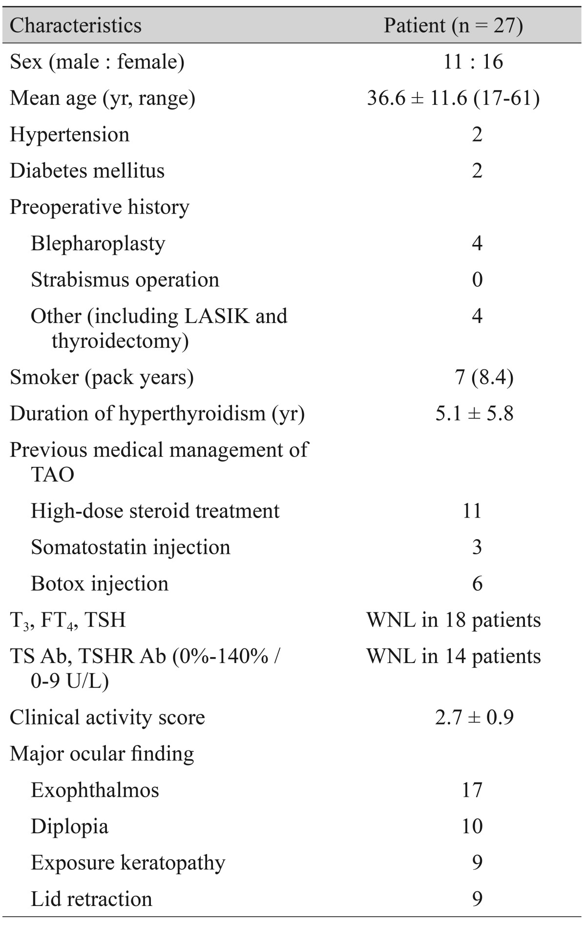
The 27 patients comprised 16 females and 11 males. The ages ranged from 17 to 61 years (36.6 ± 11.6 years). Two patients had a history of hypertension, and two patients had a history of diabetes mellitus. Seven patients had a history of smoking. The mean duration of thyroid disease was 5.1 ± 5.8 years. Most patients had undergone medical treatment for Graves' disease. Thyroid function test results were within normal limits for 18 patients, and thyroid antibodies (thyroid stimulating antibody, TSH receptor antibody, thyroglobulin antibody, and anti-thyroid peroxidase) were within normal limits in 14 patients at the time of operation. The mean clinical activity score was 2.7 ± 0.9 for all patients at the time of surgery. Every patient visited the endocrinology department and demonstrated stable thyroid function at the time of surgery. Eleven patients had undergone high-dose steroid treatment, three patients had received intramuscular lanreotide (Somatuline Depot; Ipsen, Paris, France) injection, and six patients had a history of botulinum toxin injection (Table 1). All patients had more than one typical clinical symptom such as congestion, dryness, and findings of exposure keratitis, diplopia, limitation of ocular motility, proptosis, eyelid retraction, or hyperemia of the conjunctiva.
Table 1. Clinical demographics of the patients who underwent customized decompression surgery.
Values are presented as number or mean ± standard deviation unless otherwise indicated.
LASIK = laser in situ keratomileusis; TAO = thyroid-associated ophthalmopathy; T3 = triiodothyronine; FT4 = free thyroxine; TSH = thyroid stimulating hormone; WNL = within normal limits; TS Ab = thyroid stimulating antibody; TSHR Ab = thyroid-stimulating hormone receptor antibody.
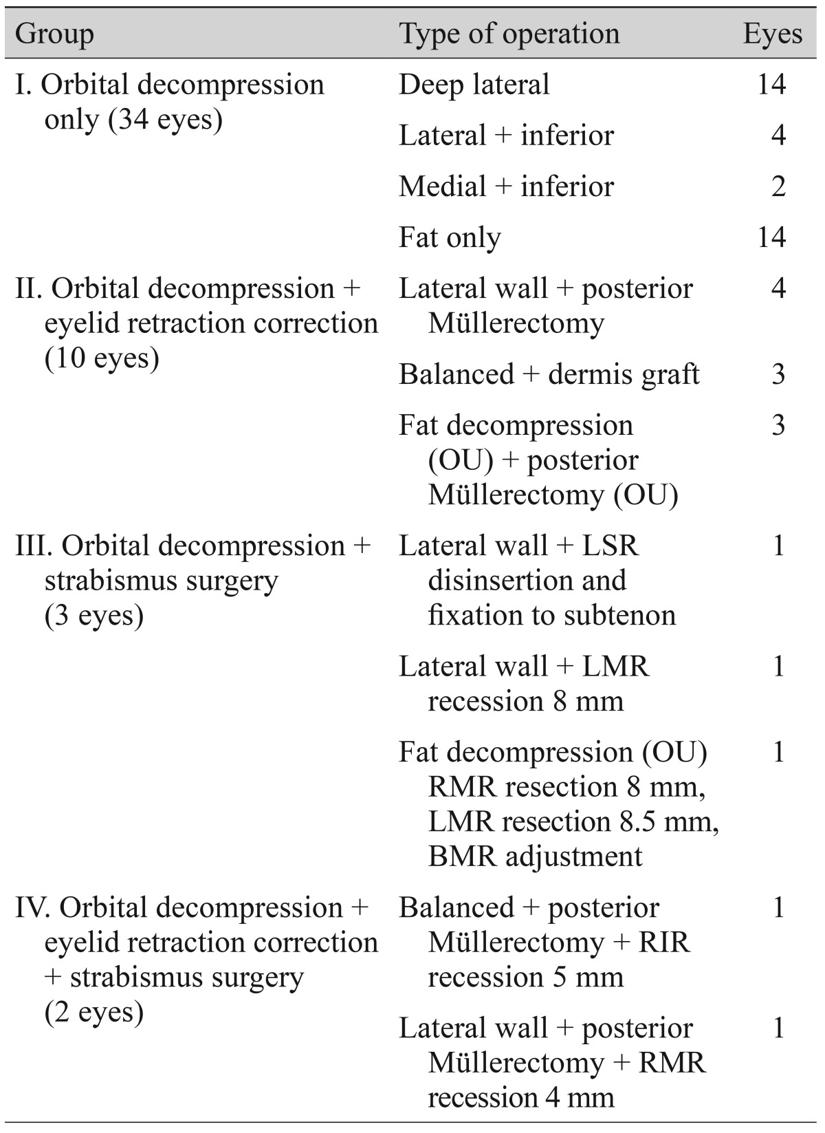
In 34 eyes, only orbital decompression surgery was performed (14 deep lateral orbital decompressions, 4 lateral and inferior orbital decompressions, 2 medial and inferior orbital decompressions, and 14 orbital fat decompressions), orbital decompression surgery with correction of eyelid retraction was performed in 10 eyes, orbital decompression with strabismus surgery was performed in 3 eyes, and orbital decompression surgery with correction of eyelid retraction and strabismus surgery was performed in 2 eyes. Patients were divided into four groups according to type of operation (group I, orbital decompression only; group II, orbital decompression with eyelid retraction correction; group III, orbital decompression with strabismus surgery; group IV, orbital decompression with eyelid retraction correction and strabismus surgery) (Table 2).
Table 2. Classification of patients according to type of operation.
OU = both eyes; LSR = left superior rectus muscle; LMR = left medial rectus muscle; RMR = right medial rectus muscle; BMR = bilateral medial rectus muscles; RIR = right inferior rectus muscle.
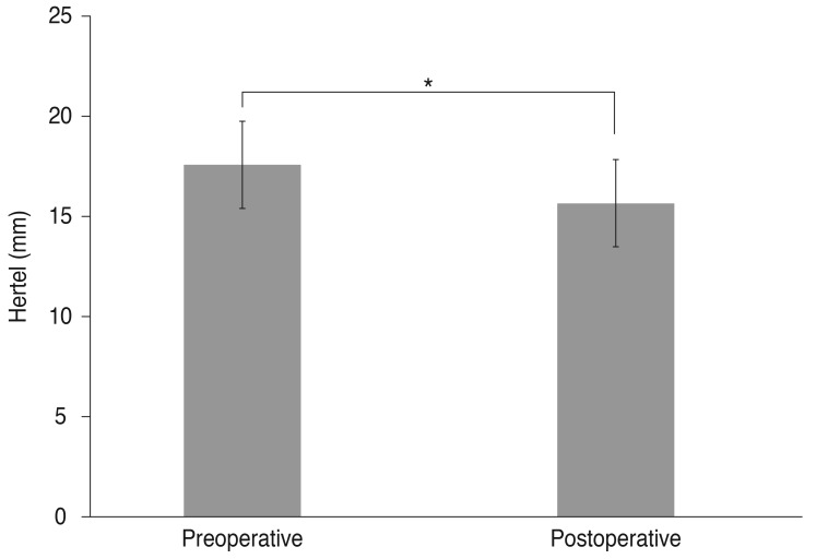
The mean Hertel values before and after decompression were 17.6 ± 2.2 mm and 15.6 ± 2.2 mm, respectively. The mean postoperative Hertel value change was 2.1 ± 2.0 mm (p = 0.00) (Fig. 2).
Fig. 2. The changes in exophthalmometric values measured with Hertel exophthalmometry following customized orbital decompression surgery. *p < 0.05.
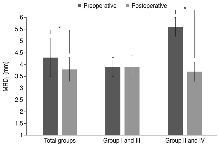
The mean MRD1 values of all patients decreased from 4.3 ± 0.8 mm to 3.8 ± 0.5 mm (p < 0.05) (Fig. 3). The mean MRD1 value of group II and IV (groups with eyelid retraction correction surgery) decreased from 5.6 ± 0.4 mm to 3.7 ± 0.4 mm (p < 0.05), and the mean MRD1 value of group I and III (groups without eyelid retraction correction surgery) decreased from 3.9 ± 0.4 mm to 3.9 ± 0.5 mm (p = 0.687).
Fig. 3. The changes in marginal reflex distance (MRD1) following customized orbital decompression surgery, and comparison of MRD1 change according to combined lid retraction surgery. Group I = orbital decompression only; group II = orbital decompression + eyelid retraction correction; group III = orbital decompression + strabismus surgery; group IV = orbital decompression + eyelid retraction correction + strabismus surgery. *p < 0.05.
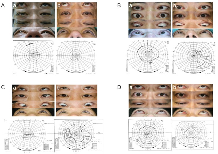
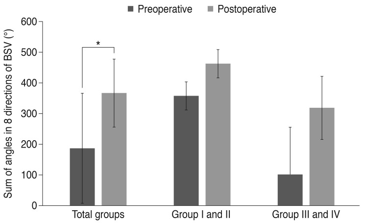
Representative data of patients in the four groups, including perioperative photos taken in upgaze, primary position, down gaze, and extension views, and BSV, are shown in Fig. 4A-4D. In BSV, the angle of the nondiplopic region increased in all patients. The mean preoperative sum of the angles in eight directions of the nondiplopic region in BSV was 186.7°, which increased to 366.7° after surgery (p < 0.05). No patients had double vision in the primary or reading position. The mean preoperative sum of the angle in eight directions of the nondiplopic region in BSV of groups III and IV (groups with strabismus surgery) was 101.3°, which increased to 318.8° (p = 0.068). The mean preoperative sum of the angles in eight directions of the nondiplopic region in BSV in groups I and II (groups without strabismus surgery) was 357.5°, which increased to 462.5° (p = 0.180) (Fig. 5).
Fig. 4. Representative data of patients in the four groups. Pictures taken in upgaze, primary position, down gaze, and extension views and binocular single-vision field preoperatively (left) and postoperatively (right). (A) A 42-year-old male underwent deep lateral decompression (right eye, OD). (Aa) shows preoperative status and (Ab) shows postoperative status. (B) A 47-year-old female underwent lateral decompression (both eyes, OU), transcaruncular orbitotomy with ethmoidectomy (OU), and lower eyelid retraction correction with dermis graft by englove lysis (OU). (Ba) is preoperative status and (Bb) is postoperative status. (C) A 49-year-old female underwent lateral and inferior decompression (left eye, OS) and left medial rectus muscle recession of 8 mm. (Ca) is preoperative status, and (Cb) is postoperative status. (D) A 37-year-old male underwent lateral orbital decompression (OD), posterior Müllerectomy (OD), and right medial rectus muscle recession of 4 mm. (Da) is preoperative status and (Db) is postoperative status.
Fig. 5. Relationships between group and the sum of the angles in eight directions of a nondiplopic region in binocular single-vision field (BSV [°]). Group I = orbital decompression only; group II = orbital decompression + eyelid retraction correction; group III = orbital decompression + strabismus surgery; group IV = orbital decompression + eyelid retraction correction + strabismus surgery. *p < 0.05.
No severe complications occurred after orbital decompression, eyelid retraction surgery, or strabismus surgery except one patient who experienced cerebrospinal leakage; the patient experienced spontaneous improvement without complications after 1 week of strict bed rest.
Discussion
The earliest surgical approach to orbital decompression, bony decompression of the lateral wall of the orbit, was performed by Dollinger in 1911 [18,19]. Recently, balanced decompression of the medial and lateral orbital walls has gained popularity because it can lessen the occurrence of postoperative strabismus [20,21,22]. In addition, the use of deep lateral decompression with intraconal fat removal has increased in an effort to minimize postoperative diplopia [14,15].
Traditional surgical treatment is a stepwise operation for management of TAO patients. Roncevic [23] insisted that strabismus operations should not be performed before orbital orbitaldecompression because the strabismus operation will only lead to temporary improvement in extraocular muscle function as it does not solve the basic cause of muscle changes. Therefore, a staged operation is generally recommended, as orbital decompression surgery-induced autoimmune inflammation and extraocular muscle and eyelid positions can occur [24]. Combined blepharoplasty and fat removal is now performed, in conjunction with lower lid retraction correction in some cases. On occasion, decompression, lid procedure, and combined muscle surgery are considered to protect the cornea and repair Bell's phenomenon in cases of very severe corneal threat.
The order of elective surgery has to be governed by the effects of various procedures on orbital structures; in principle, decisions about decompression should be first, since decompression can affect lid position and ocular deviation. The second procedure to consider is muscle surgery, since strabismus surgery can affect lid position. Finally, lid surgery is performed to address malposition, which might have been altered by the antecedent surgeries. It should be noted that the order has to be individualized depending on the technique and surgical approach.
Michel et al. [25] described eye muscle surgery performed during the same session as orbital decompression to reduce the rate of postoperative diplopia. One hundred forty-five endonasal decompressions were performed on 78 subjects, and ocular motility was corrected by recession of the medial rectus muscle in 58 of 78 cases. Twenty-six of these 58 cases underwent simultaneous operation immediately after transnasal decompression; the others were completed after a period of 2 to 3 months. A field free of double vision within margins of 20° in each direction was achieved in 31 of 58 subjects after the initial operation [25].
Moreover, Ben Simon et al. [14] evaluated 14 TAO patients who underwent deep lateral orbital wall decompression. After surgery, exophthalmos decreased by an average of 2.7 mm, and only two patients demonstrated clinically significant postoperative diplopia. They found that deep lateral wall orbital decompression with intraconal fat debulking had no statistical effect on horizontal or vertical ocular deviation as measured by automated Hess screen testing. These results are evidence that minimal to no ocular shift occurs with this type of orbital decompression [15].
Orbital medial wall and floor decompression surgeries are associated with up to 30% new-onset strabismus [16], whereas deep lateral wall decompression is associated with a much lower rate of new-onset diplopia (2% to 15%) [14,15]. Ben Simon et al. [15] published recent results that showed a new-onset primary gaze diplopia rate of only 2.6% in patients with mild to moderate TAO undergoing deep lateral wall decompression with intraconal fat debulking. In our study, after customized orbital decompression surgery, no new onset diplopia or aggravation of diplopia was noted, and no severe complications were seen.
The commonly involved extraocular muscles in TAO are the inferior, medial, superior, and lateral rectus muscles. Deep lateral decompression can be useful for TAO patients with medially shifted eyes rather than conventional medial orbital decompression or inferior decompression with fat removal for superiorly deviated eyeballs. After extraocular muscle recession in TAO, patients tend to have more prominent eyes. Therefore, we think that combined extraocular muscle surgery with decompression surgery demonstrates underestimated results in terms of exophthalmometric values.
Eyelid retraction surgery is usually reserved as the last surgery (with or without concurrent blepharoplasty) because large recessions of the vertical muscles can alter eyelid positions. Ben Simon et al. [16] compared the results of surgical outcomes between two groups, one of which underwent simultaneous orbital decompression and eyelid retraction surgery and one that underwent staged surgery, and reported similar outcomes; in fact, the study showed that eyelid retraction surgery can be performed simultaneously with deep lateral wall orbital decompression without affecting functional or cosmetic outcome. In our study, all patients had a better eyelid position after customized orbital decompression surgery. With regard to this eyelid position, we expect compression to reveal downward movements of the upper eyelid af ter decompression, so reasonable undercorrection of lid retraction during surgery would be the target of the upper eyelid. Significant functional improvement and acceptable aesthetic results were obtained in all patients.
By evaluating thyroid function and thyroid antibody tests and consulting endocrinologists, we were able to determine if thyroid disease was stable and if decompression surgery was appropriate. We were thus able to minimize orbital inflammation after decompression surgery.
There are some limitations to this study. First, the small number of patients limits valid comparison of techniques. Studies with more patients with longer follow-up would help better clarify the effects of customized orbital decompression surgery. Customized orbital decompression surgery combined with eyelid surgery or strabismus surgery in TAO can simultaneously correct postoperative exophthalmos, diplopia, and abnormal lid position; thus, this method is effective, has fewer side effects, and leads to earlier recovery.
Footnotes
This paper was presented in part at the 108th annual meeting of the Korean Ophthalmological Society, November, 2012 at Goyang, Korea.
Conflict of Interest: No potential conflict of interest relevant to this article was reported.
References
- 1.McCord CD Jr, Tanenbaum M, Nunery WR, editors. Oculoplastic surgery. 3rd ed. New York: Raven Press; 1995. p. 379. [Google Scholar]
- 2.Kazim M, Goldberg RA, Smith TJ. Insights into the pathogenesis of thyroid-associated orbitopathy: evolving rationale for therapy. Arch Ophthalmol. 2002;120:380–386. doi: 10.1001/archopht.120.3.380. [DOI] [PubMed] [Google Scholar]
- 3.Trokel S, Kazim M, Moore S. Orbital fat removal: decompression for Graves orbitopathy. Ophthalmology. 1993;100:674–682. doi: 10.1016/s0161-6420(93)31589-7. [DOI] [PubMed] [Google Scholar]
- 4.Park JH, Lee TS, Kay KM. Long-term result of fat orbital decompression. J Korean Ophthalmol Soc. 2010;51:473–478. [Google Scholar]
- 5.Kamer L, Noser H, Schramm A, et al. Anatomy-based surgical concepts for individualized orbital decompression surgery in graves orbitopathy: I. orbital size and geometry. Ophthal Plast Reconstr Surg. 2010;26:348–352. doi: 10.1097/IOP.0b013e3181c9bb52. [DOI] [PubMed] [Google Scholar]
- 6.Siracuse-Lee DE, Kazim M. Orbital decompression: current concepts. Curr Opin Ophthalmol. 2002;13:310–316. doi: 10.1097/00055735-200210000-00004. [DOI] [PubMed] [Google Scholar]
- 7.Shorr N, Seiff SR. The four stages of surgical rehabilitation of the patient with dysthyroid ophthalmopathy. Ophthalmology. 1986;93:476–483. doi: 10.1016/s0161-6420(86)33712-6. [DOI] [PubMed] [Google Scholar]
- 8.Goldberg RA, Perry JD, Hortaleza V, Tong JT. Strabismus after balanced medial plus lateral wall versus lateral wall only orbital decompression for dysthyroid orbitopathy. Ophthal Plast Reconstr Surg. 2000;16:271–277. doi: 10.1097/00002341-200007000-00004. [DOI] [PubMed] [Google Scholar]
- 9.Grenzebach UH, Schnorbus U, Buchner T, et al. Development of ocular motility following modified 3-wall decompression of the orbita in endocrine orbitopathy for functional and rehabilitative indication. Klin Monbl Augenheilkd. 2003;220:345–351. doi: 10.1055/s-2003-39423. [DOI] [PubMed] [Google Scholar]
- 10.Garrity JA, Saggau DD, Gorman CA, et al. Torsional diplopia after transantral orbital decompression and extraocular muscle surgery associated with Graves' orbitopathy. Am J Ophthalmol. 1992;113:363–373. doi: 10.1016/s0002-9394(14)76157-4. [DOI] [PubMed] [Google Scholar]
- 11.Nunery WR, Nunery CW, Martin RT, et al. The risk of diplopia following orbital floor and medial wall decompression in subtypes of ophthalmic Graves' disease. Ophthal Plast Reconstr Surg. 1997;13:153–160. doi: 10.1097/00002341-199709000-00001. [DOI] [PubMed] [Google Scholar]
- 12.Goldberg RA, Kim AJ, Kerivan KM. The lacrimal keyhole, orbital door jamb, and basin of the inferior orbital fissure: three areas of deep bone in the lateral orbit. Arch Ophthalmol. 1998;116:1618–1624. doi: 10.1001/archopht.116.12.1618. [DOI] [PubMed] [Google Scholar]
- 13.Beden U, Edizer M, Elmali M, et al. Surgical anatomy of the deep lateral orbital wall. Eur J Ophthalmol. 2007;17:281–286. doi: 10.1177/112067210701700301. [DOI] [PubMed] [Google Scholar]
- 14.Ben Simon GJ, Syed HM, Lee S, et al. Strabismus after deep lateral wall orbital decompression in thyroid-related orbitopathy patients using automated hess screen. Ophthalmology. 2006;113:1050–1055. doi: 10.1016/j.ophtha.2006.02.015. [DOI] [PubMed] [Google Scholar]
- 15.Ben Simon GJ, Wang L, McCann JD, Goldberg RA. Primary-gaze diplopia in patients with thyroid-related orbitopathy undergoing deep lateral orbital decompression with intraconal fat debulking: a retrospective analysis of treatment outcome. Thyroid. 2004;14:379–383. doi: 10.1089/105072504774193221. [DOI] [PubMed] [Google Scholar]
- 16.Ben Simon GJ, Mansury AM, Schwarcz RM, et al. Simultaneous orbital decompression and correction of upper eyelid retraction versus staged procedures in thyroid-related orbitopathy. Ophthalmology. 2005;112:923–932. doi: 10.1016/j.ophtha.2004.12.028. [DOI] [PubMed] [Google Scholar]
- 17.Chang HS, Lee D, Taban M, et al. "En-glove" lysis of lower eyelid retractors with AlloDerm and dermis-fat grafts in lower eyelid retraction surgery. Ophthal Plast Reconstr Surg. 2011;27:137–141. doi: 10.1097/IOP.0b013e3181c53d38. [DOI] [PubMed] [Google Scholar]
- 18.Lund VJ, Larkin G, Fells P, Adams G. Orbital decompression for thyroid eye disease: a comparison of external and endoscopic techniques. J Laryngol Otol. 1997;111:1051–1055. doi: 10.1017/s0022215100139313. [DOI] [PubMed] [Google Scholar]
- 19.White WA, White WL, Shapiro PE, et al. Combined endoscopic medial and inferior orbital decompression with transcutaneous lateral orbital decompression in Graves' orbitopathy. Ophthalmology. 2003;110:1827–1832. doi: 10.1016/S0161-6420(03)00566-9. [DOI] [PubMed] [Google Scholar]
- 20.Shepard KG, Levin PS, Terris DJ. Balanced orbital decompression for Graves' ophthalmopathy. Laryngoscope. 1998;108(11 Pt 1):1648–1653. doi: 10.1097/00005537-199811000-00011. [DOI] [PubMed] [Google Scholar]
- 21.Graham SM, Brown CL, Carter KD, et al. Medial and lateral orbital wall surgery for balanced decompression in thyroid eye disease. Laryngoscope. 2003;113:1206–1209. doi: 10.1097/00005537-200307000-00017. [DOI] [PubMed] [Google Scholar]
- 22.Unal M, Leri F, Konuk O, Hasanreisoglu B. Balanced orbital decompression combined with fat removal in Graves ophthalmopathy: do we really need to remove the third wall? Ophthal Plast Reconstr Surg. 2003;19:112–118. doi: 10.1097/01.IOP.0000056145.71641.F5. [DOI] [PubMed] [Google Scholar]
- 23.Roncevic R. Correction of exophthalmos and eyelid deformities in patients with severe thyroid ophthalmopathy. J Craniofac Surg. 2008;19:628–636. doi: 10.1097/SCS.0b013e31816adca2. [DOI] [PubMed] [Google Scholar]
- 24.Baldeschi L, Lupetti A, Vu P, et al. Reactivation of Graves' orbitopathy after rehabilitative orbital decompression. Ophthalmology. 2007;114:1395–1402. doi: 10.1016/j.ophtha.2006.10.036. [DOI] [PubMed] [Google Scholar]
- 25.Michel O, Oberlander N, Neugebauer P, et al. Follow-up of transnasal orbital decompression in severe Graves' ophthalmopathy. Ophthalmology. 2001;108:400–404. doi: 10.1016/s0161-6420(00)00533-9. [DOI] [PubMed] [Google Scholar]


