Abstract
Background & objectives:
Plasma amino acid levels are known to be altered in conditions like sepsis and burns which are situations of metabolic stress. Polycystic ovary syndrome (PCOS), a condition which affects a woman throughout her life, is said to be associated with metabolic stress. This study was undertaken to assess if there were significant alterations in the levels of plasma amino acids in women with PCOS.
Methods:
Sixty five women with PCOS along with the similar number of age matched normal controls were included in this study. Levels of 14 amino acids were determined using reverse phase high performance liquid chromatography.
Results:
The levels of methionine, cystine, isoleucine, phenylalanine, valine, tyrosine, proline, glycine, lysine and histidine were found to be significantly (P<0.001) lower in cases than in controls. Arginine and alanine levels were found to be significantly (P<0.001) higher in cases compared with controls.
Interpretation & conclusions:
Our findings showed significant derangement in the levels of plasma amino acids in women with PCOS which might be due to the oxidative and metabolic stress associated with it. Further studies need to be done to confirm the findings.
Keywords: Amino acids-arginine, branched chain amino acids, cysteine, oxidative stress, polycystic ovary syndrome
Polycystic ovary syndrome (PCOS) is one of the common endocrinological disorders that affects 4-12 per cent of women during their reproductive years leading to a wide spectrum of clinical manifestations1. The condition is known to have a major effect throughout life on the metabolic, reproductive and cardiovascular health of affected women. It was first described by Stein and Leventhal in 1935 as a symptom complex associated with oligomenorrhoea, hirsuitism, obesity and bilaterally enlarged polycystic ovaries2. The current diagnostic criteria for PCOS is the 2003 Rotterdam European Society of Human Reproduction and Embryology / American Society for Reproductive Medicine (ESHRE/ASRM) revised consensus according to which at least two of the three following criteria are needed for the diagnosis: (i) anovulation or oligoovulation, (ii) clinical and/or biochemical signs of hyperandrogenism, and (iii) presence of polycystic ovaries on ultrasonography and exclusion of related disorders. Although not enclosed within the diagnostic criteria, a significant feature of PCOS is insulin resistance that results in compensative hyperinsulinaemia, acanthosis nigricans, hyperandrogenism, cardiovascular risk, and type 2 diabetes mellitus3.
Amino acids function as essential precursors for synthesis of a variety of molecules of significance and also regulate key metabolic pathways and processes that are vital for the proper functioning and maintenance of homeostasis of organisms4. Besides their role as building blocks of proteins and polypeptides, amino acids are also said to have antioxidant functions. Plasma proteins are considered preventive antioxidants and act by sequestering or inactivating transition metal catalysts (e.g. transferrin, ceruloplasmin). These were also discovered to be chain-breaking antioxidants5. Normally, the levels of reactive oxygen species (ROS) and antioxidants remain in balance. Oxidative stress occurs when ROS exceeds the level of antioxidants6. Free radical attack on lipids leads to lipid peroxidation resulting in the formation of reactive aldehydes which can diffuse into the cells, and attack targets far from the site of the original event. Some of these aldehydes have been shown to react with various biomolecules like proteins, DNA, and phospholipids7.
Reproductive cells and tissues remain stable only when antioxidant and oxidant status is in balance1. Increased oxygen free radical production and decreased catalase activity have been observed in PCOS8. Palacio et al9 suggested that the oxidative status in PCOS was supported by high levels of malondialdehyde (MDA) modified proteins in serum. Oxidation of proteins can result in cleavage of the polypeptide chain and modification of amino acid side chains leading to impairment of their activity and thus result in various pathological states10.
The role of free radical mediated oxidation of amino acids in PCOS has not been explored. Hence we conducted this pilot study with the aim to find out if there were any changes in the levels of plasma amino acids in women with PCOS.
Material & Methods
This study was conducted in 65 consecutive women in the age group of 20-40 yr with clinically proven PCOS, attending the outpatients of Obstetrics and Gynecology department of Amrita Institute of Medical Sciences (AIMS), Kochi, Kerala, India. A similar number of age matched healthy women with regular menstrual cycles were included as controls. Controls were apparently normal healthy women presenting for routine health check-up in the comprehensive health check-up clinic of AIMS. We also included healthy female staff and students of AIMS who were also of the same age group. The duration of the study was for 10 months from October 2011 to August 2012. The study was conducted after the approval of the protocol by the ethical committee of AIMS - School of Medicine and informed, written consents of the participants were obtained.
Inclusion criteria: Women (20-40 yr) with complaints of any two of the following were included: oligomenorrhoea/amenorrhoea and inability to conceive, hirsuitism and polycystic ovaries in ultrasonography3. None of these women received any hormonal contraceptives, vitamin supplements or any other significant drug therapy. None of them were alcoholics or chronic smokers and did not suffer from any other illnesses.
Exclusion criteria: Women with the following conditions were excluded from the study: those with hyperprolactinaemia, androgen secreting neoplasm, congenital adrenal hyperplasia, history of diabetes mellitus, liver or pancreatic diseases, history of infections or fever in the recent past, those on steroids, antiepileptic drugs like valproate or any other significant drug therapy, any chronic illness and any history of muscle trauma in the recent past.
Anthropometric measurements of the patients namely, weight (kg) and height (metres) were taken to assess the body mass index (BMI). Patients were classified into obese and non obese based on the BMI (those having BMI ≥25 kg/m2 were classified as obese and those with BMI <25 kg/m2 as non obese)11.
Laboratory analysis: Venous blood samples (4 ml) were collected with aseptic precautions in vacutainers without anticoagulant to measure serum luteinising hormone (LH), follicle stimulating hormone (FSH), thyroid stimulating hormone (TSH), prolactin, insulin and high sensitive C-reactive protein (hsCRP). Vacutainers containing fluoride were used for collecting blood (2 ml) for measuring fasting plasma glucose and 2-h oral glucose tolerance test (OGTT). World Health Organization (WHO, 2006) criteria12 were used to determine the presence of diabetes mellitus. Fasting plasma glucose values between 110 - 126 mg/dl and 2-h post-glucose values ≥140 mg/dl but less than 200 mg/dl indicated impaired glucose tolerance while fasting plasma glucose ≥126 mg/dl or 2-h post-glucose values ≥200 mg/dl indicated frank diabetes mellitus12. Insulin resistance was evaluated by homeostatic model assessment of insulin resistance (HOMA-IR)13,14. Values more than 2.5 were consistent with insulin resistance13,14. Serum LH15, FSH15, TSH16, prolactin15 and insulin17 levels were estimated by chemiluminiscent microparticle immunoassay (Abott Architect i 2000 SR; Illinois, USA). Serum dehydroepiandrostenedione (DHEAS)18 was estimated by electrochemiluminiscence (Roche Cobas E411, Germany). Total cholesterol, HDL, LDL, VLDL, triglycerides, blood glucose and 2-h OGTT were measured using the principle of colorimetry in Beckman Coulter Olympus AU 2700, USA. The hsCRP level was measured using immunoturbidimetry (Beckman Coulter Olympus AU 2700, USA)19. Total cholesterol19, HDL19, LDL19, VLDL19, triglycerides19, 2-h OGTT20 and blood glucose20 were measured using the principle of colorimetry in Beckman Coulter Olympus AU 2700; USA.
For measuring plasma free amino acids, the patients and controls were instructed to have normal diet (avoid high protein diet) and avoid strenuous physical exercise prior to sample collection. Fourteen amino acids were measured- glycine, alanine, valine, leucine, isoleucine, tyrosine, phenylalanine, methionine, cystine, serine, histidine, arginine, lysine and proline. Venous blood samples (2 ml) were obtained under aseptic precautions in EDTA treated vacutainers. Plasma separated from the samples after centrifugation at 1500 g for 15 min was used for estimation of amino acids. Those samples that were not immediately analysed were stored at -20°C and the assay was performed within two weeks. Samples were deproteinized with sulphosalicylic acid and treated with phenyl isothiocyanate (PITC) and triethylamine (TEA) prior to injection-precolumn derivatization. Amino acids were separated using reverse phase - high performance liquid chromatography (HPLC)21 using Column Phenomenex LUNA C18 (250x4.6mm i.d; 5 μm particle size, Torrance, CA, USA).
In addition to monitoring the individual amino acids, the amino acids were also grouped as follows: (i) phenylalanine/tyrosine ratio (Phe/Tyr); (ii) glycine: branched chain amino acids (BCAA) ratio (Gly/BCAA); (iii) glycine: valine ratio (Gly/Val), and (iv) BCAA to aromatic amino acids (AAA) ratio- Fischer's ratio22.
Statistical analysis: For all the continuous variables the results are given in mean ± standard deviation. To compare the means of continuous parameters between groups, those following normal distribution, Student's independent samples t test was performed. To compare the average of continuous parameters between groups, those not following normal distribution, Mann Whitney test was performed.
Results
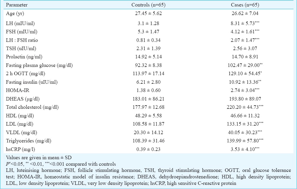
No significant difference was seen between the mean age of patients and controls. Significantly elevated LH, LH: FSH ratio, fasting plasma glucose, 2-h OGTT, fasting insulin, total cholesterol, LDL, triglycerides, VLDL and hsCRP were seen in patients with PCOS compared with controls. FSH levels were significantly (P<0.001) decreased. TSH, prolactin, DHEAS and HDL levels did not show significant difference. (Table I) Insulin resistance was calculated using the HOMA-IR [fasting plasma glucose (mg/dl) x fasting serum insulin (uIU/ml)/405] and was found to be significantly (P<0.001) increased in patients than controls. Only five cases were found to have diabetes mellitus.
Table I.
Baseline characteristics of controls and patients with PCOS

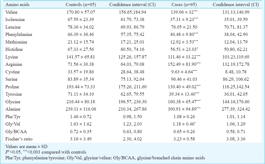
When compared with the normal controls, the levels of methionine, cystine, isoleucine, phenylalanine, valine, tyrosine, proline, glycine, lysine and histidine were found to be significantly lower in cases. The levels of arginine and alanine were significantly higher in cases than controls. Leucine and serine levels were not significantly different between cases and controls. Glycine/Valine ratio was significantly (P<0.05) decreased in cases than controls (Table II).
Table II.
Plasma amino acid levels (μmol/l) in patients with PCOS and controls

Discussion
In our study significantly lower levels of histidine, methionine, cystine, isoleucine, phenylalanine, valine, tyrosine, proline, glycine and lysine were observed in women with PCOS compared with normal controls. Histidine is considered as an anti-inflammatory amino acid and an antioxidant. Being a nucleophilic amino acid it is vulnerable to modification by lipid peroxidation derived electrophiles such as 2-alkenals, ketoaldehydes and 4-hydroxy-2-alkenals7. Thus the low levels of this amino acid found in PCOS patients may be due to its increased utilization as an antioxidant in the presence of oxidative stress. In addition, the increased concentration of free radicals might have led to its oxidation resulting in decreased levels.
Cystine is the oxidized form of cysteine which is a sulphur containing amino acid. Glutathione (GSH) is gamma glutamyl cysteinyl glycine, a tripeptide that is known for its antioxidant property23. It is synthesized by most cells and is one of the primary cellular antioxidants responsible for maintaining the proper antioxidant state within the body23. Due to the oxidative stress present in patients with PCOS increased amounts of cystine is converted to cysteine and then to GSH to meet the stressful situation. As a result, low levels of cystine are obtained.
Study done by Katayama and Mine23 revealed that BCAA such as leucine, isoleucine and valine have the capacity to upregulate the activities of the antioxidant enzymes glutathione-S-transferase (GST) and catalase. Thus the potent induction of catalase and GST may contribute to the protective effects of BCAA against oxidative stress. The low levels of isoleucine and valine obtained in our patients might be because of the increased utilization of these amino acids to combat the oxidative stress. Levels of leucine were similar in cases and controls, the reasons for the same were not known. Due to the increased oxidative stress in patients with PCOS increased amounts of methionine are subjected to its oxidised form resulting in its decreased concentration. Also, because of the high oxidative stress, regeneration of methionine may not be sufficient24. This might be the reason for low levels of methionine seen in our patients.
Singlet oxygen is an active oxygen species that can cause oxidative damage in biological systems. Proline is an effective quencher of this free radical. It has been shown that proline can reduce the levels of malondialdehyde produced during strong illumination of isolated thylakoids25. Schuessler and Schilling26 have proposed that proline residues in polypeptide chains are the site of oxygen radical-mediated cleavage of polypeptide chain. Whether proline acts against other free radicals is not known. Thus the low levels of proline observed in our study group may be due to the increased oxidative stress leading to increased utilization of proline.
Hyperphenylalaninemia has been associated with conditions like burns27 and sepsis28. Tetrahydrobiopterin (BH4) which is the coenzyme for phenylalanine hydroxylase (PAH) is chemically very labile. Hence it is easily susceptible to oxidative damage and thus causes diminution of the PAH activity29. As a result, the phenylalanine accumulates and tyrosine level decreases. In our study decreased levels of both amino acids were observed; reasons being unknown. Alanine is known to cause induction of glutathione reductase which helps to elevate the levels of GSH23. This maintains the antioxidant status of the cells. Similarly, arginine through nitric oxide scavenges free radicals and thus functions as an antioxidant30. In our study elevated levels of alanine and arginine were observed. This could probably be the body's compensatory mechanism to meet the elevated oxidative stress.
Phenylalanine/tyrosine ratio is an indicator of the catabolic state of the body22. An elevated Phe/Tyr ratio can be seen in conditions like HIV infection31, phenylketonuria, burns27, and sepsis28. In our patients due to the increased oxidative stress an elevated ratio was expected, but no significant difference was seen.
N-acetyl cysteine (NAC) is a derivative of cysteine and is considered as an antioxidant and antiapoptotic agent. Inside the cells it is said to stimulate glutathione production and thus scavenge free radicals. The NAC is also known to influence insulin receptor activity and thus increases the uptake of glucose32.
In conclusion, significant alterations in the levels of plasma amino acids were observed in patients with PCOS. Whether supplementation of amino acids can restore their levels in plasma or can improve the pathologies in PCOS like infertility, menstrual disturbances and hyperandrogenism has not yet been proved. Hence more studies with a large sample need to be performed to reach a definite conclusion.
Footnotes
Conflicts of Interest: None.
References
- 1.Lee JY, Baw CK, Gupta S, Aziz N, Agarwal A. Role of oxidative stress in polycystic ovary syndrome. Curr Womens Health Rev. 2010;6:96–107. [Google Scholar]
- 2.Stein IF, Leventhal ML. Amenorrhea associated with bilateral polycystic ovaries. Am J Obstet Gynecol. 1935;29:181–91. [Google Scholar]
- 3.Sheehan MT. Polycystic orarian syndrome: diagnosis and management. Clin Med Res. 2004;2:13–27. doi: 10.3121/cmr.2.1.13. [DOI] [PMC free article] [PubMed] [Google Scholar]
- 4.Wu G. Amino acids: metabolism, functions, and nutrition. Amino Acids. 2009;37:1–17. doi: 10.1007/s00726-009-0269-0. [DOI] [PubMed] [Google Scholar]
- 5.Wayner DD, Burton GW, Ingold KU, Barclay LR, Locke SJ. The relative contributions of vitamin E, urate, ascorbate and proteins to the total peroxyl radical-trapping antioxidant activity of human blood plasma. Biochim Biophys Acta. 1987;924:408–19. doi: 10.1016/0304-4165(87)90155-3. [DOI] [PubMed] [Google Scholar]
- 6.Agarwal A, Gupta S, Sharma RK. Role of oxidative stress in female reproduction. Reprod Biol Endocrinol. 2005;3:28. doi: 10.1186/1477-7827-3-28. [DOI] [PMC free article] [PubMed] [Google Scholar]
- 7.Uchida K. Histidine and lysine as targets of oxidative modification. Amino Acids. 2003;25:249–57. doi: 10.1007/s00726-003-0015-y. [DOI] [PubMed] [Google Scholar]
- 8.Mohan SK, Priya VV. Lipid peroxidation, glutathione, ascorbic acid, vitamin E, antioxidant enzyme and serum homocysteine status in patients with polycystic ovary syndrome. Biol Med. 2009;1:44–9. [Google Scholar]
- 9.Palacio JR, Iborra A, Ulcova-Gallova Z, Badia R, Martínez P. The presence of antibodies to oxidative modified proteins in serum from polycystic ovary syndrome patients. Clin Exp Immunol. 2006;144:217–22. doi: 10.1111/j.1365-2249.2006.03061.x. [DOI] [PMC free article] [PubMed] [Google Scholar]
- 10.Stadtman ER. Protein oxidation and aging. Science. 1992;257:1220–4. doi: 10.1126/science.1355616. [DOI] [PubMed] [Google Scholar]
- 11.Weisell RC. Body mass index as an indicator of obesity. Asia Pac J Clin Nutr. 2002;11(Suppl):S681–4. [Google Scholar]
- 12.American Diabetes Association. Diagnosis and classification of diabetes mellitus. Diabetes Care. 2010;33:S62–9. doi: 10.2337/dc10-S062. [DOI] [PMC free article] [PubMed] [Google Scholar]
- 13.da Silva R, Miranda WL, Chacra AR, Dib SA. Metabolic syndrome and insulin resistance in normal glucose tolerant Brazilian adolescents with family history of type 2 diabetes. Diabetes Care. 2005;28:716–8. doi: 10.2337/diacare.28.3.716. [DOI] [PubMed] [Google Scholar]
- 14.Rekha S, Patel ML, Gupta P, Diwakar A, Sachan P, Natu SM. Correlation between elevated homocysteine levels and insulin resistance in infertile women with or without polycystic ovary syndrome in North Indian population. Int J Med Med Sci. 2013;5:116–23. [Google Scholar]
- 15.Demers ML, Vance ML. Pituitary function. In: Burtis CA, Ashwood ER, Burns DE, editors. Tietz textbook of clinical chemistry and molecular diagnostics. 4th ed. Amsterdam, The Netherlands: Elsevier; 2006. pp. 1980–7. [Google Scholar]
- 16.Demers ML, Spencer C. The thyroid: Pathophysiology and thyroid function testing. In: Burtis CA, Ashwood ER, Burns DE, editors. Tietz textbook of clinical chemistry and molecular diagnostics. 4th ed. Amsterdam, The Netherlands: Elsevier; 2006. p. 2066. [Google Scholar]
- 17.Moriyama M, Hayashi N, Ohyabu C, Mukai M, Kawano S, Kumagai S. Performance evaluation and cross-reactivity from insulin analogs with the Architect insulin assay. Clin Chem. 2006;52:1423–6. doi: 10.1373/clinchem.2005.065995. [DOI] [PubMed] [Google Scholar]
- 18.Haymond S, Gronowsky AM. Reproductive related disorders. In: Burtis CA, Ashwood ER, Burns DE, editors. Tietz textbook of clinical chemistry and molecular diagnostics. 4th ed. Amsterdam, The Netherlands: Elsevier; 2006. pp. 2132–3. [Google Scholar]
- 19.Rifai N, Warnick GR. Lipids, lipoproteins, apolipoproteins and other cardiovascular risk factors. In: Burtis CA, Ashwood ER, Burns DE, editors. Tietz textbook of clinical chemistry and molecular diagnostics. 4th ed. Amsterdam, The Netherlands: Elsevier; 2006. pp. 942–51. [Google Scholar]
- 20.Sacks DB. Carbohydrates. In: Burtis CA, Ashwood ER, Burns DE, editors. Tietz textbook of clinical chemistry and molecular diagnostics. 4th ed. Amsterdam, The Netherlands: Elsevier; 2006. pp. 860–70. [Google Scholar]
- 21.Hariharan M, Naga S, VanNoord T. Systematic approach to the development of plasma amino acid analysis by High performance liquid chromatography with ultraviolet detection with precolumn derivatization using Phenylisothiocyanate. J Chromatography. 1993;621:15–22. doi: 10.1016/0378-4347(93)80071-b. [DOI] [PubMed] [Google Scholar]
- 22.Girish BN, Rajesh G, Vaidyanathan K, Balakrishnan V. Alterations in plasma amino acid levels in chronic pancreatitis. JOP. 2011;12:11–8. [PubMed] [Google Scholar]
- 23.Katayama S, Mine Y. Antioxidative activity of amino acids on tissue oxidative stress in human intestinal epithelial cell model. J Agric Food Chem. 2007;55:8458–64. doi: 10.1021/jf070866p. [DOI] [PubMed] [Google Scholar]
- 24.Levine RL, Moskovitz J, Stadtman ER. Oxidation of methionine in proteins: roles in antioxidant defense and cellular regulation. IUBMB Life. 2000;50:301–7. doi: 10.1080/713803735. [DOI] [PubMed] [Google Scholar]
- 25.Alia, Saradhi PP, Mohanty P. Proline enhances primary photochemical activities in isolated thylakoid membrane of Brassica juncea by arresting photoinhibitory damage. Biochem Biophys Res Commun. 1991;181:1238–44. doi: 10.1016/0006-291x(91)92071-q. [DOI] [PubMed] [Google Scholar]
- 26.Schuessler H, Schilling K. Oxygen effect in the radiolysis of proteins. Part 2. Bovine serum albumin. Int J Radiat Biol Relat Stud Phys Chem Med. 1984;45:267–81. doi: 10.1080/09553008414550381. [DOI] [PubMed] [Google Scholar]
- 27.Chang XJ, Yang CC, Hsu WS, Xu WZ, Shih TS. Serum and erythrocyte amino acid pattern: studies on major burn cases. Burns Incl Therm Inj. 1983;9:240–8. doi: 10.1016/0305-4179(83)90053-0. [DOI] [PubMed] [Google Scholar]
- 28.Sprung CL, Cerra FB, Freund HR, Schein RM, Konstantinides FN, Marcial EH, et al. Amino acid alterations and encephalopathy in the sepsis syndrome. Crit Care Med. 1991;19:753–7. doi: 10.1097/00003246-199106000-00004. [DOI] [PubMed] [Google Scholar]
- 29.Ploder M, Neurauter G, Spittler A, Schroecksnadel K, Roth E, Fuchs D. Serum phenylalanine in patients post trauma and with sepsis correlate to neopterin concentrations. Amino Acids. 2008;35:303–7. doi: 10.1007/s00726-007-0625-x. [DOI] [PubMed] [Google Scholar]
- 30.Tripathi P, Misra MK. Therapeutic role of L-arginine on free radical scavenging system in ischemic heart diseases. Indian J Biochem Biophys. 2009;46:498–502. [PubMed] [Google Scholar]
- 31.Zangerle R, Kurz K, Neurauter G, Kitchen M, Sarcletti M, Fuchs D. Increased blood phenylalanine to tyrosine ratio in HIV-1 infection and correction following effective antiretroviral therapy. Brain Behav Immun. 2010;24:403–8. doi: 10.1016/j.bbi.2009.11.004. [DOI] [PubMed] [Google Scholar]
- 32.Fulghesu AM, Ciampelli M, Muzg G, Belosi C, Selvaggi L, Ayala GF, et al. N-acetyl cysteine treatment improves insulin sensitivity in women with polycystic ovarian syndrome. Fertil Steril. 2002;77:1128–35. doi: 10.1016/s0015-0282(02)03133-3. [DOI] [PubMed] [Google Scholar]


