Abstract
Objective:
The study was aimed to evaluate the precision of Elekta four-dimensional (4D) cone beam CT (CBCT)-based automatic dual-image registrations using different landmarks for clipbox for radiation treatment of lung cancer.
Methods:
30 4D CBCT scans from 15 patients were studied. 4D CBCT images were registered with reference CT images using dual-image registration: a clipbox registration and a mask registration. The image registrations performed in clinic using a physician-defined clipbox, were reviewed by physicians, and were taken as the standard. Studies were conducted to evaluate the automatic dual registrations using three kinds of landmarks for clipbox: spine, spine plus internal target volume (ITV) and lung (including as much of the lung as possible). Translational table shifts calculated from the automatic registrations were compared with those of the standard.
Results:
The mean of the table shift differences in the lateral direction were 0.03, 0.03 and 0.03 cm, for clipboxes based on spine, spine plus ITV and lung, respectively. The mean of the shift differences in the longitudinal direction were 0.08, 0.08 and 0.08 cm, respectively. The mean of the shift differences in the vertical direction were 0.03, 0.03 and 0.03 cm, respectively.
Conclusion:
The automatic registrations using three different landmarks for clipbox showed similar results. One can use any of the three landmarks in 4D CBCT dual-image registration.
Advance in knowledge:
The study provides knowledge and recommendations for application of Elekta 4D CBCT image registration in radiation therapy of lung cancer.
INTRODUCTION
Cone beam CT (CBCT), which can provide visualization of bony anatomy and soft tissues, is being used in radiation therapy as image guidance to ensure the accuracy of treatment delivery.1,2 Image registrations between planning CT and CBCT, which use volumetric landmarks, have been used to correct patient setup errors.3–6 In an image registration, a volume of interest defined with a solid box in the images is called clipbox. Information within the clipbox is used for image registration. There have been studies7–9 to compare the precision of automatic image registrations using different landmarks for clipbox in radiation treatment of lung cancer, and it has been reported that using different landmarks for clipbox in automatic image registrations led to different precisions. These studies,7–9 however, were based on three-dimensional (3D) CBCT. Recently, four-dimensional (4D) CBCT became available in clinics.10–12 4D CBCT has been used to quantify localization precision and intrafraction variability of lung tumour position and validate planning target volume (PTV) margins,10 to serve as the gold standard for comparing different 3D CBCT registration techniques11 and to quantify interfraction and intrafraction tumour motion.12 In an Elekta X-ray volume imaging (XVI) 4D CBCT system (Elekta Oncology Systems Ltd, Crawley, UK), 4D CBCT data sets are created with respiratory correlated CBCT reconstruction. The breathing signal necessary for respiratory correlation is extracted from two-dimensional projection data of diaphragm motion.13 Dual-image registration is an option for the 4D CBCT image registration, which includes two image registration processes or steps: a clipbox registration followed by a mask registration. The clipbox registration is the same as those in 3D CBCT, while the mask registration is a new feature of Elekta XVI CBCT system, which is a soft-tissue registration using a soft-tissue volume called mask. A mask can be created from any structure contour in planning CT by a user, with or without a margin. In our clinic, a mask is created from PTV with a fixed margin. In the mask registration, only the volume of the reference planning CT within the mask is used for soft-tissue registration. In the automatic dual-image registration, the clipbox is thus the only variable which could be defined differently by different users.
The aim of the current study was to examine whether automatic 4D CBCT dual-image registration using different landmarks for clipbox will lead to different precisions and, therefore, to provide knowledge or recommendations for the application of Elekta 4D CBCT dual-image registration in radiation treatment of lung cancer. To the authors' knowledge, no similar studies had been reported.
METHODS AND MATERIALS
30 4D CBCT scans (i.e., 30 study cases) from 15 patients with lung cancer were studied. The scans were selected randomly from the lung patients' daily CBCT. Tumour information is listed in Table 1.
Table 1.
Tumour location, classification and volumes
| Study (scan) ID | Location | Classification | Internal target volume (cm3) | Planning target volume (cm3) |
|---|---|---|---|---|
| 1, 2 | RUL | Peripheral | 3.45 | 15.69 |
| 3, 4 | RML | Central | 1.78 | 5.43 |
| 5, 6 | RUL | Peripheral | 4.16 | 37.89 |
| 7, 8 | RUL | Peripheral | 6.28 | 47.99 |
| 9, 10 | LLL | Peripheral | 63.73 | 145.99 |
| 11, 12, 13 | RUL | Peripheral | 17.12 | 34.89 |
| 14, 15, 16, 17, 18, 19, 20 | RUL | Peripheral | 38.28 | 61.25 |
| 21 | LLL | Peripheral | 3.86 | 21.22 |
| 22 | LUL | Peripheral | 2.44 | 10.34 |
| 23 | RLL | Peripheral | 0.82 | 8.60 |
| 24 | LLL | Peripheral | 2.76 | 61.94 |
| 25 | RUL | Peripheral | 12.18 | 43.69 |
| 26 | RLL | Peripheral | 13.94 | 27.60 |
| 27 | LLL | Peripheral | 5.13 | 11.76 |
| 28 | LUL | Peripheral | 3.70 | 27.73 |
| 29 | RUL | Peripheral | 17.46 | 49.59 |
| 30 | LLL | Peripheral | 14.26 | 53.80 |
ID, identification number; LLL, left lower lobe; LUL, left upper lobe; RLL, right lower lobe; RML, right middle lobe; RUL, right upper lobe.
For treatment planning, patients were scanned on a GE LightSpeed 4D CT scanner (General Electric, Fairfield, CT), with a Varian Real-time Position Management™ system (Varian® Medical Systems, Palo Alto, CA). 10-phase images were reconstructed from the 4D CT scan. Gross target volume (GTV) was contoured on each phase, and an internal target volume (ITV) was generated by including the GTVs in 10-phase images. A PTV was created from the ITV with a 5- to 10-mm margin. Average-intensity-projection (AveIP) images, which were reconstructed from the 4D CT scan, were used for treatment planning and were sent to CBCT system as a reference image for image registration.
Prior to treatment, the patients were scanned on an Elekta XVI CBCT system using a 4D module—Symmetry v. R4.5. Symmetry is a module that includes 4D CBCT image acquisition, in-line reconstruction and automated anatomically correlated 4D registration to find a time-weighted average position of the tumour. A Vac-Lok™ bag (Civco Medical Solutions, Coralville, IA) was used for patient immobilization during CT scan, CBCT scan and treatment. 4D CBCT images were registered with reference CT images using dual-image registration. The 4D CBCT system, including the dual-image registration, had been commissioned with phantom tests when the 4D CBCT system was initially installed. In the study, a mask was defined as PTV plus 5-mm margin. Automatic soft-tissue registration between the mask volume of the reference planning CT (AveIP images obtained from 4D CT scan) and those of 10-phase 4D CBCT was performed. The position of the target was identified by the image registration software in each breathing phase in 4D CBCT, and the difference of target position in the planning CT relative to the target position in each 4D CBCT phase was calculated as tumour position error. Position errors between the planning CT and 4D CBCT over 10 phases were calculated, and the average position errors were used for table shifts to correct patient setup. The registrations were reviewed by physicians, which might be manually adjusted by physicians. The final registration results which were reviewed by physicians were taken as the standard in the study.
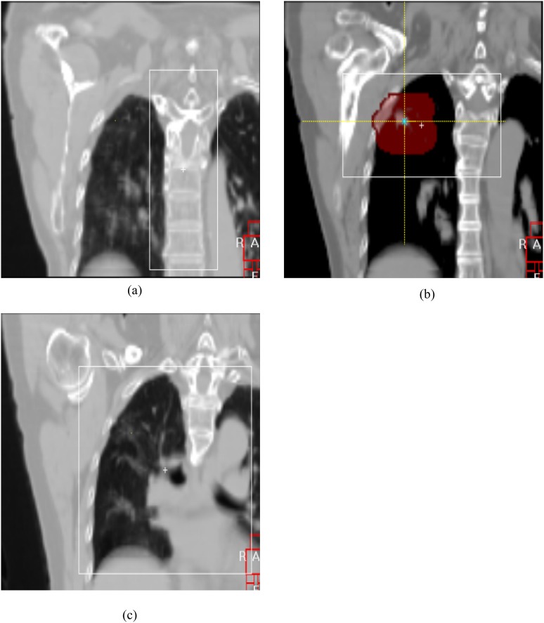
In the study, the automatic dual registrations using three kinds of landmarks for clipbox: spine, spine plus ITV and lung (including as much of the lung as possible) were examined. The study was focused on translational shifts. Figure 1 shows examples of three types of clipboxes used in the study. Translational table shifts calculated from the automatic registrations were compared with those of the standard which was the registration reviewed by physicians.
Figure 1.
Examples of the three kinds of clipboxes using different landmarks: (a) spine; (b) spine plus internal target volume; and (c) lung.
RESULTS AND DISCUSSION
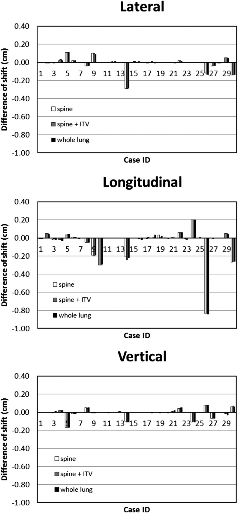
Table 2 lists actual table shifts (i.e., the standard in the study, which was reviewed by physicians) and shifts calculated from automatic dual-image registrations using spine, spine plus ITV and whole lung, for clipboxes, respectively. Table 3 lists the differences of table shifts between the actual and the automatic dual-image registrations calculated using the different landmarks for clipbox. Figure 2 shows the differences of translational table shifts between those obtained from automatic dual registrations using different landmarks for clipbox and the standard, in lateral, longitudinal and vertical directions. The horizontal axis shows study case number. In each case, the differences of table shifts were similar among the automatic registrations using different clipboxes; they all had the same directions of deviation and similar magnitudes.
Table 2.
Actual table shifts (i.e., the standard in the study, which was reviewed by physicians) and shifts calculated from automatic dual-image registrations using spine, spine plus internal target volume (ITV) (spine + ITV) and whole lung for clipbox, respectively
| Study ID | Actual (standard) |
Spine |
Spine + ITV |
Whole lung |
||||||||
|---|---|---|---|---|---|---|---|---|---|---|---|---|
| Lat (cm) | Long (cm) | Vert (cm) | Lat (cm) | Long (cm) | Vert (cm) | Lat (cm) | Long (cm) | Vert (cm) | Lat (cm) | Long (cm) | Vert (cm) | |
| 1 | 0.83 | 0.94 | −0.02 | 0.83 | 0.93 | −0.02 | 0.83 | 0.93 | −0.02 | 0.83 | 0.93 | −0.02 |
| 2 | 0.49 | 0.99 | −0.25 | 0.48 | 1.04 | −0.25 | 0.48 | 1.04 | −0.25 | 0.48 | 1.03 | −0.25 |
| 3 | −0.41 | 0.09 | −0.55 | −0.41 | 0.07 | −0.56 | −0.42 | 0.08 | −0.55 | −0.41 | 0.07 | −0.54 |
| 4 | −0.19 | −0.03 | −0.35 | −0.17 | −0.05 | −0.33 | −0.16 | −0.05 | −0.33 | −0.17 | −0.06 | −0.33 |
| 5 | −0.33 | 0.85 | −0.68 | −0.22 | 0.88 | −0.85 | −0.22 | 0.89 | −0.85 | −0.22 | 0.89 | −0.85 |
| 6 | −0.37 | 0.87 | −0.80 | −0.35 | 0.88 | −0.82 | −0.35 | 0.88 | −0.82 | −0.35 | 0.88 | −0.82 |
| 7 | 0.03 | −0.05 | 0.02 | 0.03 | −0.06 | 0.02 | 0.03 | −0.05 | 0.02 | 0.03 | −0.05 | 0.02 |
| 8 | −0.16 | 0.04 | −0.04 | −0.20 | −0.01 | 0.01 | −0.20 | −0.01 | 0.00 | −0.20 | −0.01 | 0.01 |
| 9 | 0.51 | 0.53 | −0.29 | 0.61 | 0.34 | −0.30 | 0.61 | 0.33 | −0.30 | 0.60 | 0.34 | −0.30 |
| 10 | 1.79 | 0.52 | −0.16 | 1.79 | 0.22 | −0.16 | 1.79 | 0.22 | −0.16 | 1.79 | 0.23 | −0.16 |
| 11 | −0.08 | −1.19 | −0.16 | −0.08 | −1.19 | −0.16 | −0.08 | −1.19 | −0.16 | −0.08 | −1.19 | −0.17 |
| 12 | −0.23 | −0.35 | 0.59 | −0.22 | −0.35 | 0.59 | −0.23 | −0.35 | 0.59 | −0.22 | −0.35 | 0.59 |
| 13 | 1.16 | −0.22 | −0.85 | 1.16 | −0.22 | −0.84 | 1.16 | −0.22 | −0.84 | 1.16 | −0.22 | −0.85 |
| 14 | 0.29 | 1.89 | 0.25 | 0.00 | 1.68 | 0.15 | 0.00 | 1.65 | 0.14 | 0.00 | 1.67 | 0.14 |
| 15 | −0.27 | 1.23 | −0.11 | −0.27 | 1.23 | −0.11 | −0.26 | 1.23 | −0.11 | −0.26 | 1.23 | −0.11 |
| 16 | 0.59 | 0.99 | 0.06 | 0.60 | 0.98 | 0.06 | 0.59 | 0.99 | 0.06 | 0.59 | 0.97 | 0.06 |
| 17 | 0.00 | 0.33 | 0.25 | 0.00 | 0.33 | 0.25 | −0.01 | 0.32 | 0.25 | −0.01 | 0.34 | 0.25 |
| 18 | 1.40 | 1.54 | −0.11 | 1.41 | 1.54 | −0.12 | 1.40 | 1.55 | −0.11 | 1.39 | 1.57 | −0.11 |
| 19 | 0.69 | 1.03 | 0.35 | 0.69 | 1.06 | 0.35 | 0.69 | 1.04 | 0.34 | 0.69 | 1.05 | 0.35 |
| 20 | −0.03 | 0.15 | −0.13 | −0.03 | 0.16 | −0.13 | −0.03 | 0.15 | −0.13 | −0.03 | 0.14 | −0.14 |
| 21 | 0.36 | −0.28 | 0.97 | 0.36 | −0.27 | 0.98 | 0.36 | −0.27 | 0.98 | 0.35 | −0.28 | 0.99 |
| 22 | −0.41 | 0.41 | 0.38 | −0.39 | 0.47 | 0.42 | −0.39 | 0.47 | 0.42 | −0.40 | 0.47 | 0.43 |
| 23 | −0.50 | 0.76 | 0.02 | −0.50 | 0.75 | 0.02 | −0.50 | 0.74 | 0.02 | −0.50 | 0.76 | 0.02 |
| 24 | −1.64 | 0.09 | 0.46 | −1.64 | 0.29 | 0.35 | −1.64 | 0.29 | 0.36 | −1.64 | 0.29 | 0.35 |
| 25 | 0.52 | −0.65 | 0.72 | 0.52 | −0.65 | 0.72 | 0.52 | −0.64 | 0.72 | 0.52 | −0.65 | 0.72 |
| 26 | −0.05 | 0.29 | 0.01 | −0.17 | −0.54 | 0.09 | −0.18 | −0.54 | 0.09 | −0.19 | −0.55 | 0.09 |
| 27 | 0.68 | 0.17 | −0.16 | 0.64 | 0.17 | −0.23 | 0.64 | 0.16 | −0.23 | 0.64 | 0.17 | −0.23 |
| 28 | −0.04 | −0.28 | 0.17 | −0.05 | −0.28 | 0.17 | −0.05 | −0.28 | 0.17 | −0.04 | −0.28 | 0.17 |
| 29 | −0.08 | −0.66 | −0.16 | −0.03 | −0.61 | −0.18 | −0.03 | −0.61 | −0.18 | −0.03 | −0.62 | −0.19 |
| 30 | −0.42 | 0.05 | 0.17 | −0.56 | −0.22 | 0.23 | −0.56 | −0.21 | 0.24 | −0.56 | −0.21 | 0.23 |
ID, identification number; Lat, lateral direction; Long, longitudinal direction; Vert, vertical direction.
Table 3.
Differences of table shifts between the actual (i.e., the standard in the study, which was reviewed by physicians) and the automatic dual-image registrations calculated using spine, spine plus internal target volume (ITV) (spine + ITV) and whole lung for clipbox, respectively
| Study ID | Spine |
Spine + ITV |
Whole lung |
||||||
|---|---|---|---|---|---|---|---|---|---|
| Lat (cm) | Long (cm) | Vert (cm) | Lat (cm) | Long (cm) | Vert (cm) | Lat (cm) | Long (cm) | Vert (cm) | |
| 1 | 0.00 | −0.01 | 0.00 | 0.00 | −0.01 | 0.00 | 0.00 | −0.01 | 0.00 |
| 2 | −0.01 | 0.05 | 0.00 | −0.01 | 0.05 | 0.00 | −0.01 | 0.04 | 0.00 |
| 3 | 0.00 | −0.02 | −0.01 | −0.01 | −0.01 | 0.00 | 0.00 | −0.02 | 0.01 |
| 4 | 0.02 | −0.02 | 0.02 | 0.03 | −0.02 | 0.02 | 0.02 | −0.03 | 0.02 |
| 5 | 0.11 | 0.03 | −0.17 | 0.11 | 0.04 | −0.17 | 0.11 | 0.04 | −0.17 |
| 6 | 0.02 | 0.01 | −0.02 | 0.02 | 0.01 | −0.02 | 0.02 | 0.01 | −0.02 |
| 7 | 0.00 | −0.01 | 0.00 | 0.00 | 0.00 | 0.00 | 0.00 | 0.00 | 0.00 |
| 8 | −0.04 | −0.05 | 0.05 | −0.04 | −0.05 | 0.04 | −0.04 | −0.05 | 0.05 |
| 9 | 0.10 | −0.19 | −0.01 | 0.10 | −0.20 | −0.01 | 0.09 | −0.19 | −0.01 |
| 10 | 0.00 | −0.30 | 0.00 | 0.00 | −0.30 | 0.00 | 0.00 | −0.29 | 0.00 |
| 11 | 0.00 | 0.00 | 0.00 | 0.00 | 0.00 | 0.00 | 0.00 | 0.00 | −0.01 |
| 12 | 0.01 | 0.00 | 0.00 | 0.00 | 0.00 | 0.00 | 0.01 | 0.00 | 0.00 |
| 13 | 0.00 | 0.00 | 0.01 | 0.00 | 0.00 | 0.01 | 0.00 | 0.00 | 0.00 |
| 14 | −0.29 | −0.21 | −0.10 | −0.29 | −0.24 | −0.11 | −0.29 | −0.22 | −0.11 |
| 15 | 0.00 | 0.00 | 0.00 | 0.01 | 0.00 | 0.00 | 0.01 | 0.00 | 0.00 |
| 16 | 0.01 | −0.01 | 0.00 | 0.00 | 0.00 | 0.00 | 0.00 | −0.02 | 0.00 |
| 17 | 0.00 | 0.00 | 0.00 | −0.01 | −0.01 | 0.00 | −0.01 | 0.01 | 0.00 |
| 18 | 0.01 | 0.00 | −0.01 | 0.00 | 0.01 | 0.00 | −0.01 | 0.03 | 0.00 |
| 19 | 0.00 | 0.03 | 0.00 | 0.00 | 0.01 | −0.01 | 0.00 | 0.02 | 0.00 |
| 20 | 0.00 | 0.01 | 0.00 | 0.00 | 0.00 | 0.00 | 0.00 | −0.01 | −0.01 |
| 21 | 0.00 | 0.01 | 0.01 | 0.00 | 0.01 | 0.01 | −0.01 | 0.00 | 0.02 |
| 22 | 0.02 | 0.06 | 0.04 | 0.02 | 0.06 | 0.04 | 0.01 | 0.06 | 0.05 |
| 23 | 0.00 | −0.01 | 0.00 | 0.00 | −0.02 | 0.00 | 0.00 | 0.00 | 0.00 |
| 24 | 0.00 | 0.20 | −0.11 | 0.00 | 0.20 | −0.10 | 0.00 | 0.20 | −0.11 |
| 25 | 0.00 | 0.00 | 0.00 | 0.00 | 0.01 | 0.00 | 0.00 | 0.00 | 0.00 |
| 26 | −0.12 | −0.83 | 0.08 | −0.13 | −0.83 | 0.08 | −0.14 | −0.84 | 0.08 |
| 27 | −0.04 | 0.00 | −0.07 | −0.04 | −0.01 | −0.07 | −0.04 | 0.00 | −0.07 |
| 28 | −0.01 | 0.00 | 0.00 | −0.01 | 0.00 | 0.00 | 0.00 | 0.00 | 0.00 |
| 29 | 0.05 | 0.05 | −0.02 | 0.05 | 0.05 | −0.02 | 0.05 | 0.04 | −0.03 |
| 30 | −0.14 | −0.27 | 0.06 | −0.14 | −0.26 | 0.07 | −0.14 | −0.26 | 0.06 |
ID, identification number; Lat, lateral direction; Long, longitudinal direction; Vert, vertical direction.
Figure 2.
Comparison of automatic dual registrations using different landmarks for clipbox: differences of translational table shifts in lateral, longitudinal and vertical directions, between automatic dual registrations and the standard (difference = automatic registration − standard). ITV, internal target volume. ID, identification number.
Table 4 lists the mean and range of the differences (absolute values) of table shifts in lateral, longitudinal and vertical directions. The mean values of the shift differences in the lateral direction were 0.03 cm (range: 0–0.29 cm; standard deviation: 0.06 cm), 0.03 cm (range: 0–0.29 cm; standard deviation: 0.06 cm) and 0.03 cm (range: 0–0.29 cm; standard deviation: 0.06 cm), for clipboxes based on spine, spine plus ITV and lung, respectively. The mean values of the shift differences in the longitudinal direction were 0.08 cm (range: 0–0.83 cm; standard deviation: 0.17 cm), 0.08 cm (range: 0–0.83 cm; standard deviation: 0.17 cm) and 0.08 cm (range: 0–0.84 cm; standard deviation: 0.17 cm), respectively. The mean values of the shift differences in the vertical direction were 0.03 cm (range: 0–0.17 cm; standard deviation: 0.04 cm), 0.03 cm (range: 0–0.17 cm; standard deviation: 0.04 cm) and 0.03 cm (range: 0–0.17 cm; standard deviation: 0.04 cm), respectively. A one-way analysis of variance was performed to evaluate the table shift differences, which showed that the quantities were similar among registrations using different clipboxes. The p-values were much larger than the threshold value 0.05, and were 0.999, 0.999 and 0.987 for the lateral, longitudinal and vertical direction comparisons, respectively.
Table 4.
Mean and ranges of table shift differences (absolute values) between automatic registrations and the standard, in lateral, longitudinal and vertical directions
| Clipbox type | Mean (minimum, maximum) [cm] |
||
|---|---|---|---|
| Lateral | Longitudinal | Vertical | |
| Spine | 0.03 (0, 0.29) | 0.08 (0, 0.83) | 0.03 (0, 0.17) |
| Spine + internal target volume | 0.03 (0, 0.29) | 0.08 (0, 0.83) | 0.03 (0, 0.17) |
| Whole lung | 0.03 (0, 0.29) | 0.08 (0, 0.84) | 0.03 (0, 0.17) |
The results showed that most of the cases had small shift differences between the automatic registrations and the standard. In the lateral and vertical directions, all the cases had shift differences (from the standard) within 3 mm. In the longitudinal direction, 97% of the cases had shift differences within 3 mm.
The results showed that using different landmarks for clipbox resulted in similar results in the automatic dual registrations of 4D CBCT images, which are different from the published study of 3D CBCT,9 where using different landmarks led to different registration precisions for peripheral and central lung tumours, e.g. using spine as a landmark for clipbox caused larger errors in 3D CBCT image registration of central tumours.9 In our study, Cases 3 and 4 are of central lung tumours, and the others are of peripheral lung tumours. In Cases 3 and 4, no significant differences were observed among automatic dual-image registrations using different landmarks including spine for clipboxes. The table shift difference of the registration using spine clipbox was only approximately 0.02 cm. The results showed that compared with 3D CBCT, 4D CBCT improved registrations. The clipbox registration made gross alignments and the mask made fine adjustments. It was indicated that the mask registrations, which were conducted after clipbox registration, played an important role in the automatic dual registrations in ensuring target match, and the dual-image registration was robust. The technique is helpful in assessing if a margin is adequate or a smaller margin can be used. To the authors' best knowledge, there are no similar studies for evaluating 4D CBCT image registration.
Among all the cases, the maximum shift difference occurred in Case 26, which was 0.83 cm. In that case, the tumour had significant motion in the longitudinal direction. The physician made an adjustment on the automatic registration results for a better match. The result implies that although overall automatic dual-image registrations presented satisfactory precisions, manual adjustments may be needed in some cases.
CONCLUSION
The automatic registrations using three different landmarks for clipbox showed similar results, which overall had good agreements with those of the standard. The results indicate that one can use any of the three landmarks in automatic dual-image registrations. Review of automatic registrations by a physician is recommended.
Contributor Information
Jun Li, Email: Jun.Li@jefferson.edu.
Amy Harrison, Email: jun.li@jefferson.edu.
Yan Yu, Email: jun.li@jefferson.edu.
Ying Xiao, Email: jun.li@jefferson.edu.
Maria Werner-Wasik, Email: jun.li@jefferson.edu.
Bo Lu, Email: jun.li@jefferson.edu.
REFERENCES
- 1.Guckenberger M, Meyer J, Wilbert J, Baier K, Mueller G, Wulf J, et al. Cone-beam CT based image-guidance for extracranial stereotactic radiotherapy of intrapulmonary tumours. Acta Oncol 2006; 45: 897–906. doi: 10.1080/02841860600904839 [DOI] [PubMed] [Google Scholar]
- 2.Worm ES, Hansen AT, Petersen JB, Muren LP, Præstegaard LH, Høyer M. Inter- and intrafractional localisation errors in cone-beam CT guided stereotactic radiation therapy of tumours in the liver and lung. Acta Oncol 2010; 49: 1177–83. doi: 10.3109/0284186X.2010.498435 [DOI] [PubMed] [Google Scholar]
- 3.Guckenberger M, Meyer J, Vordermark D, Baier K, Wilbert J, Flentje M. Magnitude and clinical relevance of translational and rotational patient setup errors: CBCT study. Int J Radiat Oncol Biol Phys 2006; 65: 934–42. doi: 10.1016/j.ijrobp.2006.02.019 [DOI] [PubMed] [Google Scholar]
- 4.Purdie TG, Bissonnette JP, Franks K, Bezjak A, Payne D, Sie F, et al. Cone-beam computed tomography for on-line image guidance of lung stereotactic radiotherapy: localization, verification, and intrafraction tumor position. Int J Radiat Oncol Biol Phys 2007; 68: 243–52. doi: 10.1016/j.ijrobp.2006.12.022 [DOI] [PubMed] [Google Scholar]
- 5.Grills IS, Hugo G, Kestin LL, Galerani AP, Chao KK, Wloch J, et al. Image-guided radiotherapy via daily online cone-beam CT substantially reduces margin requirements for stereotactic lung radiotherapy. Int J Radiat Oncol Biol Phys 2008; 70: 1045–56. doi: 10.1016/j.ijrobp.2007.07.2352 [DOI] [PubMed] [Google Scholar]
- 6.Bissonnette JP, Purdie TG, Higgins JA, Li W, Bezjak A. Cone-beam computed tomographic image guidance for lung cancer radiation therapy. Int J Radiat Oncol Biol Phys 2009; 73: 927–34. doi: 10.1016/j.ijrobp.2008.08.059 [DOI] [PubMed] [Google Scholar]
- 7.Higgins J, Bezjak A, Franks K, Le LW, Cho BC, Payne D, et al. Comparison of spine, carina, and tumor as registration landmarks for volumetric image-guided lung radiotherapy. Int J Radiat Oncol Biol Phys 2009; 73: 1404–13. doi: 10.1016/j.ijrobp.2008.06.1926 [DOI] [PubMed] [Google Scholar]
- 8.Lavoie C, Higgins J, Bissonnette JP, Le LW, Sun A, Brade A, et al. Volumetric image guidance using carina vs spine as registration landmarks for conventionally fractionated lung radiotherapy. Int J Radiat Oncol Biol Phys 2012; 84: 1086–92. doi: 10.1016/j.ijrobp.2012.02.012 [DOI] [PubMed] [Google Scholar]
- 9.Li HS, Kong LL, Zhang J, Li BS, Chen JH, Zhu J, et al. Evaluation of the geometric accuracy of anatomic landmarks as surrogates for intrapulmonary tumours in image-guided radiotherapy. Asian Pac J Cancer Prev 2012; 13: 2393–8. doi: 10.7314/APJCP.2012.13.5.2393 [DOI] [PubMed] [Google Scholar]
- 10.Sonke JJ, Rossi M, Wolthaus J, van Herk M, Damen E, Belderbos J. Frameless stereotactic body radiotherapy for lung cancer using four-dimensional cone beam CT guidance. Int J Radiat Oncol Biol Phys 2009; 74: 567–74. doi: 10.1016/j.ijrobp.2008.08.004 [DOI] [PubMed] [Google Scholar]
- 11.Sweeney RA, Seubert B, Stark S, Homann V, Müller G, Flentje M, et al. Accuracy and inter-observer variability of 3D versus 4D cone-beam CT based image-guidance in SBRT for lung tumours. Radiat Oncol 2012; 7: 81. doi: 10.1186/1748-717X-7-81 [DOI] [PMC free article] [PubMed] [Google Scholar]
- 12.Bissonnette JP, Franks KN, Purdie TG, Moseley DJ, Sonke JJ, Jaffray DA, et al. Quantifying interfraction and intrafraction tumor motion in lung stereotactic body radiotherapy using respiration correlated cone beam computed tomography. Int J Radiat Oncol Biol Phys 2009; 75: 688–95. doi: 10.1016/j.ijrobp.2008.11.066 [DOI] [PubMed] [Google Scholar]
- 13.Sonke JJ, Zijp L, Remeijer P, van Herk M. Respiratory correlated cone beam CT. Med Phys 2005; 32: 1176–86. doi: 10.1118/1.1869074 [DOI] [PubMed] [Google Scholar]


