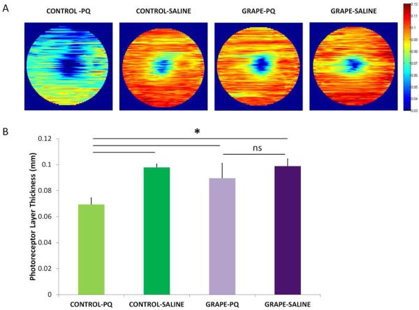
Figure 3.
A. The structural integrity of the retina was imaged using OCT. Representative retina thickness heat maps are shown. Orange-red indicates thicker retinas and blue-green indicates thinner retinas. The grape-supplemented diet preserved the retina in the paraquat injected eyes (Grape-PQ) whereas the retina degenerated and was thinner in mice fed the control diet (Control-PQ). (B) Quantification of photoreceptor layer thickness. OCT measurements demonstrated that the nuclear layer plus inner and outer segments were significantly thinner after paraquat injection in mice fed the control diet (compare Control-PQ and Control-Saline). This decrease was absent in paraquat-injected mice fed the grape diet (compare Grape-PQ and Grape-Saline), and mice on the grape diet had thicker retinas than mice on the control diet (paraquat-injected mice, n=8; saline-injected mice, n=5). There was minimal variation of degeneration within each treatment group. Mean ± SD shown. *p<0.05.

