Abstract
Carcinoid tumors of the duodenum are relatively rare. Although they were considered benign lesions, they are now classified malignant, occasionally with poor prognosis. We report a case of esophageal cancer with a synchronous multiple carcinoid of the duodenal bulb. An upper endoscopy visualized with esophageal scan disclosed a stenotic lesion in the lower esophagus and revealed multiple 4-–5-mm-diameter masses which were on the fore wall of the duodenal bulb. The postoperative pathology report confirmed the diagnosis of esophageal squamous cancer and duodenal bulb carcinoid.
Keywords: Esophageal cancer, Duodenal bulb carcinoid, Synchronous cancers, Esophageal cancer
Introduction
Duodenal bulb carcinoid is a rare neuroendocrine neoplasia. We report a case of an esophageal cancer with a synchronous multiple carcinoid of duodenal bulb in a 47-year-old man. Esophageal cancer with a synchronous duodenal bulb carcinoid is extremely rare, with no cases reported in the literature. A case report is presented, with discussion of carcinoid tumors and management when occurring synchronously with non-carcinoid tumors. We gave the patient endoscopic submucosal resection. After 3 years of follow-up, the patient had no cancer metastasis.
Case Report
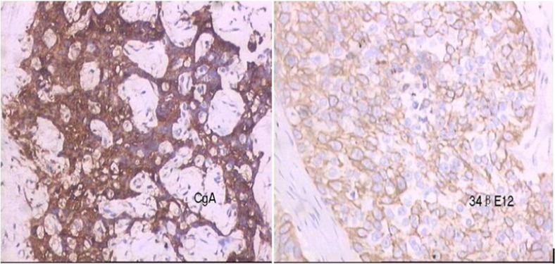
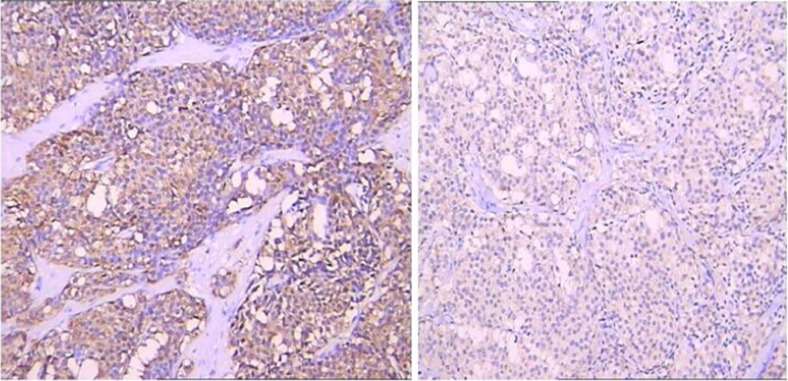
A 47-year-old male was admitted to our hospital with suspected esophageal cancer in 4 January 2010 with difficulty in eating. An upper endoscopy visualized with esophageal scan disclosed a stenotic lesion in the lower esophagus and revealed some 4-–5-mm-diameter multiple masses, which were on the fore wall of the duodenal bulb. Moreover, the computed tomography (CT) of abdominal scan revealed some 4-–5-mm sizes slightly contrast-enhanced duodenal bulb soft tissue images without metastasis to the regional lymph nodes or the liver. The biopsy specimen from the lesions diagnosed esophageal squamous cancer and duodenal bulb carcinoid (Fig. 1). In 22 January 2010, under the general anesthesia, the patient underwent a transthoracic surgical operation. The patient was confirmed to have poorly differentiated squamous cell carcinoma in the lower esophagus mucous membrane, tumor-infiltrated esophagus muscular layer, and with metastasis to the lymph nodes of the esophagus (2/5) by postoperative pathology. Immunohistochemistry results demonstrated creatine kinase (+++), 34pE12 (+++), proliferate cell nuclear antigen (PCNA) (++), DNA topoisomerase (++), glutathione S-transferase (GST) (++), P-glycoprotein (++), and Bcl-2 (+−++) (Fig. 1). In 23 March 2010, preoperative endoscopic ultrasonography (EUS) revealed some hypoechoic masses measuring 4–5 mm in diameter, originating from the submucosal layer. In 25 March 2010, the patient underwent endoscopic submucosal dissection (ESD) for duodenal bulb carcinoid. Three masses measuring 3–4 mm in diameter was observed in the operation. All of them were intact resected. Pathological diagnosis showed duodenal bulb submucosal carcinoid. Immunohistochemistry revealed 35pH11 (+), Ki-67 (5 %), neuron-specific enolase (+), synaptophysin (+), and chromogranin A (+) (Fig. 2). The patient was discharged after 6 days. After 3 years of follow-up, gastroscopy, abdominal CT scan, magnetic resonance imaging (MRI) of the head, and bone scintigraphic imaging showed no metastases. This patient is still in a follow-up period.
Fig. 1.
The biopsy specimen from duodenal bulb and esophagus specimen. An immunohistochemical examination revealed that the tumor was positive for chromogranin A in duodenal bulb specimen from (×200) (left). Immunohistochemical examination of resected esophagus specimen reveals 34pE12 (×200) (right)
Fig. 2.
The specimen of duodenal bulb immunohistochemistry for synaptophysin showing diffuse positive staining in tumor cells (×200) (left). Immunohistochemistry for neuron-specific enolase showing positive staining in tumor cells (×200) (right)
Discussion
Carcinoid tumors of the gastrointestinal tract arise from neuroendocrine cells that line the tract. They can occur anywhere in a variety of anatomic locations, but less commonly they can occur in the duodenal bulb. Histologically, they are usually easy to recognize, but in uncertain cases, immunohistochemistry detects unspecific neuroendocrine markers, such as neuron-specific enolase, chromogranins, synaptophysin, serotonin, and neurohormonal peptides [1]. Actually, duodenal bulb carcinoids are very difficult to diagnose because they are typically asymptomatic and small.
A retrospective review by Gerstle et al. of 69 patients with carcinoids in their gastrointestinal tracts found that 29 (42 %) had synchronous tumors and three (4 %) had metachronous tumors [2]. Another series from France reviewed 270 cases of gastrointestinal tract carcinoid and 21 were associated with synchronous tumors [3]. Therefore, some scholars consider that carcinoid tumors are associated with an increased incidence of secondary primary malignancies. So we should pay attention to gastrointestinal tract tumors to avoid missing diagnosis and misdiagnosis.
A case of esophageal squamous cancer with a synchronous multiple carcinoid of the duodenal bulb is an extremely rare case that has not been previously described in the literature. In treatment terms, it might be impossible for the patient to endure the stress of all of these operative procedures at once. Therefore, we planned to perform staged treatment with prioritizing consideration. First, we performed esophagectomy. After 8 weeks, ESD was performed for the patient. Carcinoid survival rate depends upon the site, but in general, the management and prognosis depend on the non-carcinoid malignancy. After 3 years of follow-up, gastroscopy and CT were normal. This patient is still in a follow-up period.
In conclusion, this is the first time a duodenal bulb carcinoid in association with an esophageal squamous cancer has been described. It is important to be aware of the possibility of synchronous cancers, especially in those patients presenting with gastrointestinal tract tumors. It is advised that patients presenting with any neoplasm in the gastrointestinal tract should undergo a thorough exploration of the gastrointestinal tract at initial surgery to exclude this [4]. But in general, the prognosis and management depend on the non-carcinoid malignancy.
Footnotes
Xu Feng Deng and Quan Xing Liu contributed equally to this work.
References
- 1.Plockinger U, Wiedenmann B. Neuroendocrine tumours of the gastrointestinal tract. Z Gastroenterol. 2004;42(6):517–527. doi: 10.1055/s-2004-812697. [DOI] [PubMed] [Google Scholar]
- 2.Gerstle JT, Kauffman GJ, Koltun WA. The incidence, management, and outcome of patients with gastrointestinal carcinoids and second primary malignancies. J Am Coll Surg. 1995;180(4):427–432. [PubMed] [Google Scholar]
- 3.Berner M. Digestive carcinoids and synchronous malignant tumors. Helv Chim Acta. 1993;59(5–6):757–766. [PubMed] [Google Scholar]
- 4.Tse V, et al. Concurrent colonic adenocarcinoma and two ileal carcinoids in a 72-year-old male. Aust N Z J Surg. 1997;67(10):739–741. doi: 10.1111/j.1445-2197.1997.tb07124.x. [DOI] [PubMed] [Google Scholar]


