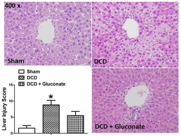
Figure 5.
Histologic assessment by H & E stained, paraffin sectioned, rat livers using light microscopy for a sham liver (upper left), a liver after DCD (upper right), and a liver after DCD in a gluconate treated donor (lower right). Rat livers were obtained 60 min after reperfusion on the isolated perfused liver device (IPL) after DCD with 24 h of cold storage in University of Wisconsin solution. Graph inset: Summary of liver injury scores in the 3 groups, values are mean ± SD from 6 rats per group. * P<0.05, relative to the fresh and DCD + Gluconate groups.

