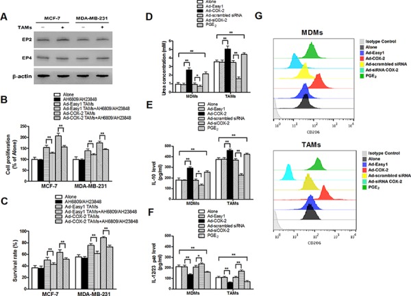
Figure 3. COX-2 is essential for macrophages polarized to M2 phenotype.

A. The expression of EP2 and EP4 in breast cancer cells co-cultured with or without TAMs was detected by Western blot. β-actin was used as an internal loading control. B. and C. Inhibiting PGE2 signal pathway partly attenuated cell proliferation and drug resistance induced by COX-2 in TAMs. Breast cancer cells were first treated with or without EPs antagonists AH6809 (5 μM) and AH23848 (10 μM) for 12 h before co-cultured with or without TAMs transfected with adenoviral COX-2. Cell proliferation (B) and cell apoptosis (C) induced by ADM were measured by CCK-8 kit and flow cytometry respectively. D. Arginase activity (urea concentration) in macrophages transfected with adenoviral COX-2 or siRNA COX-2 or treated with PGE2 (1 μM) was analyzed by microplate reader. E. and F. Expression of IL-10 and IL-12/23 in macrophages was detected by ELISA. G. Expression of CD206 in macrophages was analyzed by flow cytometry. All the experiments were performed thrice in triplicate. Mean ± SD, *p < 0.05 and **p < 0.01.
