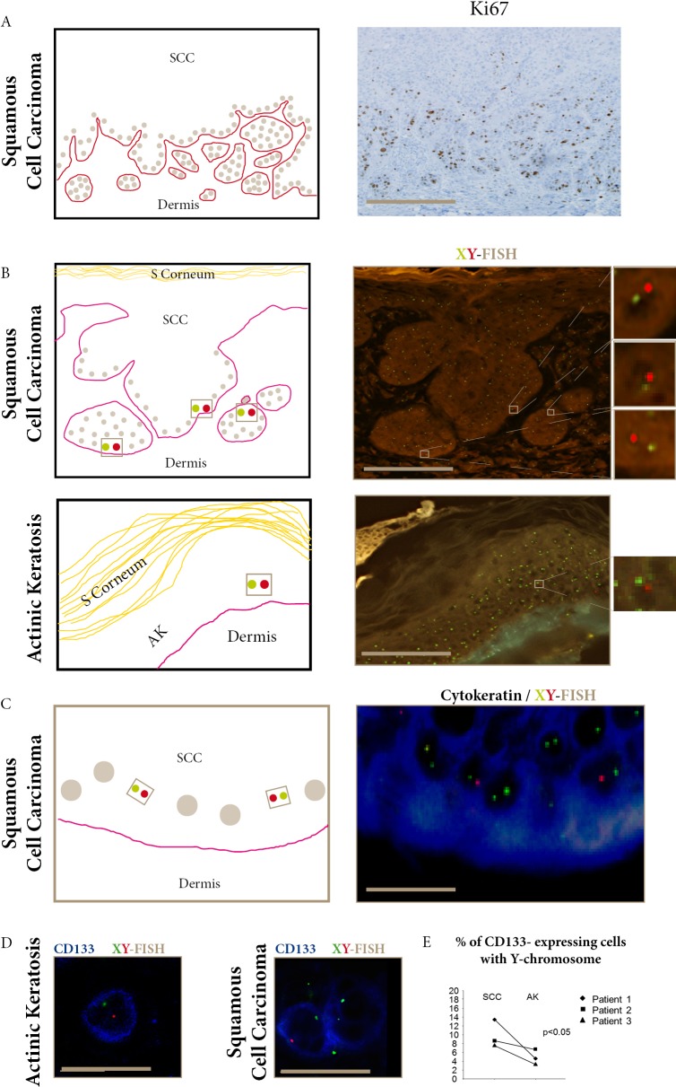
Figure 1. Donor- derived cells in SCC and AK express CD133.
A. Ki67 staining of proliferative epidermal cells in the basal layer and invasive areas of SCC (bar=100μm). B. FISH-X (green) Y (red) show donor-derived XY cells in SCC invasive areas and AK epidermal basal layers (bars=100μm). C. Combined FISH-X (green) Y (red) and AE1/AE3 (blue) show donor-derived XY in cytokeratin-expressing cells in SCC basal layer (bar=15μm). D. Combined FISH-X (green) Y (red) and CD133 (blue) show donor-derived XY in CD133 expressing cells in AK and SCC (bar=15μm). E. The percentage of CD133-expressing cells with the Y-chromosome detected using droplet digital PCR is significantly higher in SCC than in AK in the three female recipients of male kidney-transplants. (p<0.05, Khi-square test).

