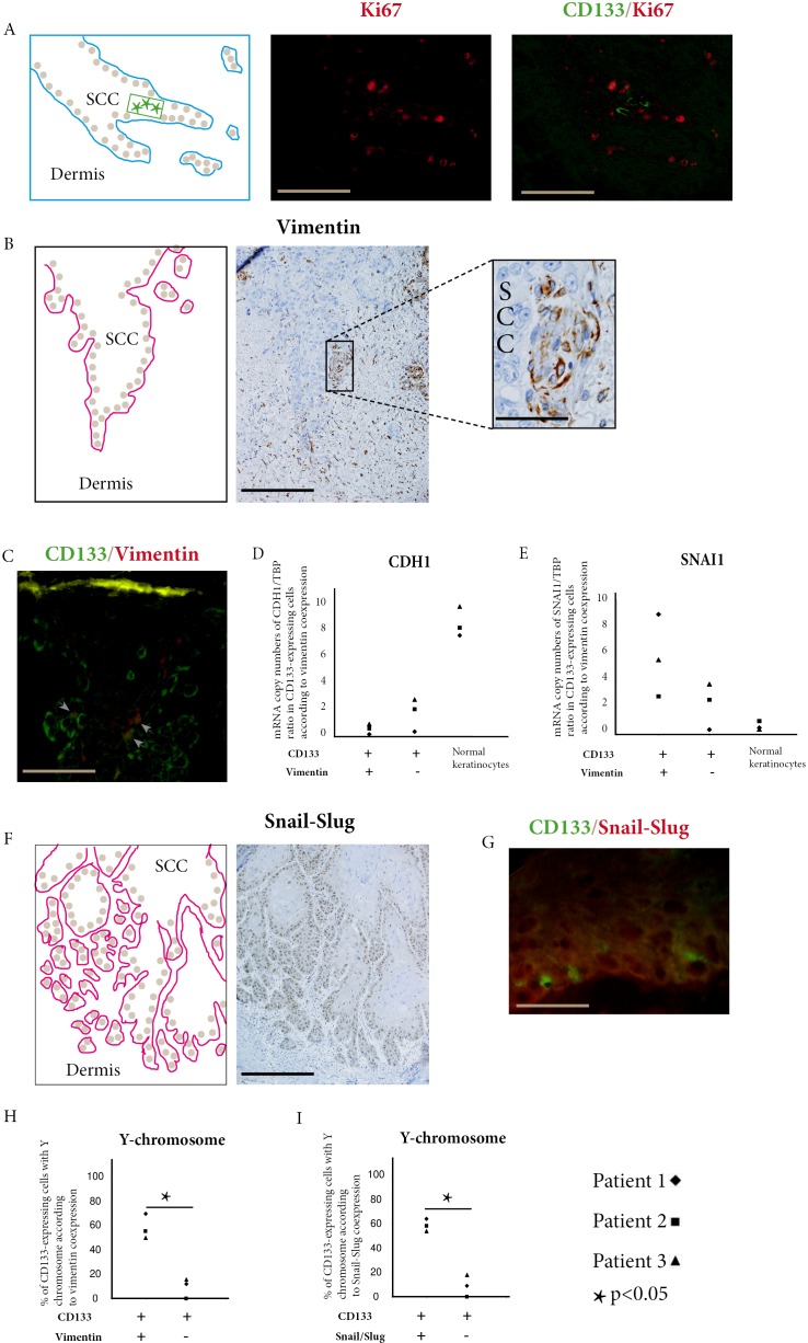
Figure 2. Donor-derived stem cells express EMT markers in squamous cell carcinoma.
A. Combined CD133 (green) and Ki67 (red) immunofluorescent stainings show CD133 expressing cells in SCC invasive areas. These cells do not co-express Ki67 (bar=100μm). B. Immunoperoxydase staining of vimentin within cells of SCC basal layer (bar=100μm, higher magnification bar=25μm). C. Combined CD133 (green) and vimentin (red) immunofluorescence stainings show double positive cells (arrow heads) in SCC outer cell layers (bar=25μm). D. In SCCs of the three kidney-transplant recipients studied, CDH1 (E-cadherin) is under-expressed in cells co-expressing CD133 and vimentin compared with cells only expressing CD133, and with normal keratinocytes. E. In the same patients, SNAI1 (Snail1) is overexpressed in cells co-expressing CD133 and vimentin compared with cells only expressing CD133, and with normal keratinocytes. F. Immunoperoxydase staining of Snail-Slug within cells of SCC basal layer and invasive areas (bar= 150μm). G. Combined CD133 (green) and Snail-Slug (red) immunofluorescent stainings show CD133 expressing cells in SCC outer cell layers. (bar=25μm). H. In SCCs of the three kidney-transplant recipients studied, cells co-expressing CD133 and vimentin have more Y-chromosome detected by droplet digital PCR than cells only expressing CD133 (p<0.05, Chi-square test). I. In SCCs of the three kidney-transplant recipients studied, cells co-expressing CD133 and Snail-Slug have more Y-chromosome detected by droplet digital PCR than cells only expressing CD133 (p<0.05, Chi-square test).

