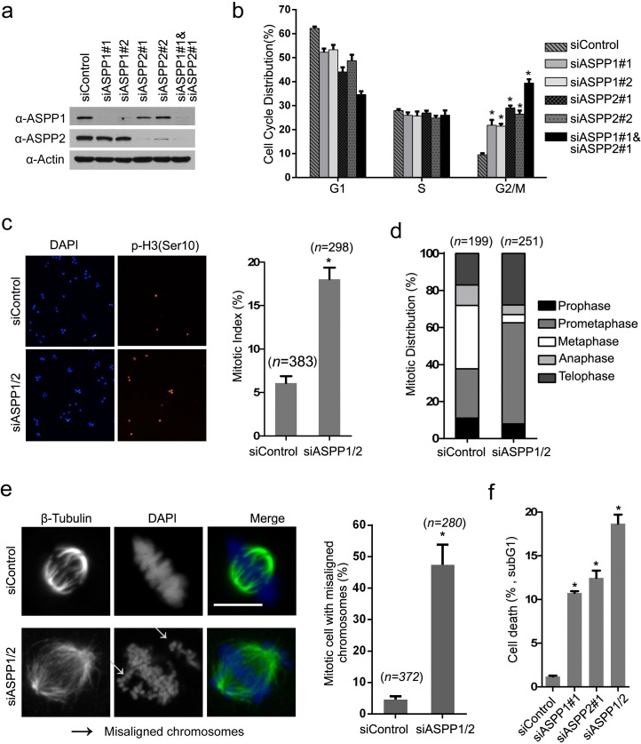
Figure 2. ASPP1/2 are required for proper mitotic progression.
a. Depletion of ASPP1/2 by siRNAs in HeLa cells. HeLa cells were transfected with the indicated siRNAs. After 48 hr, cell lysates were prepared for WB analyses with their indicated antibodies. b. ASPP1/2 co-depletion causes G2/M arrest. The cell-cycle distributions of HeLa cells transfected with indicated siRNAs for 48 hr were determined by flow cytometry. Error bars, SEM. *p<0.01 from triplicates. c. ASPP1/2 co-depletion increases the mitotic index in HeLa cells. HeLa cells were transfected with control or ASPP1/2 siRNAs as indicated. After 48 hr, cells were fixed and stained for the p-H3 (Ser10) antibody. Quantification of cells with anti-p-H3 (Ser10) staining is shown at the right (n= number of counted cells). d. Mitotic stages were quantified by DNA and spindle morphology in the mitotic population of control or ASPP1/2 co-depleted HeLa cells. e. ASPP1/2 co-depletion increases the number of mitotic cells with misaligned chromosomes. HeLa cells were transfected with control or ASPP1/2 siRNAs. After 48 hr, cells were fixed and stained with the anti-β-tubulin (green) antibody and DAPI (blue). White arrows indicate misaligned chromosomes. Scale bar = 10μm. Quantification of cells with misaligned chromosomes is shown on the right. f. ASPP1/2 depletions lead to increases in cell death. HeLa cells were transfected with control or ASPP1/2 siRNAs. After 72hr, the cell death was measured by flow cytometry using the propidium iodide staining assay.

