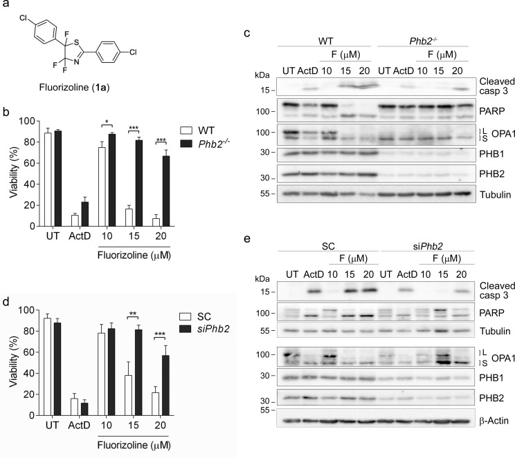
Figure 1. The presence of PHBs is required for fluorizoline-induced apoptosis.
a. Chemical structure of fluorizoline (diaryl trifluorothiazoline compound 1a). b., c. Cre recombinase was transduced in WT and Phb2fl/fl (Phb2−/−) MEFs for 72 h. Then cells were untreated (UT) or treated with either 0.15 μg/mL Actinomycin D (ActD) or increasing doses of fluorizoline (F) for 24 h. d., e. Phb2fl/fl MEFs were transfected with scramble (SC) or Phb2 (siPhb2) siRNA for 72 h. Afterwards, cells were treated with either 0.15 μg/mL Actinomycin D (ActD) or increasing doses of fluorizoline (F) for 24 h. b., d. Viability was measured by flow cytometry and it is expressed as the mean ± SEM (n ≥ 3) of the percentage of non-apoptotic cells (annexin V-negative). *p < 0.05, **p < 0.01, ***p < 0.001. c., e. Protein levels were analyzed by western blot. Tubulin and β-Actin were used as a loading control. These are representative images of at least three independent experiments.

