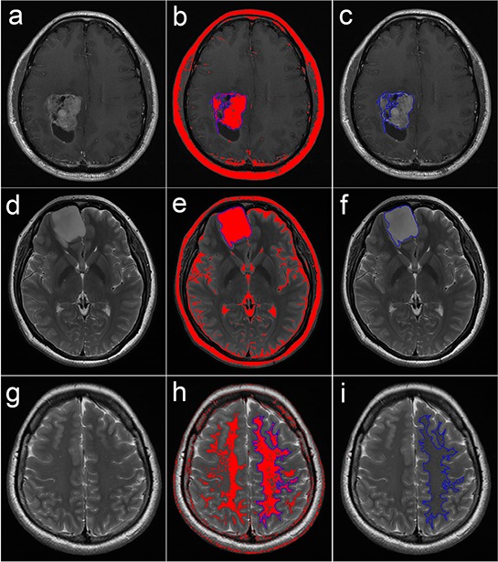
Figure 5. Semi-automated delineation of the ROIs over the solid region of the tumor and the NAWM.

a-c. Delineation over a solid enhancing tumor on transverse contrast-enhanced T1-FLAIR; d-f. delineation over a non-enhancing tumor on transverse T2-FSE; and g-i. delineation over a contralateral NAWM on transverse T2-FSE. When a proper threshold range of signal intensity was set, the corresponding pixels in the range were colored red, and then the wand tool was used to automatically delineate the connective pixels as the ROI (in blue).
ROI: region of interest; NAWM: normal-appearing white matter; T1-FLAIR: T1 fluid-attenuated inversion recovery; T2-FSE: T2 fast spin echo.
