
Figure 6. Fatostain alone or in combination with docetaxel inhibits PCa tumor growth in a subcutaneous xenograft mouse model.
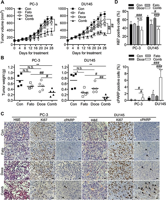
A. PC-3 and DU145 cells were subcutaneously injected in the flanks of male athymic mice. Tumor volume was monitored for 4 weeks after treatment with fatostatin and/or docetaxel. Each point represents the mean ± SEM of the measured tumor volume (N = 5/group). **P < 0.01, ***P < 0.001, compared with the control group; #P < 0.05, ##P < 0.01, compared with the combination group. Con, control; Fato, fatostatin; Doce, docetaxel; Comb, combination. B. Subcutaneous PC-3 and DU145 tumors were weighed. N.S., no significance; *P < 0.05, **P < 0.01, compared with the control group; #P < 0.05, ##P < 0.01, compared with the combination group. C. Representative H&E and IHC staining of Ki67 and cleaved PARP (cPARP; black arrow) in PC-3 and DU145 tumor sections collected from control, fatostatin-, docetaxel- and combination-treated groups. Cell nuclei were counterstained with hematoxylin (blue). Scale bar = 50 μm. D. Quantitative analysis of Ki67 and cPARP was performed and reported as the percentage of Ki67 or cPARP positive cells in PCa xenograft tumors. N.S., no significance; *P < 0.05, **P < 0.01, ***P < 0.001, compared with the control group; #P < 0.05, ###P < 0.001, compared with the combination group.