
Figure 1. Experimental procedure and macroscopic and histological observation of the AOM/DSS murine model.
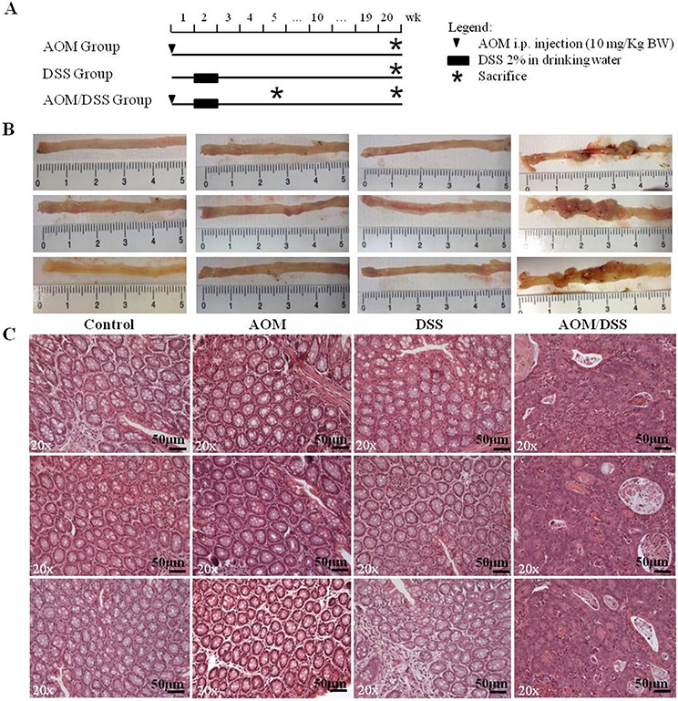
A. Schematic experimental procedure for groups treated with AOM-alone and/or DSS. Control group (untreated littermate controls) not represented. B. Macroscopic observation of the distal regions of colons from control, AOM-, DSS- and AOM/DSS-treated mice at the end of the 20th week (only 3 of 6 animals per group are shown). Evident macroscopic lesions detectable only in AOM/DSS-treated colons. C. Hematoxylin/eosin staining of tumors and normal colons. Colon mucosae of AOM-only and DSS-only treated mice show the same histological characteristics of the control group. Adenocarcinomas with a high degree of dysplasia are detectable in AOM/DSS-treated mice. 20x original magnification. Scale bar, 50 μm.