Abstract
The aim of this study was to genetically characterize clinical isolates from patients diagnosed with Mycobacterium avium lung disease and to investigate the clinical significance. Multi-locus sequencing analysis (MLSA) and pattern of insertion sequence analysis of M. avium isolates from 92 Korean patients revealed that all isolates were M. avium subspecies hominissuis. In hsp65 sequevar analysis, codes 2, 15, and 16 were most frequently found (88/92) with similar proportions among cases additionally two isolates belonging to code N2 and an unreported code were identified, respectively. In insertion element analysis, all isolates were IS1311 positive and IS900 negative. Four of the M. avium subsp. hominissuis isolates did not harbor IS1245 and 1 of the M. avium isolates intriguingly harbored DT1, which is thought to be a M. intracellulare-specific element. M. avium subsp. hominissuis harboring ISMav6 is prevalent in Korea. No significant association between clinical manifestation and treatment response has been found in patients with the hsp65 code type and ISMav6, indicating that no specific strain/genotype among M. avium subsp. hominissuis organisms was a major source of M. avium lung disease. Interestingly, the presence of ISMav6 was correlated with greater resistance to moxifloxacin. Conclusively, the genotype of Korean M. avium subsp. hominissuis isolates is not a disease determinant responsible for lung disease and specific virulent factors of M. avium subsp. hominissuis need to be investigated further.
Introduction
A rise in the incidence of pulmonary disease caused by nontuberculous mycobacteria (NTM) has been reported worldwide [1, 2]. Mycobacterium avium complex (MAC) is the most frequent etiology of NTM lung disease [3]. MAC initially included two species, M. avium and M. intracellulare. M. avium is the most clinically significant species for humans and animals within the MAC and is divided into four subspecies: M. avium subsp. avium, M. avium subsp. hominissuis, M. avium subsp. paratuberculosis, and M. avium subsp. silvaticum [4, 5].
Although subspecies of M. avium in different geographic regions or populations may have different levels of virulence due to co-evolutionary processes, consequently leading to varying epidemiological dominance, most cases of M. avium human disease are due to M. avium subsp. hominissuis. Recently, lymphadenitis patients in France were found to be infected by only M. avium subsp. hominissuis among M. avium subspecies [6]. More recently, a subspecies identification analysis of M. avium clinical strains in the USA showed M. avium subsp. hominissuis to be the dominant M. avium subspecies (92.6%), followed by M. avium subsp. avium (7.4%) [7]. All German M. avium strains isolated from children and adults were identified as M. avium subsp. hominissuis [8].
Many studies have emphasized the importance of taxonomy in distinguishing species and subspecies of MAC because non-sequencing methods or 16S rRNA sequencing frequently fails to distinguish closely related species [9, 10]. Multi-locus sequencing analysis (MLSA) has been suggested as the new standard method for identifying Mycobacterium species that are not well discriminated by 16S rRNA gene sequences alone [11–14].
The presence and distribution of various insertion sequences (IS) among M. avium subspecies have provided an unprecedented opportunity to define the genomic differences between M. avium subspecies as well as to develop molecular typing methods with sufficient discriminatory power to differentiate M. avium subspecies and isolates [15].
At our institution, the rpoB-PCR restriction fragment length polymorphism (RFLP) analysis [PRA] method was used for species identification and diagnosis of MAC lung disease until 2009 [16–18]. To gain better insight into M. avium lung disease in Korea, we used sequencing-based methods for subspecies identification and genotyping and compared clinical characteristics and treatment outcomes according to genotype. Furthermore, we investigated patterns of antibiotic resistance according to mycobacterial genotype as well as the presence or absence of ISMav6.
Materials and Methods
Study subjects
Clinical isolates from 92 patients with newly diagnosed M. avium lung disease from Jan. 2008 to Dec. 2009 at Samsung Medical Center (Seoul, Korea) were collected and stored. The data in the present study are part of an ongoing prospective observational cohort study investigating NTM lung disease (ClinicalTrials.gov Identifier: NCT00970801). The study protocol for isolates collection and genotyping analysis was approved by the institutional review board of the Samsung Medical Center (IRB approval 2008-09-016), and written informed consent was obtained from all participants. All patients met the diagnostic criteria for NTM lung disease [3]. All patients were immunocompetent and none of the patients tested positive for human immunodeficiency virus. All isolates were collected before initiating antibiotic treatment for NTM lung disease. Additionally, M. avium species initially identified by PRA based on the rpoB gene at the time of diagnosis as previously described were used for subsequent analysis.
Drug susceptibility test
Drug susceptibility testing was performed at the Korean Institute of Tuberculosis. The minimum inhibitory concentrations (MICs) of clarithromycin (CLR) and moxifloxacin (MXF) were determined using the broth microdilution method as described by the Clinical and Laboratory Standards Institute (CLSI) [19]. Isolates with CLR MIC of ≥32 μg/ml and MXF MIC of ≥4 μg/ml were considered to be resistant, according to the guidelines of the CLSI [19].
Molecular characterization of M. avium clinical isolates by MLSA
M. avium strains were propagated in Middlebrook 7H9 broth (Difco Laboratories, Detroit, MI, USA) supplemented with 10% (vol/vol) oleic acid-albumin-dextrose-catalase (OADC; BD Diagnostics, Sparks, MD, USA). Mycobacterial DNA was extracted using a DNeasy Blood and Tissue Kit according to the manufacturer’s instructions (Qiagen, Valencia, CA, USA). MLSA including hsp65, rpoB, and 16S rRNA fragments was carried out using PCR primer sets as described previously [20–22]. The PCR products of target genes were subjected to sequence analysis. The nucleotide sequences of these genes were compared with data reported by BLAST analysis (http://www.ncbi.nlm.nih.gov) against sequences from M. avium subspecies type and related strains. M. avium subsp. avium ATCC 25291, M. avium subsp. hominissuis 104, M. avium subsp. paratuberculosis K-10, and M. avium subsp. silvaticum ATCC 49884 were used as reference strains. For phylogenetic analysis, sequences were trimmed using the CLUSTAL-W multiple sequence alignment program [23]. Phylogenetic trees were obtained from DNA sequences utilizing the neighbor-joining method and Kimura’s two parameter distance correction model with 1,000 bootstrap replications supported by MEGA 6.0 software [24].
hsp65 code analysis
hsp65 code analysis was performed as previously described [25]. hsp65 gene PCR products were subjected to sequence analysis. The nucleotide sequences of the hsp65 gene were compared with data reported by BLAST analysis (http://www.ncbi.nlm.nih.gov) against the M. avium type and related strains. hsp65 codes were classified according to previously reported papers [25–27].
Insertion sequences element analysis
Multiplex PCR was performed to detect three target genes, IS900, IS1311, and DT1 using previously described methods [28]. A previously described primer set was used for the IS1245 insertion element [29]. The presence of ISMav6 was determined by PCR followed by sequencing analysis using a previously described primer set [26]. PCR products of insertion elements were sequenced and the existence of a specific insertion element in each strain was confirmed. DNA isolated from M. abscessus ATCC 19977, M. tuberculosis H37Rv ATCC 27294, and M. gastri ATCC 15754 were used as negative controls for each primer set in each PCR run.
Statistics
All data are presented as median and interquartile range for continuous variables and as number (percentage) for categorical variables. Data were compared using the Mann-Whitney U test for continuous variables and the chi-squared or Fisher’s exact test for categorical variables. The Mantel-Haenszel test for categorical variables was used to compare each hsp65 code or the presence of ISMav6 across resistant levels (susceptible, intermediate, resistant) of each drug [30]. Statistical analyses were performed using SAS 9.1 (SAS Institute Inc., Cary, NC, USA) and a P-value less than 0.05 was considered statistically significant.
Results
Subspecies identification of M. avium clinical isolates by MLSA
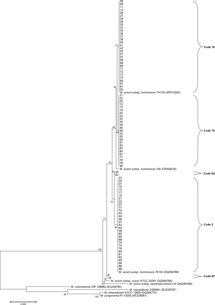
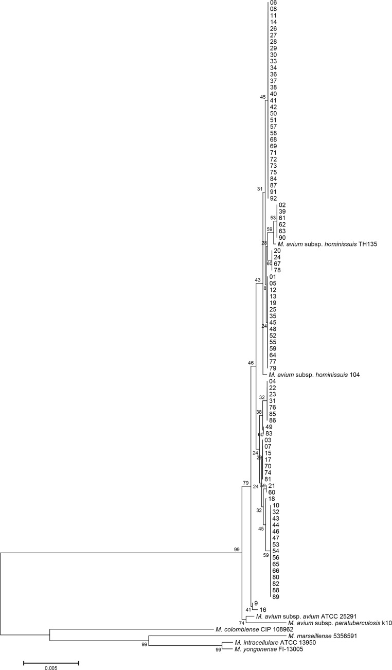
Isolates from 92 patients diagnosed with M. avium lung disease were re-identified. The 16S rRNA sequences of M. avium subsp. hominissuis are identical to those of the M. avium subsp. avium, M. avium subsp. paratuberculosis, and M. avium subsp. silvaticum. All isolates were identified as M. avium based on 16S rRNA sequencing. The rpoB sequences of 76 isolates were identical to those of the M. avium subsp. hominissuis type strain (GenBank accession no. CP000479) and clinical strain (GenBank accession no. AP012555) (S1 Fig). A total of 10 isolates showed a 1-bp mismatch (99.9% similarity) in a 711-bp fragment of the rpoB gene of M. avium subsp. hominissuis type strain (GenBank accession no. CP000479). The other 6 isolates were identical to those of the M. avium subsp. avium type strain (GenBank accession no. GQ153306). However, the nearly complete hsp65 sequences of all isolates, except 4 isolates, were identical only to those of the M. avium subsp. hominissuis type strain (GenBank accession no. CP000479) and clinical strains (GenBank accession nos. AP012555 and DQ284766) (Fig 1). Each pair of the 4 isolates were 99.8% (1413/1416) and 99.7% (1412/1416) similar to the hsp65 sequences of the M. avium subsp. hominissuis type strain (GenBank accession no. CP000479). The hsp65 sequences of M. avium subsp. hominissuis were different (6-bp mismatches) from those of the M. avium subsp. avium type strain (GenBank accession no. DQ284768). Phylogenetic analysis based on the concatenated hsp65 and rpoB sequences from all isolates and from those of closely related species within the MAC showed that all isolates belong to M. avium subsp. hominissuis (Fig 2). Therefore, all isolates were identified as M. avium subsp. hominissuis using MLSA.
Fig 1. hsp65 sequence-based phylogenetic tree using the neighbor-joining method with Kimura’s two-parameter distance correction model.
Bootstrap analyses determined from 1,000 replicates are indicated at the nodes. Bar, 0.5% difference in nucleotide sequence. GenBank accession numbers are given in parentheses.
Fig 2. Phylogenetic tree based on concatenated rpoB and hsp65 sequences using the neighbor-joining method with Kimura’s two-parameter distance correction model.
Bootstrap analyses determined from 1,000 replicates are indicated at the nodes. Bar, 0.5% difference in nucleotide sequence. GenBank accession numbers are shown in Fig 1 and S1 Fig.
Distribution of hsp65 codes in M. avium subsp. hominissuis strains
In total, the 92 isolates were classified into five different hsp65 sequevars. There were no isolates classified to hsp65 code 4, the M. avium subsp. avium sequevar. Four of these sequevars were well recognized as M. avium subsp. hominissuis type and clinical strains and 1 sequevar code was newly identified in this study. The new sequevar was coded N7 (following the code name given in the previous paper [N1-N3] [26] and accepted paper [N4-N6]) and two isolates were classified as code N7. The distribution of hsp65 sequevars in the 92 isolates is shown in Table 1. The major codes were 2, 15, and 16.
Table 1. Identification of the novel hsp65 sequevar code and distribution of hsp65 codes and insertion elements in this study.
| hsp65 codea | Nucleotide at the indicated base pair position (hsp65)b | n (%) | n (%) of isolates | ||||||||||||
|---|---|---|---|---|---|---|---|---|---|---|---|---|---|---|---|
| 324 | 645 | 927 | 1128 | 1218 | 1269 | 1272 | 1350 | 1536 | IS900 | IS1245 | IS1311 | DT1 | ISMav6 | ||
| Code 2 | T | C | C | C | G | G | C | G | G | 32 (35) | 0 | 32 | 32 | 0 | 15 (47) |
| Code 15 | C | • | T | • | A | • | • | • | A | 25 (27) | 0 | 22 | 25 | 0 | 21 (84) |
| Code 16 | C | • | T | • | A | • | • | • | • | 31 (34) | 0 | 31 | 31 | 0 | 18 (58) |
| Code N2 | • | • | T | • | • | • | • | • | • | 2 (2) | 0 | 1 | 2 | 0 | 1 (50) |
| Code N7c | C | • | • | • | • | • | G | • | • | 2 (2) | 0 | 2 | 2 | 1 | 1 (50) |
| Total | 92 (100) | 0 | 88 | 92 | 1 | 56 (61) | |||||||||
Detection of insertion sequence elements
All isolates were negative for IS900 (considered diagnostic for M. avium subsp. paratuberculosis) [31] and positive for IS1311 (considered diagnostic for all members of M. avium subspecies) [32]. One isolate belonging to the new sequevar N7 possessed DT1 (considered diagnostic for M. intracellulare and M. avium subsp. avium); the DT1 sequence of this isolate was similar (92% and 89% similarity) to those of the M. intracellulare clinical strain and M. avium serotype 2 (M. intracellulare) DT1 (GenBank accession nos. CP003491 and L04543), suggesting that horizontal transfer of DT1 might occur from M. intracellulare to M. avium. Among 92 isolates, 88 (96%) were positive for IS1245 (considered to be limited to M. avium subsp. avium, M. avium subsp. hominissuis, and M. avium subsp. silvaticum) [5, 33, 34] and the other 4 isolates did not harbor IS1245. Interestingly, 56 (61%) isolates possessed ISMav6. The code 15 sequevar showed a significantly higher prevalence of ISMav6 (84%) than did the other codes (Table 1).
Relatedness of clinical characteristics, treatment response, and drug susceptibility to hsp65 codes and presence/absence of ISMav6
We analyzed clinical characteristics and treatment response among 3 major codes (code 2, 15, and 16). There were no significant differences in clinical features among the 3 groups (S1 and S2 Tables). We also analyzed clinical characteristics and treatment response according to the presence of ISMav6. There were no significant differences in clinical features between those with and without ISMav6 (S3 and S4 Tables). The association of genotype and the presence of ISMav6 with drug susceptibility patterns in the M. avium subsp. hominissuis isolates was evaluated for CLR and MXF. Drug susceptibility test were performed in 72 and 71 patients for CLR and MXF, respectively. None of the hsp65 codes showed trends in drug susceptibility levels (data not shown); however, the presence of ISMav6 was correlated with greater resistance to MXF (Table 2).
Table 2. Association between presence of ISMav6 and antibiotic resistance.
| Drug susceptibility test | Detection of ISMav6 | P-value for trend test | |
|---|---|---|---|
| ISMav6 (+) | ISMav6 (-) | ISMav6 (+) vs. ISMav6 (-) | |
| CLR | 0.375 | ||
| S | 42 | 27 | |
| I | 1 | 0 | |
| R | 2 | 0 | |
| MXF | 0.003 | ||
| S | 17 | 17 | |
| I | 9 | 9 | |
| R | 18 | 1 | |
Data are presented as numbers.
Because drug susceptibility testing was not performed in all patients, the sum of the individual categories is less than 92.
Definition of abbreviations: CLR = clarithromycin; MXF = moxifloxacin; S = susceptible; I = intermediate; R = resistant.
Discussion
In this study, clinical isolates from 92 patients previously diagnosed with M. avium lung disease over a two-year period were further analyzed. Species identification was initially performed by a non-sequencing method and then species were re-identified using a sequencing method. Among the 92 isolates identified as M. avium by PRA at the time of diagnosis, all isolates were precisely identified as M. avium subsp. hominissuis.
In hsp65 sequevar analysis, the major codes were 2, 15, and 16 (35%, 27%, and 34%, respectively). Among Japanese clinical isolates, the main codes were 2 (63/146, 43%) and 15 (50/146, 34%), followed by 16 (9/146, 6%) [26]. The prevalence of code 16 in Korea was higher than in Japan.
ISMav6 is a novel IS recently reported in the genetic characterization of Japanese human clinical isolates [27]. In the present study, the prevalence of ISMav6 in Korean patients with M. avium lung disease was 61%. Interestingly, more clinical isolates with hsp65 code 15 harbored ISMav6 (84%, 21/25) than isolates with hsp65 code 2 and code 15 (47% and 58%, respectively). Also, both hsp65 code 15 and ISMav6 have rarely been reported in the literature except in Japan. In Germany, one M. avium subsp. hominissuis strain with hsp65 code 15 harboring ISMav6 was reported [8]. The high proportion of ISMav6 in M. avium subsp. hominissuis strains from Korea and Japan is thought to be a specific genetic feature. Thus, both hsp65 code 15 and ISMav6 may be related to the epidemiological diversity of M. avium clinical strains.
In general, DT1 is present in M. intracellulare and not present in M. avium subsp. hominissuis. One M. avium subsp. hominissuis isolates possessed DT1 in this study, which is a novel observation. Since a number of different IS elements have been described in various NTM species, species-specific IS elements have been revisited for MAC identification [28, 29, 35]. IS elements are mobile by nature, so there is a risk that similar elements will be found in unrelated bacteria because of mobility to or from MAC organisms. For example, natural occurrence of horizontal transfer of M. avium-specific IS1245 to M. kansasii has been reported [36]. Thus, the use of insertion sequences for species-specific markers should be more carefully conducted because it may influence molecular diagnosis and, consequently, treatment outcomes.
Kikuchi et al. reported that a variable number of tandem repeats (VNTR)-genotyping of 37 M. avium clinical isolates was associated with the progression of M. avium lung disease in Japan [37]. However, our study, which included more than 100 clinical isolates, did not identify any association between the M. avium VNTR genotype and disease progression of M. avium lung disease [38]. In the present study, disease progression was defined as when patients with M. avium lung disease require antibiotic treatment due to worsening symptoms, deteriorating chest radiograph features and microbiological findings within 2 years of diagnosis [39].
There was no difference in clinical characteristics and treatment response according to hsp65 sequevar codes and ISMav6, in agreement with previous VNTR-based observations that there was no association between the genotype and clinical characteristics of Korean patients [38]. Interestingly, the presence of ISMav6 was associated with drug resistance to MXF in this study. Tatano et al. reported an association between the VNTR genotype and susceptibility to quinolones and EMB [40]. Dvorska et al. found no relationship between IS1311 and IS1245-based RFLP genotypes and drug susceptibility in MAC isolates [41]. These findings suggest that some genetic factors may influence the acquisition of drug resistance and ISMav6 may be a genetic factor associated with drug resistance. As far as we know, this is the first study to suggest an association between genotypes according to hsp65 codes and ISMav6 and clinical features with drug susceptibility. Our results indicate that specific genotypes among M. avium subsp. hominissuis organisms are not predominantly responsible for M. avium lung disease in Korea and further analysis of ISMav6 (i.e. the location of ISMav6 in the genome of M. avium isolates) will help identify relationships between genetic features and drug susceptibility.
The present study has some limitations. First, this study was conducted at a single center and was performed on a referral basis with final analysis of only a small number of Korean patients; therefore, caution should be used when attempting to generalize our findings. Second, this study was preliminary because we did not investigate the specific genes associated with drug resistance. Thus, further precise drug resistance typing of rpoB and gyrA/B with a large number of isolates will provide a better understanding of the association between M. avium subsp. hominissuis genotypes and drug resistance.
Nevertheless, to the best of our knowledge, this is the first report to investigate the link between ISMav6 and drug resistance to MXF in M. avium subsp. hominissuis strains from Korean patients. Future studies of informative and valuable genetic factors related to M. avium lung disease should be conducted in both the pathogen and host.
Supporting Information
Bootstrap analyses determined from 1,000 replicates are indicated at the nodes. Bar, 0.5% difference in nucleotide sequence. GenBank accession numbers are given in parentheses.
(TIF)
(DOC)
(DOC)
(DOC)
(DOC)
Abbreviations
- MAC
Mycobacterium avium complex
- MLSA
multi-locus sequencing analysis
- IS
insertion sequences
- RFLP
restriction fragment length polymorphism
- PRA
PCR restriction fragment length polymorphism analysis
- NTM
non-tuberculous mycobacteria
- CLM
clarithromycin
- RIF
rifampin
- EMB
ethambutol
- MXF
moxifloxacin
- MIC
minimum inhibitory concentration
- CLSI
Clinical and Laboratory Standards Institute
- VNTR
variable number of tandem repeats
Data Availability
All relevant data are within the paper and its Supporting Information files.
Funding Statement
This study was supported by a grant of the Korean Health Technology R&D Project through the Korea Health Industry Development Institute (KHIDI), funded by the Ministry of Health & Welfare, Republic of Korea (HR14C0006 and HI15C2778, http://www.htdream.kr/, SJS and WJK) and by the Basic Science Research Program through the National Research Foundation of Korea (NRF) funded by the Ministry of Science, ICT, and Future Planning (NRF-2015R1A2A1A01003959, http://www.nrf.re.kr/, WJK). The funders had no role in study design, data collection and analysis, decision to publish, or preparation of the manuscript.
References
- 1.Brode SK, Daley CL, Marras TK. The epidemiologic relationship between tuberculosis and non-tuberculous mycobacterial disease: a systematic review. Int J Tuberc Lung Dis. 2014;18: 1370–1377. 10.5588/ijtld.14.0120 [DOI] [PubMed] [Google Scholar]
- 2.Koh WJ, Chang B, Jeong BH, Jeon K, Kim SY, Lee NY, et al. Increasing recovery of nontuberculous mycobacteria from respiratory specimens over a 10-year period in a tertiary referral hospital in South Korea. Tuberc Respir Dis (Seoul). 2013;75: 199–204. [DOI] [PMC free article] [PubMed] [Google Scholar]
- 3.Griffith DE, Aksamit T, Brown-Elliott BA, Catanzaro A, Daley C, Gordin F, et al. An official ATS/IDSA statement: diagnosis, treatment, and prevention of nontuberculous mycobacterial diseases. Am J Respir Crit Care Med. 2007;175: 367–416. [DOI] [PubMed] [Google Scholar]
- 4.Thorel MF, Krichevsky M, Levy-Frebault VV. Numerical taxonomy of mycobactin-dependent mycobacteria, emended description of Mycobacterium avium, and description of Mycobacterium avium subsp. avium subsp. nov., Mycobacterium avium subsp. paratuberculosis subsp. nov., and Mycobacterium avium subsp. silvaticum subsp. nov. Int J Syst Bacteriol. 1990;40: 254–260. [DOI] [PubMed] [Google Scholar]
- 5.Mijs W, de Haas P, Rossau R, Van der Laan T, Rigouts L, Portaels F, et al. Molecular evidence to support a proposal to reserve the designation Mycobacterium avium subsp. avium for bird-type isolates and 'M. avium subsp. hominissuis' for the human/porcine type of M. avium. Int J Syst Evol Microbiol. 2002;52: 1505–1518. [DOI] [PubMed] [Google Scholar]
- 6.Despierres L, Cohen-Bacrie S, Richet H, Drancourt M. Diversity of Mycobacterium avium subsp. hominissuis mycobacteria causing lymphadenitis, France. Eur J Clin Microbiol Infect Dis. 2012;31: 1373–1379. 10.1007/s10096-011-1452-2 [DOI] [PubMed] [Google Scholar]
- 7.Tran QT, Han XY. Subspecies identification and significance of 257 clinical strains of Mycobacterium avium. J Clin Microbiol. 2014;52: 1201–1206. 10.1128/JCM.03399-13 [DOI] [PMC free article] [PubMed] [Google Scholar]
- 8.Kolb J, Hillemann D, Mobius P, Reetz J, Lahiri A, Lewin A, et al. Genetic characterization of German Mycobacterium avium strains isolated from different hosts and specimens by multilocus sequence typing. Int J Med Microbiol. 2014;304: 941–948. 10.1016/j.ijmm.2014.06.001 [DOI] [PubMed] [Google Scholar]
- 9.Schweickert B, Goldenberg O, Richter E, Gobel UB, Petrich A, Buchholz P, et al. Occurrence and clinical relevance of Mycobacterium chimaera sp. nov., Germany. Emerg Infect Dis. 2008;14: 1443–1446. 10.3201/eid1409.071032 [DOI] [PMC free article] [PubMed] [Google Scholar]
- 10.Wallace RJ Jr., Iakhiaeva E, Williams MD, Brown-Elliott BA, Vasireddy S, Vasireddy R, et al. Absence of Mycobacterium intracellulare and presence of Mycobacterium chimaera in household water and biofilm samples of patients in the United States with Mycobacterium avium complex respiratory disease. J Clin Microbiol. 2013;51: 1747–1752. 10.1128/JCM.00186-13 [DOI] [PMC free article] [PubMed] [Google Scholar]
- 11.Dai J, Chen Y, Dean S, Morris JG, Salfinger M, Johnson JA. Multiple-genome comparison reveals new loci for Mycobacterium species identification. J Clin Microbiol. 2011;49: 144–153. 10.1128/JCM.00957-10 [DOI] [PMC free article] [PubMed] [Google Scholar]
- 12.Macheras E, Roux AL, Bastian S, Leao SC, Palaci M, Sivadon-Tardy V, et al. Multilocus sequence analysis and rpoB sequencing of Mycobacterium abscessus (sensu lato) strains. J Clin Microbiol. 2011;49: 491–499. 10.1128/JCM.01274-10 [DOI] [PMC free article] [PubMed] [Google Scholar]
- 13.Hashemi-Shahraki A, Bostanabad SZ, Heidarieh P, Titov LP, Khosravi AD, Sheikhi N, et al. Species spectrum of nontuberculous mycobacteria isolated from suspected tuberculosis patients, identification by multi locus sequence analysis. Infect Genet Evol. 2013;20: 312–324. 10.1016/j.meegid.2013.08.027 [DOI] [PubMed] [Google Scholar]
- 14.Jang MA, Koh WJ, Huh HJ, Kim SY, Jeon K, Ki CS, et al. Distribution of nontuberculous mycobacteria by multigene sequence-based typing and clinical significance of isolated strains. J Clin Microbiol. 2014;52: 1207–1212. 10.1128/JCM.03053-13 [DOI] [PMC free article] [PubMed] [Google Scholar]
- 15.Rindi L, Garzelli C. Genetic diversity and phylogeny of Mycobacterium avium. Infect Genet Evol. 2014;21: 375–383. 10.1016/j.meegid.2013.12.007 [DOI] [PubMed] [Google Scholar]
- 16.Koh WJ, Jeong BH, Jeon K, Lee NY, Lee KS, Woo SY, et al. Clinical significance of the differentiation between Mycobacterium avium and Mycobacterium intracellulare in M. avium complex lung disease. Chest. 2012;142: 1482–1488. 10.1378/chest.12-0494 [DOI] [PubMed] [Google Scholar]
- 17.Koh WJ, Jeong BH, Jeon K, Lee NY, Lee KS, Woo SY, et al. Clinical significance of the differentiation between Mycobacterium avium and Mycobacterium intracellulare in M avium complex lung disease. Chest. 2012;142: 1482–1488. 10.1378/chest.12-0494 [DOI] [PubMed] [Google Scholar]
- 18.Jeong BH, Jeon K, Park HY, Kim SY, Lee KS, Huh HJ, et al. Intermittent antibiotic therapy for nodular bronchiectatic Mycobacterium avium complex lung disease. Am J Respir Crit Care Med. 2015;191: 96–103. 10.1164/rccm.201408-1545OC [DOI] [PubMed] [Google Scholar]
- 19.Clinical and Laboratory Standards Institute. Susceptibility Testing of Mycobacteria, Nocardiae, and Other Aerobic Actinomycetes; Approved Standard-Second Edition (M24-A2). Wayne, PA: Clinical and Laboratory Standards Institute; 2011. [PubMed]
- 20.Adekambi T, Colson P, Drancourt M. rpoB-based identification of nonpigmented and late-pigmenting rapidly growing mycobacteria. J Clin Microbiol. 2003;41: 5699–5708. [DOI] [PMC free article] [PubMed] [Google Scholar]
- 21.Ben Salah I, Adekambi T, Raoult D, Drancourt M. rpoB sequence-based identification of Mycobacterium avium complex species. Microbiology. 2008;154: 3715–3723. 10.1099/mic.0.2008/020164-0 [DOI] [PubMed] [Google Scholar]
- 22.Harmsen D, Dostal S, Roth A, Niemann S, Rothganger J, Sammeth M, et al. RIDOM: comprehensive and public sequence database for identification of Mycobacterium species. BMC Infect Dis. 2003;3: 26 [DOI] [PMC free article] [PubMed] [Google Scholar]
- 23.Larkin MA, Blackshields G, Brown NP, Chenna R, McGettigan PA, McWilliam H, et al. Clustal W and Clustal X version 2.0. Bioinformatics. 2007;23: 2947–2948. [DOI] [PubMed] [Google Scholar]
- 24.Tamura K, Stecher G, Peterson D, Filipski A, Kumar S. MEGA6: Molecular Evolutionary Genetics Analysis version 6.0. Mol Biol Evol. 2013;30: 2725–2729. 10.1093/molbev/mst197 [DOI] [PMC free article] [PubMed] [Google Scholar]
- 25.Turenne CY, Semret M, Cousins DV, Collins DM, Behr MA. Sequencing of hsp65 distinguishes among subsets of the Mycobacterium avium complex. J Clin Microbiol. 2006;44: 433–440. [DOI] [PMC free article] [PubMed] [Google Scholar]
- 26.Iwamoto T, Nakajima C, Nishiuchi Y, Kato T, Yoshida S, Nakanishi N, et al. Genetic diversity of Mycobacterium avium subsp. hominissuis strains isolated from humans, pigs, and human living environment. Infect Genet Evol. 2012;12: 846–852. 10.1016/j.meegid.2011.06.018 [DOI] [PubMed] [Google Scholar]
- 27.Ichikawa K, Yagi T, Moriyama M, Inagaki T, Nakagawa T, Uchiya K, et al. Characterization of Mycobacterium avium clinical isolates in Japan using subspecies-specific insertion sequences, and identification of a new insertion sequence, ISMav6. J Med Microbiol. 2009;58: 945–950. 10.1099/jmm.0.008623-0 [DOI] [PubMed] [Google Scholar]
- 28.Shin SJ, Lee BS, Koh WJ, Manning EJ, Anklam K, Sreevatsan S, et al. Efficient differentiation of Mycobacterium avium complex species and subspecies by use of five-target multiplex PCR. J Clin Microbiol. 2010;48: 4057–4062. 10.1128/JCM.00904-10 [DOI] [PMC free article] [PubMed] [Google Scholar]
- 29.Johansen TB, Djonne B, Jensen MR, Olsen I. Distribution of IS1311 and IS1245 in Mycobacterium avium subspecies revisited. J Clin Microbiol. 2005;43: 2500–2502. [DOI] [PMC free article] [PubMed] [Google Scholar]
- 30.Bewick V, Cheek L, Ball J. Statistics review 8: Qualitative data—tests of association. Crit Care. 2004;8: 46–53. [DOI] [PMC free article] [PubMed] [Google Scholar]
- 31.Englund S, Ballagi-Pordany A, Bolske G, Johansson KE. Single PCR and nested PCR with a mimic molecule for detection of Mycobacterium avium subsp. paratuberculosis. Diagn Microbiol Infect Dis. 1999;33: 163–171. [DOI] [PubMed] [Google Scholar]
- 32.Roiz MP, Palenque E, Guerrero C, Garcia MJ. Use of restriction fragment length polymorphism as a genetic marker for typing Mycobacterium avium strains. J Clin Microbiol. 1995;33: 1389–1391. [DOI] [PMC free article] [PubMed] [Google Scholar]
- 33.Guerrero C, Bernasconi C, Burki D, Bodmer T, Telenti A. A novel insertion element from Mycobacterium avium, IS1245, is a specific target for analysis of strain relatedness. J Clin Microbiol. 1995;33: 304–307. [DOI] [PMC free article] [PubMed] [Google Scholar]
- 34.Oliveira RS, Sircili MP, Oliveira EM, Balian SC, Ferreira-Neto JS, Leao SC. Identification of Mycobacterium avium genotypes with distinctive traits by combination of IS1245-based restriction fragment length polymorphism and restriction analysis of hsp65. J Clin Microbiol. 2003;41: 44–49. [DOI] [PMC free article] [PubMed] [Google Scholar]
- 35.Bartos M, Hlozek P, Svastova P, Dvorska L, Bull T, Matlova L, et al. Identification of members of Mycobacterium avium species by Accu-Probes, serotyping, and single IS900, IS901, IS1245 and IS901-flanking region PCR with internal standards. J Microbiol Methods. 2006;64: 333–345. [DOI] [PubMed] [Google Scholar]
- 36.da Silva Rabello MC, de Oliveira RS, Silva RM, Leao SC. Natural occurrence of horizontal transfer of Mycobacterium avium- specific insertion sequence IS1245 to Mycobacterium kansasii. J Clin Microbiol. 2010;48: 2257–2259. 10.1128/JCM.02256-09 [DOI] [PMC free article] [PubMed] [Google Scholar]
- 37.Kikuchi T, Watanabe A, Gomi K, Sakakibara T, Nishimori K, Daito H, et al. Association between mycobacterial genotypes and disease progression in Mycobacterium avium pulmonary infection. Thorax. 2009;64: 901–907. 10.1136/thx.2009.114603 [DOI] [PubMed] [Google Scholar]
- 38.Kim SY, Lee ST, Jeong BH, Jeon K, Kim JW, Shin SJ, et al. Clinical significance of mycobacterial genotyping in Mycobacterium avium lung disease in Korea. Int J Tuberc Lung Dis. 2012;16: 1393–1399. 10.5588/ijtld.12.0147 [DOI] [PubMed] [Google Scholar]
- 39.Shin SJ, Choi GE, Cho SN, Woo SY, Jeong BH, Jeon K, et al. Mycobacterial genotypes are associated with clinical manifestation and progression of lung disease caused by Mycobacterium abscessus and Mycobacterium massiliense. Clin Infect Dis. 2013;57: 32–39. 10.1093/cid/cit172 [DOI] [PubMed] [Google Scholar]
- 40.Tatano Y, Sano C, Yasumoto K, Shimizu T, Sato K, Nishimori K, et al. Correlation between variable-number tandem-repeat-based genotypes and drug susceptibility in Mycobacterium avium isolates. Eur J Clin Microbiol Infect Dis. 2012;31: 445–454. 10.1007/s10096-011-1326-7 [DOI] [PubMed] [Google Scholar]
- 41.Dvorska L, Bartos M, Ostadal O, Kaustova J, Matlova L, Pavlik I. IS1311 and IS1245 restriction fragment length polymorphism analyses, serotypes, and drug susceptibilities of Mycobacterium avium complex isolates obtained from a human immunodeficiency virus-negative patient. J Clin Microbiol. 2002;40: 3712–3719. [DOI] [PMC free article] [PubMed] [Google Scholar]
Associated Data
This section collects any data citations, data availability statements, or supplementary materials included in this article.
Supplementary Materials
Bootstrap analyses determined from 1,000 replicates are indicated at the nodes. Bar, 0.5% difference in nucleotide sequence. GenBank accession numbers are given in parentheses.
(TIF)
(DOC)
(DOC)
(DOC)
(DOC)
Data Availability Statement
All relevant data are within the paper and its Supporting Information files.


