

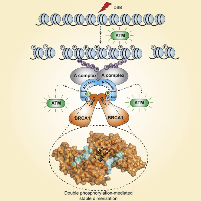
Summary
BRCA1 accumulation at DNA damage sites is an important step for its function in the DNA damage response and in DNA repair. BRCA1-BRCT domains bind to proteins containing the phosphorylated serine-proline-x-phenylalanine (pSPxF) motif including Abraxas, Bach1/FancJ, and CtIP. In this study, we demonstrate that ionizing radiation (IR)-induces ATM-dependent phosphorylation of serine 404 (S404) next to the pSPxF motif. Crystal structures of BRCT/Abraxas show that phosphorylation of S404 is important for extensive interactions through the N-terminal sequence outside the pSPxF motif and leads to formation of a stable dimer. Mutation of S404 leads to deficiency in BRCA1 accumulation at DNA damage sites and cellular sensitivity to IR. In addition, two germline mutations of BRCA1 are found to disrupt the dimer interface and dimer formation. Thus, we demonstrate a mechanism involving IR-induced phosphorylation and dimerization of the BRCT/Abraxas complex for regulating Abraxas-mediated recruitment of BRCA1 in response to IR.
Graphical Abstract
Highlights
-
•
Abraxas C terminus S404 is phosphorylated in response to ionizing radiation
-
•
Phosphorylated S404 promotes stable BRCA1 BRCT/Abraxas dimer formation
-
•
Abraxas S404 is critical for BRCA1 accumulation at DNA damage sites
-
•
BRCA1 germline mutations disrupt BRCA1-BRCT/Abraxas dimer formation
Abraxas interacts with BRCA1 through a phosphor-SPxF motif. Wu et al. show that a nearby serine 404 residue is phosphorylated in response to ionizing radiation in an ATM-dependent manner. S404 phosphorylation promotes BRCA1-BRCT/Abraxas dimer formation and is crucial for BRCA1 accumulation at DNA damage sites.
Introduction
Patients with hereditary breast and ovarian cancer (HBOC) have high germline gene-mutation rates on chromosome 17q21 tumor suppressor gene BRCA1 (breast cancer susceptibility genes 1) (Futreal et al., 1994, Hall et al., 1990, Miki et al., 1994). BRCA1 stabilizes genomic integrity by interacting with various DNA damage response (DDR) sensors, mediators, and effector proteins, thereby coordinating recognition of the DNA damage sites, cell-cycle checkpoint, DNA repair, transcription, and apoptosis/senescence. BRCA1, a large protein of 1,863 amino acids, contains an N-terminal RING domain and two C-terminal tandem BRCT domains. BRCT domains can recognize phosphorylated proteins with a phosphorylated serine-proline-x-phenylalanine (pSPxF) motif (Manke et al., 2003, Rodriguez et al., 2003, Yu et al., 2003) including Abraxas (Kim et al., 2007a, Liu et al., 2007, Wang et al., 2007), Bach1/FancJ (Cantor et al., 2001, Yu et al., 2003), and CtIP (Wong et al., 1998, Yu et al., 1998). The phosphopeptide-binding ability of BRCA1 BRCT is essential for BRCA1’s tumor suppression function (Shakya et al., 2011), where many breast and ovarian cancer related mutations occur (Clapperton et al., 2004, Couch and Weber, 1996, Friedman et al., 1994, Shattuck-Eidens et al., 1995, Shiozaki et al., 2004, Williams et al., 2004).
The two BRCA1-BRCT domains (BRCT1 and BRCT2) each contain about 100 residues and associate in a head-to-tail manner (Williams et al., 2001). Structural analysis of BRCA1-BRCT domains with pSPxF-containing phosphopeptides of Bach1 (Clapperton et al., 2004, Shiozaki et al., 2004), CtIP (Varma et al., 2005), synthetic optimized phosphopetide (Williams et al., 2004), or other binding proteins (Campbell et al., 2010, Liu and Ladias, 2013, Shen and Tong, 2008) have revealed that phosphorylated serine and phenylalanine in the pSPxF motif bind in a cleft formed at the junction of two BRCT domains in a “two-anchor” mode and the structural integrity of both binding sites is essential for peptide recognition (Glover et al., 2004, Leung and Glover, 2011, Wu et al., 2015). However, little information exists regarding the importance of the sequence surrounding the pSPxF motif. Nor is it known how the specificity is determined for BRCT binding to different pSPxF motif-containing proteins.
Abraxas mediates the interaction of BRCA1 to other components of the BRCA1-A complex, which include BRCC36, NBA1/MERIT40, BRE, and Rap80. Abraxas, a 409-residue polypeptide, contains a non-catalytic Mpr1, Pad1 N-terminal (MPN) domain at its N terminus, followed by a coiled-coil (CC) region, an unstructured region, and a BRCA1-binding pSPTF motif at the C terminus. While the N-terminal region including the MPN domain binds to Rap80, BRE, and NBA1/MERIT40, the CC domain is required for interaction with BRCC36 (Hu et al., 2011, Kim et al., 2007a, Wang and Elledge, 2007, Wang et al., 2009). Although the structures for BRCA1 BRCT in complex with other phosphopeptides have been solved previously (Campbell et al., 2010, Clapperton et al., 2004, Liu and Ladias, 2013, Shen and Tong, 2008, Shiozaki et al., 2004, Varma et al., 2005, Williams et al., 2004), the structure for BRCT/Abraxas has remained unknown.
Abraxas and the BRCA1-A complex recruit BRCA1 to DNA double-strand-break sites (DSBs) in an ATM-dependent ubiquitin-mediated signaling pathway involving E2 conjugase Ubc13, E3 ligases RNF8/RNF168, and Rap80 binding to ubiquitin lys63-linked polyubiquitin conjugates (Doil et al., 2009, Harper and Elledge, 2007, Hu et al., 2012, Huen and Chen, 2008, Kim et al., 2007b, Kolas et al., 2007, Lee and Paull, 2004, Mailand et al., 2007, Sato et al., 2009, Uziel et al., 2003, Wang, 2012, Wang and Elledge, 2007, Wu et al., 2009). Abraxas-deficient mice exhibit decreased survival and increased tumor incidence (Castillo et al., 2014). The interaction of Abraxas with BRCA1 has been shown critical for the function of Abraxas in DNA repair of DSBs and maintenance of genomic stability. Mutation of the serine residue in the pSPxF motif leads to defective DNA repair and chromosomal aberration. The importance of the Abraxas-BRCA1 interaction in tumor suppression is also suggested by identification of an Abraxas mutation in tumor in the phenylalanine residue of the pSPxF motif (F409C) (Castillo et al., 2014). Thus, structural and functional analysis of Abraxas and BRCA1 interaction is necessary to facilitate the understanding of Abraxas-mediated BRCA1 signaling in tumor suppression.
In this study, we have solved the crystal structures of BRCT with Abraxas phosphorylated peptides and revealed an ionizing radiation (IR)-induced, ATM-dependent Abraxas phosphorylation mechanism, which promotes dimerization of BRCT/Abraxas complex at the DNA damage sites. The IR-induced phosphorylation of Abraxas and the subsequent stabilization of BRCA1-BRCT dimerization are likely to comprise an important mechanism for accumulation of BRCA1 to DNA-damaged chromatin and BRCA1 mediated tumor suppression.
Results
Abraxas Is Double Phosphorylated at S404 and S406 Residues in Response to IR
Analysis of the C-terminal sequence of Abraxas reveals an additional serine (S404) located close to the pSPxF motif (Figure 1A). Double-phosphorylated Abraxas peptide containing phosphorylated S406 and S404 (GFGEYpS404RpS406PTF) has been identified to bind to BRCA1-BRCT domains in response to IR (Wang et al., 2007). We decided to investigate whether the S404 residue is important and whether it is phosphorylated upon IR. Previously, we have generated S406 phospho-specific antibody and showed that phosphorylation of S406 (pS406) occurs independently of IR (Wang et al., 2007). In view of the fact that S406 is nearby and is phosphorylated in the presence and absence of IR, we generated antibodies specifically recognizing double phosphorylated S404 and S406 (pS404pS406). The pS404pS406 specific antibody recognized Abraxas in parental, but not Abraxas knockout 293T cells and the intensity of the Abraxas band increased significantly when cells were treated with IR, indicative of IR-induced phosphorylation (Figure 1B). Mutation of either S404 (S404A), S406 (S406A), or double mutation (S404A and S406A) abolished the recognition of Abraxas by the pS404pS406 antibody (Figure 1C). Upon IR treatment, double phosphorylation of Abraxas S404 and S406 residues increased immediately (within 10 min), peaked at 1 hr, and gradually decreased to nearly basal levels at later time points (Figure 1D). Furthermore, double phosphorylation occurs in a dose-dependent manner in response to IR (Figure 1E). Since phosphorylation of S406 is not changed upon IR treatment, phosphorylation of S404 is likely to be IR-induced. As ATM plays a central role in the IR-induced signaling pathway that recruits Abraxas and the BRCA1-A complex (Harper and Elledge, 2007), we investigated whether ATM regulates the phosphorylation. Indeed, the IR-induced S404 phosphorylation is ATM dependent, treatment of an ATM inhibitor KU55933 completely abolished the IR-induced phosphorylation (Figure 1F). Other DDR kinases including ATR, DNA-PK, Chk1, or Chk2, however, did not appear to have a major effect on the IR-induced phosphorylation recognized by the pS404pS406 antibody (Figures 1G and S1).
Figure 1.

IR-Induced Double Phosphorylation of Abraxas C Terminus S404 and S406 Is ATM Dependent
(A) Abraxas-domain boundary and C-terminal sequence containing a serine residue (S404) next to the BRCA1-binding pSPxF motif (high-lighted in blue). The phosphorylation of S404 and S406 is indicated as P.
(B) Double phosphorylation of S404 and S406 residues at the Abraxas C terminus in response to IR in 293T cells and 293T/Abraxas KO cells. The lysates from cells treated with 10 Gy IR followed by incubation at 37°C for 1 hr were used for western blot with anti-pS404pS406 antibody (“∗” non-specific band).
(C) IR-induced double phosphorylation of S404 and S406 is abolished in Abraxas mutants (S404A, S406A, or double mutant, DM) (“∗” non-specific band). The FLAG- and HA-tagged Abraxas WT or mutants were expressed in 293T cells. The lysates from cells treated with 10 Gy IR and incubated at 37°C for 1 hr were used for immunoprecipitation with anti-FLAG beads and western blot with antibodies against pS404pS406, pS406, or HA.
(D) IR-induced double phosphorylation of S404 and S406 occurs immediately after IR treatment. The time points were taken after cells were treated with 4 Gy IR followed by incubation at 37°C.
(E) IR-induced phosphorylation occurs in a dose-dependent manner.
(F) ATM regulates IR-induced phosphorylation. The cells were incubated with ATM kinase inhibitor KU55933 (10 μM) for 2 hr before exposure to 4 Gy IR and subsequent incubation at 37°C for 1 hr.
(G) ATR is not involved in IR-induced double phosphorylation. The ATR inhibitor VE-821 at indicated concentrations was used for treating cells for 2 hr before cells were exposed to 4 Gy IR (see also Figure S1).
Crystal Structures of BRCA1 BRCT Domains in Complex with Single and Double-Phosphorylated Abraxas Peptides
Since S404 is separated by just one residue from S406, which is part of the pSPxF motif, we hypothesized that an additional mechanism exploiting phosphorylation of S404 might regulate Abraxas interaction with BRCA1. In order to test this, we used structural information to allow comparison of the interactions between BRCT and Abraxas in single and double-phosphorylated states. We solved crystal structures of BRCA1-BRCT in complex with both single (1p) and double phosphorylated (2p) synthesized Abraxas peptides (Ab). The BRCT-Ab1p (GFGEYSRpSPTF) complex crystal was solved at 3.5 Å resolution with no clear main-chain electron density for the N-terminal GFGE region of the peptide, indicating that this region is flexible. BRCT-Ab1p_short without the GFGE residues (YSRpSPTF) complex was then crystalized and solved at 2.5 Å resolution. The structure of BRCT domains in complex with the 2p Ab (Ab2p: GFGEYpSRpSPTF) was solved at 3.5 Å resolution. However, the BRCT-Ab2p_short (YpSRpSPTF) crystal did not diffract to a high resolution. We therefore used BRCT-Ab1p_short and BRCT-Ab2p structures (Figures 2A and S2A) for analysis. Statistics of these two structures are shown in Table 1.
Figure 2.

Crystal Structures of BRCT in Complex with Single and Double Phosphorylated Abraxas Peptide
(A) Crystal structure of BRCT-Ab1p_short. The BRCT domains are in yellow, and the Ab1p_short peptide is in wheat color.
(B–D) Show the detailed interactions between phosphopeptide and BRCT domains. The polar interaction is indicated in dashed lines.
(E) Crystal structure of BRCT-Ab2p. The BRCT is in blue, and the Ab2p peptide is in cyan.
(F and G) Show the interface between BRCT and Ab in both BRCT-Ab1p_short and BRCT-Ab2p structures. The 2Fo-Fc electron density (σ = 1.0) is shown for Abs.
(H) Superimposition of BRCT-Ab2p, BRCT-Ab1p_short, BRCT-Bach1 (PDB code: 1T29), and BRCT-CtIP (PDB code: 1Y98). The BRCT domains are shown in a gray surface representation. Ab2p, Ab1p_short, Bach1, and CtIP are in blue, yellow, green, and purple, respectively. The pSPxF motif is indicated in the image.
(I) Sequence alignment of Abraxas C terminus. The BRCT-binding motif is indicated by a blue line, and the black arrows indicate the half conserved residues (see also Figures S2 and S3).
Table 1.
The Statistics of Structures
| Crystals | BRCT-Ab1p_short | BRCT-Ab2p |
|---|---|---|
| X-ray source | Diamond Light Source Beamline I03 | Diamond Light Source Beamline I04-1 |
| Wavelength (Å) | 0.9793 | 0.9200 |
| Space group | P3221 | P212121 |
| Cell dimensions a, b, c (Å) and α, β, γ (°) | 63.8, 63.8, 93.4, 90, 90, and 120 | 86.8, 183.7, 190.5, 90, 90, and 90 |
| Resolution (Å) | 47.6-2.5 | 95.3-3.5 |
| Rsyma | 0.051 (0.498)b | 0.096 (0.701) |
| I / σ | 24 (5.1) | 16.4 (2.6) |
| Wilson B factor | 58.6 | 97.2 |
| Completeness (%) | 99.9 (99.8) | 99.8 (98.8) |
| Redundancy | 9.3 (9.4) | 7.4 (7.8) |
| Refinement | ||
| Resolution (Å) | 47.6-2.5 (2.6-2.5) | 95.3-3.5 (3.7-3.5) |
| No. unique reflections | 8,000 | 39,170 |
| Rcrystc | 0.215 (0.356) | 0.235 (0.319) |
| Rfreed | 0.252 (0.374) | 0.298 (0.341) |
| No. protein atoms | 1,701 | 14,092 |
| No. copy number of complex in ASU | 1 | 8 |
| Ramachandran favored (%) | 97.7 | 97.1 |
| Average B-factor (Å2) | 66.68 | 114.6 |
| Rmsds | ||
| Bond lengths (Å) | 0.007 | 0.007 |
| Bond angles (°) | 0.823 | 0.900 |
Rsym = Σh|Ih − < I > |/ΣhIh, where Ih is the intensity of reflection h and < I > is the mean intensity of all symmetry-related reflections.
The statistics in parenthesis are for the highest resolution shell.
Rcryst = Σ||Fobs|−|Fcalc||/Σ|Fobs|, Fobs and Fcalc are observed and calculated structure factor amplitudes.
Rfree as for Rcryst using a random subset of the data (about 10% for BRCT-Ab1p_short and 5% for BRCT-Ab2p) excluded from the refinement.
As in structures solved previously (Campbell et al., 2010, Clapperton et al., 2004, Liu and Ladias, 2013, Shen and Tong, 2008, Shiozaki et al., 2004, Varma et al., 2005, Williams et al., 2001, Williams et al., 2004), two BRCT domains of BRCA1 (BRCT1 and BRCT2) associate in a head-to-tail manner in both structures. In each domain, a four-stranded parallel β sheet is surrounded by three α helices with α1 and α3 on one side of the β sheet and α2 on the other side. Helices α2 (from BRCT1), α′1, and α′3 (from BRCT2) form the hydrophobic interface, and the two domains are further linked by extra helix αL (Figures 2A and 2E). The pSPTF motif from Abraxas binds to the BRCT domains in a similar two-anchor mode using pS406 and F409. Residues P407 and T408 do not make major interactions with the BRCT domains. The phosphate group of Abraxas S406 interacts with the side chains of BRCT K1702 and S1655, as well as the main chain of G1656 (Figure 2B). The side chain of F409 in Abraxas inserts into the BRCT hydrophobic pocket created by L1701, F1704, N1774, M1775, and L1839 (Figure 2C). As F409 is the terminal residue for Abraxas, an extra salt bridge is present between the main chain carboxyl group of F409 with the BRCT domain residue R1699 (Figure 2D). This extra interaction was seen in previous structures using tetrapeptides pSPTF (Campbell et al., 2010).
A notable difference between BRCT-Ab2p and BRCT-Ab1p_short structure is the conformation of the Y403S404R405 region (Figures 2E–2G). Extra electron density corresponding to the phosphate group of pS404 and the side chain of Y403 is observed only in BRCT-Ab2p. Unlike pS406, the pS404 phosphate group is oriented away from the BRCT domains into the solvent region, thus avoiding contact with G1656, L1657, and T1658 (Figure 2G). In BRCT-Ab2p, the Y403 side chain is positioned to interact through a hydrophobic interaction with BRCT P1659 at the N terminus of BRCT1 α1. The extra interaction could explain the increased proximity of α1 toward the N terminus of the Abraxas phosphopeptide in BRCT-Ab2p compared to BRCT-Ab1p_short (Figure S3A). Superimposition of all available BRCA1 BRCT related crystal structures also showed that α1 movement toward the phosphopeptide is most prominent in BRCT-Ab2p (Figure S2B). It indicates that one of the roles of pS404 is to fix the side chain of Y403, which is conserved in higher organisms (Figure 2I), such that a trans peptide bond can form and collision is avoided. Superimposition of the BRCT/Abraxas structures with the BRCT/Bach1 (PDB: 1T29) (Shiozaki et al., 2004) and BRCT/CtIP structures (PDB: 1Y98) (Varma et al., 2005) shows similar pSPxF-motif binding. However, compared to Bach1 and CtIP, the N-terminal sequence of Abraxas in both BRCT-Ab1p and BRCT-Ab2p structures exits on the opposite side, close to the α1 of BRCT1 domain (Figure 2H). Interestingly, a similar side chain arrangement was also seen in BRCA1 BRCT bound with “optimized peptide” (GAAYDIpSQVFPFAKKK) (PDB: 1T2V) (Williams et al., 2004) (Figure S3C), in which the tyrosine residue (Y) at −3 position (phosphorylated serine in pSxxF motif as 0 position) and negatively charged residues glutamic acid (E) or aspartic acid (D) at −2 position were shown more favored for interaction with BRCA1 BRCT domains (Manke et al., 2003, Rodriguez et al., 2003).
Dimerization of BRCT-Abraxas Complex in Crystal Structures
There are eight copies of BRCT-Ab2p in the asymmetric unit (ASU), and each BRCT-Ab2p appears to dimerize through the same interface either within the ASU or between ASUs (Figure S2B). The dimerization of the 1:1 BRCT-Ab2p complex results in a 2:2 BRCT/Abraxas complex dimer. Dimerization involves α1 and β2 of the BRCT1 domain and the Ab2p (Figures 3A–3D) burying about 1,880 Å2 area. In the dimer interface, two of the α1 helices from each BRCT-Ab2p complex form isologous interactions burying a hydrophobic patch formed by F1662, M1663, and Y1666 with aromatic side chains stacking on each other (Figure 3B). Interestingly, BRCA1 germline mutations of F1662 (F1662S) and M1663 (M1663K) have been identified in germline cancer patients as recorded in the Breast Cancer Information Core database (Szabo et al., 2000). Extensive hydrogen bonds also form between the two equivalent antiparallel β2 strands (residues T1,675–L1,679) (Figure 3D). The two-fold symmetry axis within the BRCA1/Abraxas dimer lies perpendicular to the two β strands. The cross interaction between the two BRCT/Abraxas complexes is also mediated by the ionic interaction between Abraxas and BRCT α1 of the opposite BRCT/Abraxas complex. The negative surface patch, generated by the phosphate group of pS404 and side chain of E402 at the N terminus of Ab2p peptide, leads to cross interaction with BRCT K1671 (Figure 3C). The phosphate group of pS406 also contributes to the dimer formation by interacting with the R1670 residue of the opposite BRCT (Figure 3C). Although a similar dimer interface was observed for BRCT-Ab1p_short structure, the cross interaction between the negative surface patch (formed by the phosphate group of pS404 and the side chain of E402) and BRCT K1671 is completely lacking.
Figure 3.

2p Ab Induces Dimerization of BRCT-Ab2p Complex
(A) Crystal structure of BRCT-Ab2p complex dimer viewed from three different directions with a two-fold axis. The dimer interface is within the dashed circles and zoomed in (B)–(D).
(B) Dimer interface between two BRCT α1 helices.
(C) Interaction between BRCT α1 helix and Ab2p.
(D) Dimer interface between two BRCT β2 strands. The polar interactions between labeled residues are shown in black dashed lines. The key residues are indicated in the image.
(E) Surface representation of BRCA1-1p_short (yellow) and BRCT-Ab2p dimer (blue and pink). The Abraxas pS406 binding region is indicated in the dashed circle.
(F) Gel filtration BRCT in complex with Ab1p, Ab2p, Bach1, and CtIP phosphopeptides at a concentration of 40 μM (1 mg/ml). The regions for dimer complex (2:2 complex) and monomer complex (1:1 complex) are high lined in yellow and gray shades. The elution positions for void and protein markers aprotinin (Mw = 6,500 Da), ribonuclease A (Mw = 13,700 Da), carbonic anhydrase (Mw = 29,000 Da), Ovalbumin (Mw = 44,000 Da), and Conalbumin (Mw = 75,000 Da) are indicated.
(G) The molecular weight of BRCT and its complexes with phosphopeptide, measured using native mass spectrometry.
(H) The native mass spectra of BRCT-Ab1p, BRCT-Ab2p, BRCT-Bach1, and BRCT-CtIP complexes tested at 15 μM (see also Figures S3 and S4).
Compared to the monomeric BRCT/Abraxas complex, where pS406 is half surrounded by BRCT1 and half exposed to the solvent, the dimerization of BRCT/Abraxas allows the second BRCT1 to reduce further the accessibility of pS406 to solvent (Figure 3E).
BRCT/2p Abraxas Complex Forms a Dimer In Vitro
To examine whether the BRCT/Abraxas complex exists as a dimer in vitro, we first tested whether dimers form in solution using size exclusion chromatography at protein concentration (1 mg/ml), much lower than the concentration used for crystallization (30 mg/ml). The elution profiles of BRCT-Ab1p and BRCT-Ab2p were different. The BRCT-Ab2p elution peak appeared to the left of the BRCT-Ab1p peak (Figure 3E), suggesting a larger hydrodynamic radius and a possible higher order BRCT-Ab2p complex. The controls show that BRCT does not interact with unphosphorylated peptide or phosphopeptide containing only phosphorylated S404 (Figure S3A). Under the same condition, the BRCT-Bach1 and BRCT-CtIP form complexes similar to that of BRCT-Ab1p (Figure 3F). Standard protein markers were also run and the positions of their elution peaks are indicated in Figure 3F. The size of BRCT-Ab2p is roughly double that of BRCT-Ab1p according to the protein markers.
We tested whether a higher concentration of BRCT-Ab1p leads to dimer formation as we observed in crystals. Indeed, the BRCT-Ab1p peak shifts left to the BRCT-Ab2p peak position once the concentration is increased (Figure S3E), indicating the tendency of BRCT-Ab1p to form higher order complexes at high protein concentrations as observed in the crystal structure. It is likely that at high concentrations, the BRCT-Ab1p complex is packed in a conformation that is not stable at low protein concentrations without a contribution from phosphorylated S404. Therefore stable dimerization of two BRCT/Abraxas complexes is unique for BRCT-Ab2p.
To confirm dimer formation, we also measured the exact molecular weight of peak fractions eluted from gel filtration using nano-electrospray mass spectrometry analysis under native conditions (Figures 3G and 3H). BRCT-Ab1p is shown to exist predominantly as a 1:1 complex with a small portion forming a 2:2 dimer. In contrast, the majority of BRCT-Ab2p is detected as 2:2 complexes, indicating a much more stable dimer. BRCT/Bach1 and BRCT/CtIP are detected only as 1:1 complexes. Consistent with the finding that higher protein concentration facilitates dimer formation (Figure S3E), the proportion of BRCT-Ab1p forming dimer increases significantly when the protein concentration is increased from 15 μM to 75 μM (Figure S3F). Small angle X-ray scattering (SAXS) experiments of the BRCT-Ab1p and BRCT-Ab2p also show similar results (Figure S4). Thus, our results indicate that only double-phosphorylated Abraxas C-terminal peptide induces stable dimerization of BRCT/Abraxas complexes in vitro.
Mutagenesis Studies of BRCT-Ab2p Dimer Interface Reveal the Importance of S404 Phosphorylation and Residues of BRCA1 Germline Mutations for Stable BRCT/Abraxas Dimer Formation
In order to test the dimer interface, we have generated various mutants for both BRCT domains and Abraxas (summarized in Figure 4C with peptide sequence indicated) based on the crystal structure. As shown in a simplified graph of the dimer interface (Figure 4A), three regions of interactions appear to contribute to formation of the dimer interface: (1) the N-terminal hydrophobic region of BRCT α1-α1; (2) extensive hydrogen bonds formed by β2-β2; and (3) the N-terminal region of Ab2p including the phosphorylated S404 interaction with BRCT α1. The interacting residues in α1-α1 and Ab2p-α1 are shown in Figure 4B.
Figure 4.

Mutagenesis Studies of the Interface of BRCT-Ab2p Complex Dimer
(A) Simplified BRCT-Ab2p dimer interface containing three regions observed in BRCT-Ab2p crystal.
(B) Detailed interactions mediated through BRCT α1.
(C) Summary of BRCT and Abraxas mutants. The complexes tested are grouped into four and highlighted in different colors.
(D–G) Gel filtrations of BRCT and Abraxas mutants. The same color codes are used as in (C).
By size exclusion chromatography, we first tested the importance of S404 phosphorylation. Mutations of Abraxas S404 to proline or aspartic acid were generated. While BRCT-Ab1p (S404P) leads to 1:1 complex formation, BRCT-Ab1p (S404D) can maintain the 2:2 complex dimer as BRCT-Ab2p (Figure 4D). This confirms that S404 phosphorylation is essential for dimerization. A previous report using optimized peptide containing aspartic acid in the equivalent position to Abraxas S404 was reported to not form a dimer in solution (Williams et al., 2004). We reasoned that the difference between the optimized peptide and Ab2p is in the N-terminal region (G399F400G401E402) of Ab2p. We demonstrated that BRCT-Ab2p_short without GFGE still forms a dimer indicating that GEGE is not absolutely required for dimerization when Y403 and pS404 are present. However, when we analyzed BRCT-Ab2p in solution, we found that while F400D did not affect dimer formation, E402R, as well as BRCT K1671E or R1670E, partially destabilizes dimer formation. Double mutation of BRCT(K1671E)-Ab2p(E402R) further destabilized the complex, leading to elution at a position close to that of 1:1 stoichiometry (Figure 4E). These results indicate that although GFGE is not absolutely required for the dimer formation, it contributes to the dimer stabilization when it is present.
We also tested the importance of the BRCT P1659 interaction with Abraxas Y403 to the stability of the BRCT-Ab2p dimer. In the presence of the N-terminal region (GFGE) of Ab2p, mutation of Abraxas Y403A did not destabilize the dimer complex formation in either BRCT-Ab2p(Y403A) or BRCT(P1659G)-Ab2p(Y403A). But when the N-terminal region of Abraxas was absent, the dimer complex of BRCT-Ab2p(Y403A_short) became unstable. This destabilization was further enhanced in BRCT(P1659G)-Abraxas(Y403A)_short complex, which was eluted near the 1:1 complex (Figure 4F). Together, these results are consistent with the crystal structure analysis showing that Ab2p promotes dimer formation in two different ways: (1) phosphorylated S404 fixes the side chain of Y403, which generates additional interaction with BRCT K1671 and (2) the phosphorylated group of pS404 and E402 form a negative surface region that leads to cross interaction with BRCT K1671.
We then evaluated the contribution of the hydrophobic interactions between the α helices (α1-α1) and the extensive hydrogen bonds between the two antiparallel β strands (β2-β2) in the two protomers that comprise the dimer (Figure 4G). Our results indicate that the α1-α1 interaction contributes more significantly toward the stabilization of the dimer interface than the β2-β2 interaction. The BRCT mutant N1678A, which disrupted the β2-β2 interaction (Figure 3D), reduced hydrogen bonds between the side chain of N1678 and the nearby residue T1675, but did not destabilize BRCT-Ab2p dimer formation. In contrast, F1662S and M1663K, two BRCA1 germline mutations identified in cancer patients (Szabo et al., 2000), led to complete disruption of the dimer formation as the elution peaks of these two mutants in complex with Ab2p moved to the 1:1 complex position. BRCT Y1666A mutant did not result in complete disruption of the dimer and the peak in the elution profile is located between that for the 2:2 and 1:1 complexes. These results support our observation from the crystal structure that F1662S and M1663K have a much more significant effect on disruption of the dimer interface because these two residues are located at the point of the isologous dimer interface, while Y1666A is further away.
Abraxas S404 Is Important for Cellular Resistance to IR and Accumulation of BRCA1 at the DNA Damage Site
Since IR-induced phosphorylation of S404 appears to promote stable BRCT/Abraxas dimer formation, the S404 residue is likely to be critical for the function of Abraxas in response to IR. We first tested whether S404 is important for the cellular response to IR. In an IR sensitivity assay, both S404A and S406A mutants of Abraxas were unable to fully rescue the increased sensitivity of Abraxas knockdown cells as the wild-type Abraxas did (Figures 5A 5B, and S5), suggesting that phosphorylation of S404 plays a role in the cellular resistance to IR. Abraxas recruits BRCA1 to DNA damage sites in response to IR. We thus examined the role of S404 phosphorylation in BRCA1 accumulation at the DNA damage sites. The percentage of cells containing more than ten BRCA1 IR-induced foci (IRIF), as well as the intensity of the foci, were significantly decreased in Abraxas knockdown cells. While the defect of Abraxas knockdown cells in BRCA1 IRIF formation could be rescued by expression of wild-type Abraxas, it could not be rescued by expression of Abraxas S406A or S404A mutant (Figures 5C–5E and S5). Consistently, when we examined the chromatin-bound BRCA1 levels in response to IR, we found that both the S404A and S406A mutants failed to accumulate BRCA1 to damaged chromatin as the wild-type Abraxas does (Figure 5F). As a control, the total expression level of BRCA1 was not affected in Abraxas knockdown cells or the cells complemented with expression of either wild-type or mutant Abraxas. Thus, phosphorylation of S404 is likely to play an important role in BRCA1 accumulation to DNA damage sites and in cellular resistance to IR.
Figure 5.

Phosphorylation of S404 and S406 Are Both Important for Cellular Resistance to IR and BRCA1 Accumulation at DNA Damage Sites
(A) Generation of Abraxas knockdown U2OS cells complemented with expression of small hairpin (sh)RNA-resistant HA-tagged WT, S404A, or S406A mutants of Abraxas.
(B) Increased cellular sensitivity to IR of Abraxas-deficient cells expressing mutants of Abraxas. The colony-survival assay was carried out for cells treated with 4 Gy IR. The data are presented as means ± SD. The data analyses are processed by ANOVA and the statistical significance was determined by Tukey’s multiple comparisons test (∗p < 0.02). There were three independent experiments that were performed (additional data are presented in Figure S5).
(C) Representative images of BRCA1 IRIF in Abra1 shRNA knockdown cells complemented with vector, WT, or mutants of Abraxas in response to 10 Gy IR followed by 2 hr incubation at 37°C.
(D) The percentage of cells containing more than ten BRCA1 IRIF foci was quantified. The data are presented as means ± SD. The data analyses are processed by ANOVA and the statistical significance was determined by Tukey’s multiple comparisons test (∗p < 0.0001). At least three independent experiments were performed. More than 300 cells were counted for each experiment. Additional data for quantification at different time points post IR are presented in Figure S5.
(E) Quantification of the intensity of BRCA1 IR induced foci (IRIF). The data are presented as means ± SD (n > 50). The statistical analysis was carried out by Student’s t test (∗p < 0.0002).
(F) BRCA1 accumulation at damaged chromatin depends on both S404 and S406 residues. The Orc2 was used as a marker for chromatin-bound fraction. The cells were treated with 10 Gy IR followed by 2 hr incubation at 37°C. The cellular fractionation was carried out and the chromatin fraction was analyzed (see also Figure S5).
Abraxas-Dependent BRCA1 Dimerization In Vivo
We tested whether BRCA1 forms dimers in vivo and whether the stable dimer formation is dependent on Abraxas. We co-expressed differentially Myc- or FLAG-tagged BRCA1 full-length protein in control (Ctrl) cells or Abraxas knockout (KO) cells. In the co-immunoprecipitation experiment with lysates prepared from cells treated with IR, immunoprecipitated FLAG-BRCA1 interacts with Myc-tagged BRCA1, indicating that BRCA1 indeed dimerizes in vivo. The dimerization was decreased in Abraxas KO cells, indicating the dependency of dimerization on Abraxas (Figure 6A). Similarly, a construct containing only the BRCA1-BRCT domains also dimerizes when co-expressed in cells and the dimerization is decreased in Abraxas KO cells (Figure 6B).
Figure 6.

Abraxas Promotes BRCA1 BRCT Dimerization In Vivo
(A) Abraxas-dependent BRCA1 dimerization in vivo. The differentially Myc- and FLAG-tagged BRCA1 full-length constructs were transiently transfected into parental 293T (Ctrl) or Abraxas KO cells. The lysates from cells treated with 10 Gy IR followed by 1 hr incubation at 37°C were used for FLAG-immunoprecipitation. The intensity of individual bands was quantified by densitometric analysis using NIH ImageJ software. The normalized value (IPed_mycBRCA1/Input_mycBRCA1) was shown in the bar graph.
(B) Abraxas-dependent BRCA1-BRCT domains dimerization in vivo. The differentially Myc- and HA-tagged BRCA1-BRCT domains constructs were transiently transfected into parental 293T (Ctrl) or Abraxas KO cells. The lysates from cells treated with 10 Gy IR followed by 1 hr incubation at 37°C were used for HA-immunoprecipitation. The band intensity was quantified with NIH imageJ software. The normalized value (IPed_mycBRCT/Input_mycBRCT) was shown in the bar graph.
(C and D) BRCA1 germline mutations F1662S or M1663K decrease BRCA1 dimerization in vivo. Myc-tagged BRCA1 full-length (WT-FL) and HA-tagged BRCA1 BRCT (WT-BRCT) or Myc-tagged mutant full-length (F1662S or M1663K) and HA-tagged BRCT triple mutant (TM, F1662S/M1663K/R1670E) were co-expressed in cells. The lysates from cells treated with 10 Gy IR followed by 1 hr incubation at 37°C were prepared for either Myc- immunoprecipitation (C) or reciprocal IP with HA- immunoprecipitation (D). The normalized value was shown in the bar graph.
(E) Abraxas dimerize/oligomerize in vivo independent of binding to BRCA1. The differentially tagged GFP- and HA-FLAG-tagged WT or GFP-tagged and HA-FLAG-tagged Abra1 mutants (S404A or S406A) were co-expressed in cells. The anti-GFP immunoprecipitation was carried out with lysates from 293T cells treated or not treated with 10 Gy IR followed by 1 hr incubation at 37°C. The images are from the same blot.
(F) Abraxas dimerizes/oligomerizes, in vivo, through the CC domain. The immunoprecipitations were carried out with lysates prepared from cells co-expressing HA-FLAG-tagged WT Abra1 or HA-FLAG-tagged CC domain deletion mutant (ΔCC) and GFP-tagged WT or ΔCC (see also Figure S6).
We then tested whether the germline mutations F1662S and M1663K interfere with BRCA1 dimerization in vivo. We compared the interaction of a Myc-tagged full-length BRCA1 and a HA-tagged wild-type BRCT fragment with that of the F1662S or M1663K mutant of BRCA1 and a mutant BRCA1 BRCT fragment with three residues localized in the dimer interface mutated (F1662S/M1663K/R1670E). Both the Myc-immunoprecipitation (Figure 6C) and reciprocal HA- immunoprecipitation (Figure 6D) experiments showed that the interaction/dimerization of BRCA1 and BRCT was decreased with mutation of the critical residues at the dimer interface F1662S or M1663K. Thus, BRCA1 germline mutations interfere with stable dimer formation in vivo.
To understand dimerization of the BRCT/Abraxas complex in vivo, we examined whether Abraxas forms a dimer in which the phosphorylated C-termini of Abraxas in complex with BRCT could be in close vicinity for dimerization. We expressed both GFP-tagged Abraxas and HA-FLAG-tagged Abraxas in cells and tested whether the differentially tagged Abraxas molecules interact with each other using the immunoprecipitation assay. We found that wild-type Abraxas, as well as the S404A and S406A mutant, interact with differentially tagged counterpart, indicating that Abraxas dimerizes/oligomerizes in vivo independently of its binding to BRCA1 (Figure 6E). We then investigated what region of Abraxas mediates the dimerization by examining various deletion mutants of Abraxas. Deletion of the CC domain abolished the self-interaction of either wild-type or mutant Abraxas (Figures 6F and S6). Thus, in vivo, Abraxas dimerizes/oligomerizes through the CC domain.
Discussion
BRCA1 accumulation to DNA damage sites is a crucial step for BRCA1’s function in DNA damage repair, and BRCT domains of BRCA1 are important for the tumor suppressor function of BRCA1. IR-induced ubiquitination at DNA damages sites generates docking sites for the recruitment of the Abraxas/BRCA1-A complex and accumulation of BRCA1 at sites of damage. Our data provide evidence for an IR-induced, ATM-dependent mechanism specific to Abraxas-mediated recruitment of BRCA1. In such a model (Figure 7), IR-induced phosphorylation of S404 next to the pSPxF induces stable dimer formation of the BRCA1 BRCT/Abraxas complex.
Figure 7.

A Model Showing IR-Induced Phosphorylation of Abraxas Promotes Dimerization of BRCA1 at Sites of DNA Damage for BRCA1 Accumulation and Cellular Response to IR
In the absence of IR, two BRCA1 bound to BRCA1-A complex do not form stable dimer. Extra phosphorylation at Abraxas S404 induced by IR leads to the stable dimerization of BRCA1 at the DNA damaged site. The BRCT-Abraxas interaction is indicated in a dashed circle for the “pair-hugging” interaction model. The BRCT domains are represented by gray circles and Abraxas phosphopeptides by pink lines. The line with double arrows indicates interaction for dimerization (P indicates the phosphorylation).
The crystal structural analysis of BRCT in complex with Abraxas phosphorylated peptides revealed that, although both Ab1p and Ab2p bind to BRCT domains through the same pSPxF motif, the phosphorylation of S404 in Ab2p induces stable dimerization of the BRCT/Abraxas complex. The dimer interface locates to the BRCT1 of BRCA1 tandem BRCT domains. As expected from previous reports, BRCT1 also provides the interaction site for pS406 of the pSPxF motif, while the side chain of F of the motif inserts into a hydrophobic pocket created mainly by BRCT2 domain. The dimer surface formed between two BRCT1 domains does not directly influence the interaction between the pSPxF motif and the BRCT domains. Although the hydrophobic α1 interface observed in the BRCT-Ab2p dimer was buried in a similar manner to that of the BRCT-Ab1p or other BRCA1 BRCT related crystal structures (Wu et al., 2015), this interface alone is not strong enough to form a stable dimer in solution as we observed for BRCT domain only, BRCT-Ab1p, BRCT-Bach1, or BRCT-CtIP. In contrast, under the same condition, the stable-dimer state of BRCT-Ab2p is triggered by the phosphorylation of S404. A phosphorylation-mimetic point mutant S404D stabilizes BRCT/Abraxas dimer formation in solution in a similar way to phosphorylated S404, further supporting the conclusion that phosphorylation of S404 promotes dimer formation. The impact of phosphorylation of Abraxas S404 is the following: (1) the highly charged phosphate group of pS404 faces away from BRCT domains, resulting in stabilization of the interaction of the side chain of Y403 with BRCT P1659 located at the N terminus of α1. This interaction causes the shift of α1 closer to the N terminus of the phosphopeptide; and (2) the negatively charged side chains of pS404 and E402 provide extra ionic interaction sites with K1671 of BRCT. All together, this leads to a stable BRCT-Ab2p complex dimer formation. Owing to the symmetric pairing shape, we describe the BRCT-Ab2p dimerization interaction as a “pair-hugging” interaction mode, in which the Abraxas phosphopeptide acts as an arm wrapping around the other BRCT domain therefore stabilizing the interaction (Figure 7).
The IR-induced phosphorylation of Abraxas S404 and the subsequent stable BRCA1-BRCT dimerization are likely to comprise an important mechanism for cellular response to IR since mutation of S404 leads to decreased BRCA1 accumulation to DNA damaged chromatin and increased cellular sensitivity to IR. IR-induced phosphorylation of Abraxas S404 may facilitate the accumulation of BRCA1 at DNA damage sites by stabilizing the BRCA1 protein dimerization, forming more stable higher order complexes at sites of damage. In addition, S404 phosphorylation may further facilitate the interaction of Abraxas and BRCA1 by reducing the dissociation of pS406, so prolonging the Abraxas interaction with BRCT domains or limiting the accessibility of pS406 by other proteins such as phosphatase. Alternatively, induced dimerization of BRCA1 BRCT by phosphorylation of S404 of Abraxas could increase the local concentration of BRCA1 at damaged chromatin, which is likely critical for efficient DNA damage signaling and repair.
Many tumor-derived truncation and missense mutations have been identified in the BRCA1 BRCT domains. While some of these mutations have been shown to either destabilize the protein fold of the BRCT domains or disrupt the binding surface to pSPxF-containing phosphopeptides (Cantor et al., 2001, Clapperton et al., 2004, Coquelle et al., 2011, Manke et al., 2003, Shiozaki et al., 2004, Williams and Glover, 2003, Williams et al., 2004, Yu et al., 2003), resulting in cancer predisposition, the function of a large number of BRCT mutations is still unknown (Easton et al., 2007). Our analyses reveal that germline mutations F1662S and M1663K disrupt the ability of BRCT to dimerize, in vitro and in vivo, providing a structural explanation for the possible role of these mutations in inactivating BRCA1 tumor suppressor function. Future study is needed to further characterize the effect of these mutations in the function of BRCA1 in DNA repair and damage signaling.
How is the dimerization of BRCT-Abraxas achieved in vivo? Since the phosphorylation of S406 is not IR-dependent, Abraxas binds to BRCA1 through the pSPxF motif even in the absence of DNA damage (Wang et al., 2007). The dimerization of Abraxas through the CC domain could potentially position the two BRCT tandem domains that interact at the C terminus of Abraxas into close vicinity, leading to an unstable dimer of the BRCT-Abraxas complex in the absence of DNA damage. In response to IR, IR-induced phosphorylation of S404 leads to an increase of affinity between the phosphate group and the residues at the dimer surface, which consequently results in a much more stable dimer complex of BRCA1 BRCT/Abraxas. Since the CC domain of Abraxas also appears to dimerize with the CC domain of BRCC36 (Wang and Elledge, 2007), it is likely that, in the BRCA1-A complex, Abraxas and BRCC36 form an oligomeric bundle through the CC domain present on each of the Abraxas and BRCC36 molecules. Future structural analysis of the BRCA1-A complex is needed to further understand the multimerization of Abraxas and BRCC36 of the BRCA1-A complex. Nevertheless, IR-induced phosphorylation at Abraxas S404 appears to function as a regulatory switch, which leads to stable dimerization of two nearby BRCT domains.
The phosphorylation-induced BRCT dimerization is observed only in the BRCT/Abraxas complex. We demonstrate that, in addition to the pSPxF-binding motif, IR-induced phosphorylation of a nearby S404 residue can further regulate the interaction of BRCT and Abraxas. Thus, amino acid sequences outside the pSPxF motif may confer specificity in regulation of the BRCT binding to phosphorylated proteins. Indeed, in addition to phosphorylated S404, the N-terminal region of Ab (GFGE402Y403pS404RpSPVF) also contributes to stable BRCT/Abraxas dimer formation. The E402 residue cooperates with the phosphorylated S404 in stabilizing the dimer formation; the side chain of Y403 is fixed to interact with BRCT P1659 when S404 is phosphorylated. Thus, the unique sequence feature outside of the pSPxF motif ensures that stable dimer formation occurs only with BRCT/Abraxas, but not with other BRCT complexes. Since Abraxas-BRCA1 interaction has been shown critical for DNA repair and maintenance of genomic stability, the additional regulatory mechanism uncovered in this study in regulating the interaction of Abraxas and BRCA1 further highlights the importance of this interaction in BRCA1 signaling and tumor suppression.
In summary, our study reveals a phosphorylation-dependent mechanism in Abraxas-mediated recruitment and accumulation of BRCA1 at DNA damage sites, deepening our understanding of BRCA1 and Abraxas tumor suppressor function and related cellular signaling. Our study also provides structural insights that will assist the design of small molecules modulating BRCA1-Abraxas interaction in the future.
Experimental Procedures
Cell Lines, Culture, and Antibodies
U2OS cells were cultured in McCoy’s 5A medium supplemented with 10% fetal bovine serum (FBS). 293T cells were grown in Dulbecco’s modified Eagles medium (DMEM) supplemented with 10% FBS. Details of generation of Abraxas knockdown or KO cells are described in the Supplemental Information. Antibodies used are described in the Supplemental Information.
Immunofluorescence
Cells were fixed with 3.6% formaldehyde for 10 min, permeabilized with 0.5% Triton X-100 solution, and incubated with primary antibodies for 1 hr at 37°C followed by appropriate Alexa 488-conjugated (green; Invitrogen) and Alexa 555-conjugated (red; Invitrogen) secondary antibodies. Additional information is included in the Supplemental Information.
Cell Lysis and Immunoprecipitation
Cell lysates were prepared from 293T cells or 293T Abra1-gene KO cells either untreated or treated with 10 Gy IR followed by incubation at 37°C for 1 hr. FLAG immunoprecipitation (IP) was performed with lysates prepared from cells either treated or untreated with 10 Gy IR followed by 1 hr incubation at 37°C. Western blots were carried out using indicated antibodies. 293T cells were incubated with kinase inhibitors for 2 hr at indicated concentrations. Cells were then either exposed to 4 Gy IR or untreated. After 1 hr incubation, cells were lysed and Abraxas pS404pS406 levels were determined by western blot. Additional information is included in the Supplemental Information.
Clonogenic Survival Assay
Stable U2OS cell lines were seeded at low density in 10 cm dishes and irradiated with 4 Gy ionizing irradiation using a 137Cs source. The cells were then cultured at 37°C for 14 days to allow colonies to form. Colonies were stained with 2% methylene blue and 50% ethanol for 10 min.
Chromatin Fractionation
Abraxas knockdown cells complemented with vector, wild-type (WT), S404A, S406A, and double mutant (S404A and S406A) were irradiated at 10 Gy and incubated for 1 hr at 37°C. Cells were then subjected for chromatin fractionation followed by detection with indicated antibodies. Details are described in the Supplemental Information.
BRCT Construct, Purification, Crystallization, and Data Collection
Construction and purification of BRCA1 BRCT and mutants are described in the Supplemental Information. Purified BRCT was mixed with Abs at a 1:3 molar ratio as has been previously reported (Shiozaki et al., 2004) and incubated at 4°C for 30 min. Final protein concentration was 30 mg/ml. Crystallization was set up using the hanging-drop vapor diffusion method with drops containing 1 μl of protein sample and 1 μl of crystallization solution. Crystals appeared after 3–4 days. BRCT-Ab2p was crystallized in 0.1 M HEPES (pH 7.0), 60 mM ammonium sulfate, and 5% (w/v) PEG 4000. BRCT-Ab1p_short was crystallized in 1 M lithium chloride, 0.1 Tris (pH 8.0), and 20% (w/v) PEG 6000. X-ray diffraction data were processed by XDS (Kabsch, 2010) and Scala (Winn et al., 2011). The phases for the structure factors were obtained through molecular replacement using Phaser module in Phenix 1.8.4-1496 (Adams et al., 2010). More detail of protein crystal and structure determination can be found in Supplemental Information.
Size Exclusion Chromatography for BRCT with Abraxas, Bach1, and CtIP Phosphopeptides
BRCT protein and Abraxas, Bach1, and CtIP peptides were mixed in a 1:3 molar ratio to a final concentration of 1 mg/ml (about 40 μM) in 500 μl loops. Gel filtration was performed in Buffer A using Superdex 75 10/300 column (GE Healthcare life) with a flow rate of 0.5 ml/min. For studying the high protein concentration effects on BRCT-Abraxas complex formation, a final concentration of 10 mg/ml was used. Protein markers (GE Gel Filtration LMW Calibration Kit) were run following the kit protocol.
Native Mass Spectrometry
Samples were diluted to 15 or 75 μM protein concentration in 300 mM ammonium acetate (pH 7.6) and further buffer exchanged into 300 mM ammonium acetate using Bio-Spin 6 (Bio-Rad) column. The desalted samples were loaded into the in-house prepared gold-coated glass capillaries (Hernández and Robinson, 2007). Nano-electrospray mass spectrometric analyses were performed under native conditions on a hybrid quadrupole time-of-flight mass spectrometer previously modified for high mass transmission (Sobott et al., 2002). Typically the following instrumental conditions were used: capillary voltage 1.3 kV, sample cone 200 V, and collision cell energy 5 V.
Author Contributions
Q.W., B.W., and T.L.B. initiated this project. Q.W. performed most of the in vitro work and solved structures. A.P., D.S., and B.W. designed the in vivo experiments. D.S. generated Abraxas KO cells and analyzed Abraxas double phosphorylation in response to IR and in the presence of kinase inhibitors, as well as Abraxas dimerization in vivo. A.P. generated Abraxas knockdown cells, examined the Abraxas mutants in rescuing the defects of Abraxas-deficient cells, and Abraxas-dependent BRCA1 dimerization in vivo. S.M. and C.V.R. performed and analyzed the native mass spectrometry experiment. T.O. conducted SAXS experiment. E.L.B. made and purified the BRCT P1659G mutant. T.K.F. and B.X. generated constructs expressing Myc-tagged BRCA1 F1662S and M1663K mutants. Q.W., A.P., B.W., and T.L.B. wrote the paper together with comments from other co-authors.
Acknowledgments
We thank beamline scientists at Diamond Light Source for help during data collection of crystals and SAXS. The crystallization experiments were performed in the Crystallographic X-ray Facility at the Department of Biochemistry, University of Cambridge. We are grateful for the advice and help from Facility Manager Dr. Dimitri Chirgadze. We also thank Dr. Yanfen Hu (University of Texas Health Science Center at San Antonio) for the pFLAG-BRCA1 plasmid and Dr. Angela Pacitto (University of Cambridge) for reading the manuscript. We thank Dr. Adriana Paulucci-Holthanuzen (Department of Genetics-MD Anderson Microscopy Core Facility) for assistance with images and analysis. Q.W., T.O., and T.L.B. are funded by the Wellcome Trust (grant 093167/Z/10/Z). A.P. is an awardee of the Schissler Foundation Fellowship, the Center for Cancer Epigenetics Scholarship, and the Andrew Sowell-Wade Huggins Scholarship. This work is supported by the NIH grant (CA155025 to B.W.) with funds from the University of Texas MD Anderson Cancer Center (IRG, Center for Cancer Epigenetics, Center for Genetics, and Genomics Pilot Award). S.M. is funded by the Medical Research Council (grant 98101 to C.V.R.) and C.V.R. is a Royal Society Research Professor. T.K.F. and B.X. are supported by NIH grant (R01CA138804 to B.X.).
Published: January 14, 2016
Footnotes
This is an open access article under the CC BY license (http://creativecommons.org/licenses/by/4.0/).
Supplemental Information includes Supplemental Experimental Procedures and six figures and can be found with this article online at http://dx.doi.org/10.1016/j.molcel.2015.12.017.
Contributor Information
Bin Wang, Email: bwang3@mdanderson.org.
Tom L. Blundell, Email: tom@cryst.bioc.cam.ac.uk.
Accession Numbers
The accession numbers for the BRCT-Ab1p_short and BRCT-Ab2p data reported in this paper are PDB: 4Y2G, 4Y18.
Supplemental Information
References
- Adams P.D., Afonine P.V., Bunkóczi G., Chen V.B., Davis I.W., Echols N., Headd J.J., Hung L.W., Kapral G.J., Grosse-Kunstleve R.W.G. PHENIX: a comprehensive Python-based system for macromolecular structure solution. Acta Crystallogr. D Biol. Crystallogr. 2010;66:213–221. doi: 10.1107/S0907444909052925. [DOI] [PMC free article] [PubMed] [Google Scholar]
- Campbell S.J., Edwards R.A., Glover J.N.M. Comparison of the structures and peptide binding specificities of the BRCT domains of MDC1 and BRCA1. Structure. 2010;18:167–176. doi: 10.1016/j.str.2009.12.008. [DOI] [PubMed] [Google Scholar]
- Cantor S.B., Bell D.W., Ganesan S., Kass E.M., Drapkin R., Grossman S., Wahrer D.C.R., Sgroi D.C., Lane W.S., Haber D.A., Livingston D.M. BACH1, a novel helicase-like protein, interacts directly with BRCA1 and contributes to its DNA repair function. Cell. 2001;105:149–160. doi: 10.1016/s0092-8674(01)00304-x. [DOI] [PubMed] [Google Scholar]
- Castillo A., Paul A., Sun B., Huang T.H., Wang Y., Yazinski S.A., Tyler J., Li L., You M.J., Zou L. The BRCA1-interacting protein Abraxas is required for genomic stability and tumor suppression. Cell Rep. 2014;8:807–817. doi: 10.1016/j.celrep.2014.06.050. [DOI] [PMC free article] [PubMed] [Google Scholar]
- Clapperton J.A., Manke I.A., Lowery D.M., Ho T., Haire L.F., Yaffe M.B., Smerdon S.J. Structure and mechanism of BRCA1 BRCT domain recognition of phosphorylated BACH1 with implications for cancer. Nat. Struct. Mol. Biol. 2004;11:512–518. doi: 10.1038/nsmb775. [DOI] [PubMed] [Google Scholar]
- Coquelle N., Green R., Glover J.N.M. Impact of BRCA1 BRCT domain missense substitutions on phosphopeptide recognition. Biochemistry. 2011;50:4579–4589. doi: 10.1021/bi2003795. [DOI] [PMC free article] [PubMed] [Google Scholar]
- Couch F.J., Weber B.L. Mutations and polymorphisms in the familial early-onset breast cancer (BRCA1) gene. Breast Cancer Information Core. Hum. Mutat. 1996;8:8–18. doi: 10.1002/humu.1380080102. [DOI] [PubMed] [Google Scholar]
- Doil C., Mailand N., Bekker-Jensen S., Menard P., Larsen D.H., Pepperkok R., Ellenberg J., Panier S., Durocher D., Bartek J. RNF168 binds and amplifies ubiquitin conjugates on damaged chromosomes to allow accumulation of repair proteins. Cell. 2009;136:435–446. doi: 10.1016/j.cell.2008.12.041. [DOI] [PubMed] [Google Scholar]
- Easton D.F., Deffenbaugh A.M., Pruss D., Frye C., Wenstrup R.J., Allen-Brady K., Tavtigian S.V., Monteiro A.N., Iversen E.S., Couch F.J., Goldgar D.E. A systematic genetic assessment of 1,433 sequence variants of unknown clinical significance in the BRCA1 and BRCA2 breast cancer-predisposition genes. Am. J. Hum. Genet. 2007;81:873–883. doi: 10.1086/521032. [DOI] [PMC free article] [PubMed] [Google Scholar]
- Friedman L.S., Ostermeyer E.A., Szabo C.I., Dowd P., Lynch E.D., Rowell S.E., King M.-C. Confirmation of BRCA1 by analysis of germline mutations linked to breast and ovarian cancer in ten families. Nat. Genet. 1994;8:399–404. doi: 10.1038/ng1294-399. [DOI] [PubMed] [Google Scholar]
- Futreal P.A., Liu Q., Shattuck-Eidens D., Cochran C., Harshman K., Tavtigian S., Bennett L.M., Haugen-Strano A., Swensen J., Miki Y. BRCA1 mutations in primary breast and ovarian carcinomas. Science. 1994;266:120–122. doi: 10.1126/science.7939630. [DOI] [PubMed] [Google Scholar]
- Glover J.N.M., Williams R.S., Lee M.S. Interactions between BRCT repeats and phosphoproteins: tangled up in two. Trends Biochem. Sci. 2004;29:579–585. doi: 10.1016/j.tibs.2004.09.010. [DOI] [PubMed] [Google Scholar]
- Hall J.M., Lee M.K., Newman B., Morrow J.E., Anderson L.A., Huey B., King M.-C. Linkage of early-onset familial breast cancer to chromosome 17q21. Science. 1990;250:1684–1689. doi: 10.1126/science.2270482. [DOI] [PubMed] [Google Scholar]
- Harper J.W., Elledge S.J. The DNA damage response: ten years after. Mol. Cell. 2007;28:739–745. doi: 10.1016/j.molcel.2007.11.015. [DOI] [PubMed] [Google Scholar]
- Hernández H., Robinson C.V. Determining the stoichiometry and interactions of macromolecular assemblies from mass spectrometry. Nat. Protoc. 2007;2:715–726. doi: 10.1038/nprot.2007.73. [DOI] [PubMed] [Google Scholar]
- Hu X., Kim J.A., Castillo A., Huang M., Liu J., Wang B. NBA1/MERIT40 and BRE interaction is required for the integrity of two distinct deubiquitinating enzyme BRCC36-containing complexes. J. Biol. Chem. 2011;286:11734–11745. doi: 10.1074/jbc.M110.200857. [DOI] [PMC free article] [PubMed] [Google Scholar]
- Hu X., Paul A., Wang B. RAP80 recruitment to DNA double strand breaks requires binding to both sumo- and ubiquitin-conjugates. J. Biol. Chem. 2012;287:25510–25519. doi: 10.1074/jbc.M112.374116. [DOI] [PMC free article] [PubMed] [Google Scholar]
- Huen M.S.Y., Chen J. The DNA damage response pathways: at the crossroad of protein modifications. Cell Res. 2008;18:8–16. doi: 10.1038/cr.2007.109. [DOI] [PubMed] [Google Scholar]
- Kabsch W. XDS. Acta Crystallogr. D Biol. Crystallogr. 2010;66:125–132. doi: 10.1107/S0907444909047337. [DOI] [PMC free article] [PubMed] [Google Scholar]
- Kim H., Huang J., Chen J. CCDC98 is a BRCA1-BRCT domain-binding protein involved in the DNA damage response. Nat. Struct. Mol. Biol. 2007;14:710–715. doi: 10.1038/nsmb1277. [DOI] [PubMed] [Google Scholar]
- Kim H., Chen J., Yu X. Ubiquitin-binding protein RAP80 mediates BRCA1-dependent DNA damage response. Science. 2007;316:1202–1205. doi: 10.1126/science.1139621. [DOI] [PubMed] [Google Scholar]
- Kolas N.K., Chapman J.R., Nakada S., Ylanko J., Chahwan R., Sweeney F.D., Panier S., Mendez M., Wildenhain J., Thomson T.M. Orchestration of the DNA-damage response by the RNF8 ubiquitin ligase. Science. 2007;318:1637–1640. doi: 10.1126/science.1150034. [DOI] [PMC free article] [PubMed] [Google Scholar]
- Lee J.-H.H., Paull T.T. Direct activation of the ATM protein kinase by the Mre11/Rad50/Nbs1 complex. Science. 2004;304:93–96. doi: 10.1126/science.1091496. [DOI] [PubMed] [Google Scholar]
- Leung C.C.Y., Glover J.N.M. BRCT domains: easy as one, two, three. Cell Cycle. 2011;10:2461–2470. doi: 10.4161/cc.10.15.16312. [DOI] [PMC free article] [PubMed] [Google Scholar]
- Liu X., Ladias J.A.A. Structural basis for the BRCA1 BRCT interaction with the proteins ATRIP and BAAT1. Biochemistry. 2013;52:7618–7627. doi: 10.1021/bi400714v. [DOI] [PubMed] [Google Scholar]
- Liu Z., Wu J., Yu X. CCDC98 targets BRCA1 to DNA damage sites. Nat. Struct. Mol. Biol. 2007;14:716–720. doi: 10.1038/nsmb1279. [DOI] [PubMed] [Google Scholar]
- Mailand N., Bekker-Jensen S., Faustrup H., Melander F., Bartek J., Lukas C., Lukas J. RNF8 ubiquitylates histones at DNA double-strand breaks and promotes assembly of repair proteins. Cell. 2007;131:887–900. doi: 10.1016/j.cell.2007.09.040. [DOI] [PubMed] [Google Scholar]
- Manke I.A., Lowery D.M., Nguyen A., Yaffe M.B. BRCT repeats as phosphopeptide-binding modules involved in protein targeting. Science. 2003;302:636–639. doi: 10.1126/science.1088877. [DOI] [PubMed] [Google Scholar]
- Miki Y., Swensen J., Shattuck-Eidens D., Futreal P.A., Harshman K., Tavtigian S., Liu Q., Cochran C., Bennett L.M., Ding W. A strong candidate for the breast and ovarian cancer susceptibility gene BRCA1. Science. 1994;266:66–71. doi: 10.1126/science.7545954. [DOI] [PubMed] [Google Scholar]
- Rodriguez M., Yu X., Chen J., Songyang Z. Phosphopeptide binding specificities of BRCA1 COOH-terminal (BRCT) domains. J. Biol. Chem. 2003;278:52914–52918. doi: 10.1074/jbc.C300407200. [DOI] [PubMed] [Google Scholar]
- Sato Y., Yoshikawa A., Mimura H., Yamashita M., Yamagata A., Fukai S. Structural basis for specific recognition of Lys 63-linked polyubiquitin chains by tandem UIMs of RAP80. EMBO J. 2009;28:2461–2468. doi: 10.1038/emboj.2009.160. [DOI] [PMC free article] [PubMed] [Google Scholar]
- Shakya R., Reid L.J., Reczek C.R., Cole F., Egli D., Lin C.-S., DeRooij D.G., Hirsch S., Ravi K., Hicks J.B. BRCA1 tumor suppression depends on BRCT phosphoprotein binding, but not its E3 ligase activity. Science. 2011;334:525–528. doi: 10.1126/science.1209909. [DOI] [PMC free article] [PubMed] [Google Scholar]
- Shattuck-Eidens D., McClure M., Simard J., Labrie F., Narod S., Couch F., Hoskins K., Weber B., Castilla L., Erdos M. A collaborative survey of 80 mutations in the BRCA1 breast and ovarian cancer susceptibility gene. Implications for presymptomatic testing and screening. JAMA. 1995;273:535–541. [PubMed] [Google Scholar]
- Shen Y., Tong L. Structural evidence for direct interactions between the BRCT domains of human BRCA1 and a phospho-peptide from human ACC1. Biochemistry. 2008;47:5767–5773. doi: 10.1021/bi800314m. [DOI] [PMC free article] [PubMed] [Google Scholar]
- Shiozaki E.N., Gu L., Yan N., Shi Y. Structure of the BRCT repeats of BRCA1 bound to a BACH1 phosphopeptide: implications for signaling. Mol. Cell. 2004;14:405–412. doi: 10.1016/s1097-2765(04)00238-2. [DOI] [PubMed] [Google Scholar]
- Sobott F., Hernández H., McCammon M.G., Tito M.A., Robinson C.V. A tandem mass spectrometer for improved transmission and analysis of large macromolecular assemblies. Anal. Chem. 2002;74:1402–1407. doi: 10.1021/ac0110552. [DOI] [PubMed] [Google Scholar]
- Szabo C., Masiello A., Ryan J.F., Brody L.C. The breast cancer information core: database design, structure, and scope. Hum. Mutat. 2000;16:123–131. doi: 10.1002/1098-1004(200008)16:2<123::AID-HUMU4>3.0.CO;2-Y. [DOI] [PubMed] [Google Scholar]
- Uziel T., Lerenthal Y., Moyal L., Andegeko Y., Mittelman L., Shiloh Y. Requirement of the MRN complex for ATM activation by DNA damage. EMBO J. 2003;22:5612–5621. doi: 10.1093/emboj/cdg541. [DOI] [PMC free article] [PubMed] [Google Scholar]
- Varma A.K., Brown R.S., Birrane G., Ladias J.A.A. Structural basis for cell cycle checkpoint control by the BRCA1-CtIP complex. Biochemistry. 2005;44:10941–10946. doi: 10.1021/bi0509651. [DOI] [PubMed] [Google Scholar]
- Wang B. BRCA1 tumor suppressor network: focusing on its tail. Cell Biosci. 2012;2:6. doi: 10.1186/2045-3701-2-6. [DOI] [PMC free article] [PubMed] [Google Scholar]
- Wang B., Elledge S.J. Ubc13/Rnf8 ubiquitin ligases control foci formation of the Rap80/Abraxas/Brca1/Brcc36 complex in response to DNA damage. Proc. Natl. Acad. Sci. USA. 2007;104:20759–20763. doi: 10.1073/pnas.0710061104. [DOI] [PMC free article] [PubMed] [Google Scholar]
- Wang B., Matsuoka S., Ballif B.A., Zhang D., Smogorzewska A., Gygi S.P., Elledge S.J. Abraxas and RAP80 form a BRCA1 protein complex required for the DNA damage response. Science. 2007;316:1194–1198. doi: 10.1126/science.1139476. [DOI] [PMC free article] [PubMed] [Google Scholar]
- Wang B., Hurov K., Hofmann K., Elledge S.J. NBA1, a new player in the Brca1 A complex, is required for DNA damage resistance and checkpoint control. Genes Dev. 2009;23:729–739. doi: 10.1101/gad.1770309. [DOI] [PMC free article] [PubMed] [Google Scholar]
- Williams R.S., Glover J.N.M. Structural consequences of a cancer-causing BRCA1-BRCT missense mutation. J. Biol. Chem. 2003;278:2630–2635. doi: 10.1074/jbc.M210019200. [DOI] [PubMed] [Google Scholar]
- Williams R.S., Green R., Glover J.N.M. Crystal structure of the BRCT repeat region from the breast cancer-associated protein BRCA1. Nat. Struct. Biol. 2001;8:838–842. doi: 10.1038/nsb1001-838. [DOI] [PubMed] [Google Scholar]
- Williams R.S., Lee M.S., Hau D.D., Glover J.N.M. Structural basis of phosphopeptide recognition by the BRCT domain of BRCA1. Nat. Struct. Mol. Biol. 2004;11:519–525. doi: 10.1038/nsmb776. [DOI] [PubMed] [Google Scholar]
- Winn M.D., Ballard C.C., Cowtan K.D., Dodson E.J., Emsley P., Evans P.R., Keegan R.M., Krissinel E.B., Leslie A.G.W., McCoy A. Overview of the CCP4 suite and current developments. Acta Crystallogr. D Biol. Crystallogr. 2011;67:235–242. doi: 10.1107/S0907444910045749. [DOI] [PMC free article] [PubMed] [Google Scholar]
- Wong A.K., Ormonde P.A., Pero R., Chen Y., Lian L., Salada G., Berry S., Lawrence Q., Dayananth P., Ha P. Characterization of a carboxy-terminal BRCA1 interacting protein. Oncogene. 1998;17:2279–2285. doi: 10.1038/sj.onc.1202150. [DOI] [PubMed] [Google Scholar]
- Wu J., Huen M.S., Lu L.-Y.Y., Ye L., Dou Y., Ljungman M., Chen J., Yu X. Histone ubiquitination associates with BRCA1-dependent DNA damage response. Mol. Cell. Biol. 2009;29:849–860. doi: 10.1128/MCB.01302-08. [DOI] [PMC free article] [PubMed] [Google Scholar]
- Wu Q., Jubb H., Blundell T.L. Phosphopeptide interactions with BRCA1 BRCT domains: More than just a motif. Prog. Biophys. Mol. Biol. 2015;117:143–148. doi: 10.1016/j.pbiomolbio.2015.02.003. [DOI] [PMC free article] [PubMed] [Google Scholar]
- Yu X., Wu L.C., Bowcock A.M., Aronheim A., Baer R. The C-terminal (BRCT) domains of BRCA1 interact in vivo with CtIP, a protein implicated in the CtBP pathway of transcriptional repression. J. Biol. Chem. 1998;273:25388–25392. doi: 10.1074/jbc.273.39.25388. [DOI] [PubMed] [Google Scholar]
- Yu X., Chini C.C.S., He M., Mer G., Chen J. The BRCT domain is a phospho-protein binding domain. Science. 2003;302:639–642. doi: 10.1126/science.1088753. [DOI] [PubMed] [Google Scholar]
Associated Data
This section collects any data citations, data availability statements, or supplementary materials included in this article.