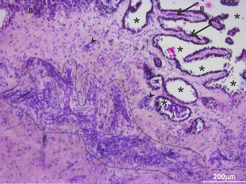
FIGURE 5. Representative haemotoxylin-eosin stained tangential limbal section.
(n = 3 sections of different donor eyes) showing the presence of a large number of stromal cells either as clusters (highlighted region, arrow head) or as individual cells adjacent to basal epithelial cells. Such cell clusters were also observed in the interpalisade region (arrow). Location of * indicates the epithelial region wherein the epithelial cells might have been lost in the donor eye; B - basal epithelium.

